ABSTRACT
Foot-and-mouth disease (FMD) is an acute epidemic that spreads rapidly among cattle and pigs. In 2014, in Korea, despite enforced vaccination, the type O Southeast Asia (SEA) topotype viruses (Mya-98 lineage) infected mainly cattle and pigs simultaneously, thereby causing enormous damage. If a vaccine that is completely protective against this FMD virus is developed and used, it can become a very important preventive measure in Asia, which is where this type of virus mainly circulates. The SEA topotype has been steadily evolving and transforming into new variations since it became epidemic in Asia. Therefore, it became necessary to develop a new vaccine that could provide protection against the FMD virus strain that was responsible for the 2014–2015 outbreak in Korea. This study aimed to develop a vaccine that would provide complete protection against the SEA topotype FMD virus to control sporadic FMD outbreaks, which occur despite the enforcement of vaccination, and to completely prevent virus shedding, thereby preventing the virus from spreading. The vaccine candidate virus developed in this study showed low pathogenicity and can be distinguished from the wild-type FMD virus strain. The developed vaccine was able to protect mice from SEA and Middle East–South Asia topotype virus strains and induced high titers of antibodies against both virus strains in pigs, thereby confirming the sufficiency of its protective function. In particular, the results of the SEA topotype virus challenge test in pigs revealed that perfect immunity was created in the vaccinated pigs, without virus shedding and viremia.
Keywords: Foot-and-mouth disease; type O; vaccine; Korea
INTRODUCTION
There are 7 serotypes of the foot-and-mouth disease virus (FMDV), of which, globally, the O serotype most frequently causes a foot-and-mouth disease (FMD) outbreak [1,2]. The O serotype circulates in most FMD outbreak areas, including Asia, Middle East, India, Africa, and South America. Given that in recent years, Asia has mainly experienced outbreaks of Southeast Asia (SEA) and Middle East–South Asia (ME-SA) topotype-mediated FMD, protection from these topotypes is urgently required [3]. The Cathay topotype strain has
Original Article
Received: Mar 29, 2019 Revised: Apr 24, 2019 Accepted: Jun 13, 2019
*Corresponding author:
Jong-Hyeon Park
Center for Foot-and-Mouth Disease Vaccine Research, Animal and Plant Quarantine Agency, 177 Hyeoksin 8-ro, Gimcheon 39660, Korea.
E-mail: [email protected]
© 2019 The Korean Society of Veterinary Science
This is an Open Access article distributed under the terms of the Creative Commons Attribution Non-Commercial License (https://
creativecommons.org/licenses/by-nc/4.0) which permits unrestricted non-commercial use, distribution, and reproduction in any medium, provided the original work is properly cited.
ORCID iDs Hye-Eun Jo
https://orcid.org/0000-0003-4920-1893 Mi-Kyeong Ko
https://orcid.org/0000-0002-9714-4232 Joo-Hyung Choi
https://orcid.org/0000-0001-6026-6095 Sung Ho Shin
https://orcid.org/0000-0001-9060-5131 Hyundong Jo
https://orcid.org/0000-0003-2739-8904 Su-Hwa You
https://orcid.org/0000-0001-9679-7445 Min Ja Lee
https://orcid.org/0000-0002-5916-5083
Hye-Eun Jo , Mi-Kyeong Ko , Joo-Hyung Choi , Sung Ho Shin ,
Hyundong Jo , Su-Hwa You , Min Ja Lee , Su-Mi Kim , Byounghan Kim , Jong-Hyeon Park
*Center for Foot-and-Mouth Disease Vaccine Research, Animal and Plant Quarantine Agency, Gimcheon 39660, Korea
New foot-and-mouth disease vaccine, O JC-R, induce complete protection to pigs against SEA topotype viruses occurred in South Korea, 2014–2015
Microbiology
Su-Mi Kim
https://orcid.org/0000-0002-9242-2731 Byounghan Kim
https://orcid.org/0000-0003-0559-0997 Jong-Hyeon Park
https://orcid.org/0000-0003-0825-8121 Funding
This study was supported by the Animal and Plant Quarantine Agency, Gimcheon, Gyeongsangbuk-do, Republic of Korea.
Conflict of Interest
The authors declare no conflicts of interest.
Author Contributions
Conceptualization: Park JH, Kim B, Jo HE, Ko MK; Data curation: Jo HE, Ko MK, Shin SH; Formal analysis: Jo HE, Lee MJ; Funding acquisition: Park JH, Kim B; Investigation: Park JH, Kim B, Jo HE; Methodology: Jo HE, Shin SH, You SH; Project administration: Lee MJ, Kim SM; Resources: Ko MK, Choi JH; Software:
Ko MK, Choi JH; Supervision: Park JH, Kim B; Validation: Jo HE; Visualization: Jo HE;
Writing - original draft: Jo HE, Park JH; Writing - review & editing: Park JH, Kim B.
also caused outbreaks, but only in a few Asian regions. Recently, all 3 topotype strains were responsible for FMD outbreaks in China and Southeast Asian countries; thus, the development of vaccines capable of protecting against all 3 topotypes has become an important issue in Asia [4]. In East Asia, of the O serotype strains, the Pan-Asia and Ind2001 genotypes of the ME-SA topotype, the Mya-98 genotype of the SEA topotype, and the Cathay topotype are causing outbreaks [3].
The vaccines developed against the SEA topotype (Mya-98), which was responsible for the 2014 outbreak in Korea, were international vaccine strains such as O Manisa + O 3039, O Primorsky, and O Campos; these vaccines were determined to be able to protect against the SEA topotype on the basis of vaccine-matching experiments and neutralization test conducted in the laboratory [5,6]. However, although these vaccine strains provided protection, some vaccine strains belonging to different topotypes induced insufficient antibody reactions after immunization or induced delayed immunity. Previously, an FMD vaccine was developed using the virus that caused the 2010 (November) outbreak in Korea; this vaccine was evaluated for its immune response against the virus strain that caused a recent outbreak, and it was observed that it has a different immune reactivity, one that induced almost no production of antibodies [7]. Therefore, this result suggested that the same SEA topotype vaccine strains do not always guarantee protection. Further, because immunoreactivity in pigs tends to be lower than that of cattle, vaccines producing perfect immune responses in cattle may not suffice in pigs [8].
As seen in Korea, where a larger number of FMD outbreaks have occurred in pigs since 2014 [6]. In this respect, vaccines that can induce a more robust immune response against the SEA topotype in pigs are expected to have roles as effective FMD vaccines in the event of an FMD recurrence, mediated by the same topotype.
In Korea, a SEA topotype FMDV outbreak lasted for 5 months—from December 2014 to April 2015 [6]—but subsequently, no virus strains similar to this SEA topotype were observed until an outbreak from January to March in 2016 [9,10]. Unlike other virus strains, we speculated that this FMDV strain would not be eradicated, but it persisted for 3–5 months after the first cases in 2014 and 2016.
Thus, the present study aimed to develop a vaccine that can provide protection from this SEA topotype virus. In addition, a virus neutralization test (VNT) was conducted in order to determine whether the vaccine developed in this study can provide protection from the ME-SA, SEA, and Cathay topotypes that are prevalent in the Asia region; moreover, animal experiments were conducted to determine if the developed vaccine can protect against other topotypes and to establish whether the vaccine can protect animals from the O/Jincheon/
SKR/2014 virus.
MATERIALS AND METHODS
Preparation of the infectious clone
An infectious complementary DNA (cDNA) plasmid, which was already secured by
removing the 3B
1B
2site and manipulating the site into 3B
3B
3, was used; in addition, an
infectious clone where the 142nd residue (C142T) in the 3C region was manipulated,
such that C was replaced by T was used. The O/Jincheon/SKR/2014 virus was used
to prepare cDNA, and sense (5′-GGAGCCGGGCAATCCAGTCCG-3′) and antisense
(5′-CTGCTTTACAGGTGCCACTATTTTC-3′) primers were used in the polymerase chain
reaction (PCR) used to amplify the P1 region. The infectious clones were produced using the same method [11].
Virus recovery and cell culture
In order to secure the FMDVs from the infectious clone, experiments were conducted using a previously described experimental method[12].
The recombinant plasmid (pO-JC-R) containing the P1 region of O/Jincheon/SKR/2014 (O JC; GenBank KX162590) was reacted with restriction enzyme Spe I (NEB, USA) for 24 h at 37°C to divide the gene into a single fragment. Thereafter, baby hamster kidney (BHK) T7-9 cells (a cell line in which T7 RNA polymerase is expressed) were transfected with the purified DNA using lipofectamine 2000 (Invitrogen, USA) and cultivated for 2–3 days; then, the O JC-R virus was secured. Thereafter, the secured viruses were multiplied through successive passages using ZZ-R cells (fetal goat tongue epithelium cells) or BHK-21 cells.
In order to produce antigens for use in vaccine preparation, the viruses were multiplied using BHK21-suspension cells, which are the cell types required to produce FMDV antigens. Sixteen h after virus infection, the viruses were inactivated by 0.003 N of binary ethylenimine for 24 h and concentrated with polyethylene glycol 6000 (81260; Sigma Aldrich, USA).
The concentrated antigen was layered on 15%–45% sucrose-density gradients and
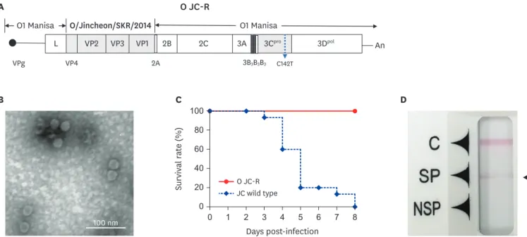
centrifuged. After ultracentrifugation, the bottom of the centrifuge tube was punctured, and 1 mL fractions were collected. As done in the previous study [12], the final inactivated antigen (FMD viral particles) was examined by using transmission electron microscopy (Fig. 1).
Differentiation from wild-type strains was confirmed by using a lateral flow device for FMD antigens (Princeton BioMeditech Corporation, USA).
D B
100 nm
O JC-R JC wild type
C
1
0 7 8
40 100
Survival r at e (%)
Days post-infection 80
20 60
2 3 4 5 6
0
A O JC-R
L An
VPg 3B
3B
3B
3O1 Manisa O/Jincheon/SKR/2014 O1 Manisa
VP2
VP4 2A
C142TVP3 VP1 2B 2C 3A 3C
pro3D
polFig. 1. Characterization of a type O FMD vaccine strain, O JC-R virus. (A) Schematic diagram of the type O JC-R FMDV genome; the 3B
1B
2replacement was performed by using the method given by Ko et al. [11] and the 142 residue (C142T) in the 3C region was mutated. (B) Electron microscopy of the FMDV vaccine strain, O JC-R.
The bar represents 100 nm. (C) Pathogenesis of O JC-R and O JC wild-type (O/Jincheon/SKR/2014) or mutated viruses in 7-days-old mice. (D) No detection of the NSP antigen in virus cultured supernatant when using the FMDV antigen rapid kit (PBM, USA) for differentiation between vaccine and wild-type virus using the previous method [11].
C, control line; SP, FMDV structural protein line; NSP, FMDV non-structural protein line; FMD, foot-and-mouth disease; FMDV, foot-and-mouth disease virus.
Preparation of experimental vaccine
The vaccine was prepared using the method described in Ko et al.'s [11] study. To briefly explain the method, 15 μg (1 dose) of purified 146S antigen of inactivated O JC-R virus was mixed with ISA206VG (Seppic, France) to a ratio of 1:1 (volume [v]/v); then, 10% aluminum hydroxide gel (Rehyragel HPA; General Chemical, USA) and saponin (0.5 μg) were added to that mixture to prepare the vaccine in a water-in-oil-in-water form.
Pathogenicity in young mice
Seven-day-old Institute of Cancer Research (ICR) mice, supplied by the Orient (Korea), were used for this experiment. The animals were kept at the Animal and Plant Quarantine Agency (APQA) and were used with the approval of the Animal Care and Use Committee of APQA (# 2017-627).
The mice were divided into 2 challenge groups (n = 12 per group) and were administered 0.1 mL of 1 × 10
550% tissue culture infective dose (TCID
50) of O JC-R virus or O JC wild-type virus by intraperitoneal (IP) injection. All mice were observed for 7 days after the challenge.
Virus challenge after vaccination in adult C57BL/6 mice
Seven-week-old C57BL/6 female mice, supplied by the KOSA BIO (Korea), were used for this experiment. The animals were kept at the APQA and were used with the approval of the Animal Care and Use Committee of APQA. The mice were challenged by IP injection of 0.1 mL of O JC or O Vet 2013 (O Vet, ME-SA/PanAsia lineage) virus at 100 50% of a lethal dose (LD
50);
the actual dose was 1 × 10
5TCID
50. All mice were observed for 7 days after the challenge.
Immunogenicity of the vaccine in pigs
For the immunogenicity test, 6 FMDV antibody–negative 3-month-old-farm pigs were used. Once the pigs (n = 4) were inoculated with the test vaccine, their blood was collected at intervals of 0, 7, 14, 21, and 28 days, and additionally at 0–14 days after the challenge to measure the presence of FMDV antibodies. Briefly, the neutralizing antibody titers in the serum were measured using the VNT specified in the Manual of Diagnostic Tests and Vaccines for Terrestrial Animals of the World Organisation for Animal Health. Serum samples were collected from the animals after vaccination and after the virus challenge.
The sera were heat inactivated at 56°C for 30 min. Then, following 1 h incubation in a serial diluted sera and virus suspension, LF-BK cells were added to the microplate and the plate incubated for a period of 3 days. The neutralizing antibody titers were calculated as log10 of the reciprocal antibody dilution to neutralize the 100 TCID
50of the virus.
Challenge test after vaccination in pigs
The experimental O JC-R vaccines were prepared with 146S antigen at different inoculation amounts of 15 μg while keeping the other compositions unchanged. The test animals were divided into 2 groups: 4 animals in the O JC-R vaccinated group (1 dose: 15 μg) and 2 animals in the control group. Blood was collected at 0, 7, 14, 21, and 28 days after vaccination, and the pigs in all groups were challenged with FMDV O/Jincheon/SKR/2014 at a titer of 10
5TCID
50on their tongues at 28 days after vaccination. After the virus challenge, the pigs in individual groups were separately raised; when clinical symptoms of FMD appeared, they were isolated.
Oral swabs and sera were collected in the period from 0 days post challenge (dpc) to 7 dpc.
The blood samples were collected by venipuncture (anterior vena cava) and placed into
Vacutainer serum tubes (BD Biosciences, USA). The oral swabs were collected using the
BD
™Universal Viral Transport Kit (BD Biosciences). The FMDV viral RNA was identified
by extracting the viral RNA from oral swab samples and performing quantitative real-time
reverse transcription (RT)-PCR. The MagNapure 96 system (Roche, Germany) was used
for the extraction of the viral RNA, and the quantitative real-time RT-PCR was conducted using the same method described in a previous experiment[11]. Clinical observations were performed daily after the challenge. An animal's clinical score was determined by the addition of points as described in a previous study [12]. Briefly, clinical scores were calculated using the following criteria, for a maximum of 15 points: 1) elevated body temperature of 40°C (1 point), > 40.5°C (2 points), and > 41°C (3 points); 2) lethargy (1 point); 3) hoof and foot vesicles (1–2 points per foot); and 4) snout, lips, and tongue vesicles (1 point for each affected area). The non-structural protein (NSP) enzyme-linked immunosorbent assay (ELISA) kit (Bionote, Korea)—an ELISA kit for the detection of FMDV NSP antibodies in serum samples of pigs and bovines—was utilized to detect NSP antibodies.
Virus detection in immunized and challenged pigs
Real-time RT-PCR was performed on the sera and swab samples of the experimental animals to detect FMDV. Swab samples were collected from the mouth and nose areas using cotton swabs.
Total cellular RNA was extracted using the MagNa pure 96 system (Roche) in accordance with the manufacturer's protocol. Real-time RT-PCR was conducted using the 1-step prime-script RT-PCR kit (Bioneer, Korea) in accordance with the manufacturer's instructions.
Statistical analysis
The statistical relationships between the inoculated groups and negative control groups were determined. The t-tests and log-rank tests were conducted by using GraphPad Prism (Ver 5.0;
GraphPad Software, USA) and GraphPad Instant (Ver 3.05; GraphPad Software).
RESULTS
Characteristics and pathogenicity of the vaccine strain
Genetic modifications were introduced into the FMD vaccine strain to differentiate it from the wild-type strain, and a mutation was introduced into the 3C region, which is the gene related to pathogenicity (Fig. 1A). The FMD vaccine virus developed in this study was confirmed to have the expected size of FMDV (25 nm) (Fig. 1B). The developed virus was subjected to a pathogenicity test in 7-days-old suckling mice. No pathogenicity was observed in the mice infected with the virus containing the C142T replacement in the 3C gene, while the wild-type O/Jincheon/SKR/2014 virus showed pathogenicity. Further, the virus strain where only 3B was replaced also retained the same pathogenicity (Fig. 1C). In the newly developed vaccine virus, 3B was changed (from 3B
1B
2to 3B
3B
3), which made it distinguishable from the wild-type strain; the developed vaccine strain could also be differentiated with wild- type virus by its unresponsiveness to NSP when tested with a lateral flow device (Fig. 1D).
Challenge test in mice after immunization
When challenged with the O/Jincheon/SKR/2014 (SEA topotype) virus 21 days after FMD vaccination, the mice vaccinated with the 1/10 to 1/640 doses, which were diluted to the dose levels for cattle and pigs, showed protection from the virus (Fig. 2A and B). When mice were inoculated with a single dose (0.1 mL; 1/10th of the dose for pig and cattle, 1.5 µg), the 50%
mouse protective dose (mPD50) was determined to be more than 128. When challenged with
O Vet 2013 virus (the ME-SA topotype), the mice showed an 80% survival rate at a 1/640 dose
with a 97 mPD50 (Fig. 2C and D). Further, when challenged with the O/Jincheon/SKR/2014
virus 7 days after immunization, the mouse group showed a survival rate of 80% at a 1/640
dose with a 97 mPD50 (Fig. 2E and F).
Challenge test in pigs after immunization
Pigs were challenged with the O/Jincheon/SKR/2014 (SEA topotype) virus 28 days after immunization with the experimental vaccine (Fig. 3). Symptoms began to appear in the unimmunized control animals 2–3 days after the challenge; the highest clinical index was observed 4–5 days after the challenge, and viremia was the severest 3 days after the challenge (Fig. 3A). Further, virus shedding levels were the highest 6–7 days after the challenge. The immunized animals showed no clinical signs and no evidence of viremia or viral shedding (Fig. 3B).
21 DPV/O JC challenge A
1 7
40 100
Per cent survival (%)
DPC 80
20 60
2 3 4 5 6
1/10 dose 1/40 dose 1/160 dose 1/640 dose Negative control 0
21 DPV/O JC challenge B
0 7
90 120
Per cent body weight (%)
DPC 110
80 100
2 3 4 5 6
1/10 dose 1/40 dose 1/160 dose 1/640 dose Negative control 70
1/10 dose 1/40 dose 1/160 dose 1/640 dose Negative control
21 DPV/O Vet challenge C
1 7
40 100
Per cent survival (%)
DPC 80
20 60
2 3 4 5 6
0
1/10 dose 1/40 dose 1/160 dose 1/640 dose Negative control
21 DPV/O Vet challenge D
0 7
90 120
Per cent body weight (%)
DPC 110
80 100
2 3 4 5 6
70
1/10 dose 1/40 dose 1/160 dose 1/640 dose Negative control
7 DPV/O JC challenge E
1 0
0
0 7
40 100
Per cent survival (%)
DPC 80
20 60
2 3 4 5 6
0
1/10 dose 1/40 dose 1/160 dose 1/640 dose Negative control
7 DPV/O JC challenge F
0 1
1
1 7
90 120
Per cent body weight (%)
DPC 110
80 100
2 3 4 5 6
70
Fig. 2. Survival and weight loss in vaccinated adult C57BL/6 mice after homologous or heterologous challenge after vaccination. The mice were vaccinated with the 1/10 to 1/640 doses (diluted to 1 dose for cattle and pigs). (A) Survival rate in O JC-R-vaccinated adult C57BL/6 mice (n = 5) after the O/Jincheon/SKR/2014 challenge at 21 DPV. (B) Body weight in O JC-R-vaccinated adult C57BL/6 mice (n = 5) after the O/Jincheon/SKR/2014 challenge at 21 DPV, (C) Survival rate in O JC-R-vaccinated adult C57BL/6 mice (n = 5) after the O Vet 2013 challenge at 21 DPV, (D) Body weight in O JC-R-vaccinated adult C57BL/6 mice (n = 5) after the O Vet 2013 challenge at 21 DPV, (E) Survival rate in O JC-R-vaccinated adult C57BL/6 mice (n = 5) after the O/Jincheon/SKR/2014 challenge at 7 DPV. (F) Body weight in O JC-R-vaccinated adult C57BL/6 mice (n = 5) after the O/Jincheon/SKR/2014 challenge at 7 DPV.
DPC, days post challenge; DPV, days post vaccination.
Antibody titers and cross-VNT results in immunized pigs
The ELISA results showed that antibody levels increased 2 weeks after the immunization with the experimental vaccine and peaked in the third week after the immunization, which was then followed by a decline in the percent inhibition value (Fig. 4A). In the VNT for the SEA topotype (Mya-98) virus, the neutralizing antibody titers were determined to be more than 1:100 in the fourth week (Fig. 4B), which was similar to the antibody titers of the ME-SA (Ind
#3–6 #4–5
0
0 1 2 3 4 5 6 7 8 9 10 0
8
16 6
5 4
Clinical scor e 2 Cop y number s of vir al RNA ( log)
4 12
3
1
A Control group (n = 2)
0
0 1 2 3 4 5 6 7 8 9 10 0
8
16 6
5 4
Clinical scor e 2
DPI DPI
Cop y number s of vir al RNA ( log)
4 12
3
1 Serum
Clinical score Swab
Serum Clinical score Swab
#JC-1 #JC-3
0
0 1 2 3 4 5 6 7 0
5
15 6
5 4
Clinical scor e 2 Cop y number s of vir al RNA ( log)
10
3
1
B Vaccination group (n = 4)
0
0 1 2 3 4 5 6 7 0
5
15 6
5 4
Clinical scor e 2
DPI DPI
Cop y number s of vir al RNA ( log)
10
3
1
#JC-5 #JC-6
0
0 1 2 3 4 5 6 7 0
5
15 6
5 4
Clinical scor e 2 Cop y number s of vir al RNA ( log)
10
3
1
0
0 1 2 3 4 5 6 7 0
5
15 6
5 4
Clinical scor e 2
DPI DPI
Cop y number s of vir al RNA ( log)
10
3
1 Serum
Clinical score Swab
Serum Clinical score Swab
Serum Clinical score Swab
Serum Clinical score Swab
Fig. 3. Clinical scores, viremia, and virus shedding in O JC-R-immunized pigs after the O/Jincheon/SKR/2014 virus challenge. (A) Virus-neutralizing antibody titers in immunized pigs for the virus challenge. The O/Jincheon/SKR/2014 virus challenge was attempted at 28 days post challenge. (B) Clinical scores and virus detection in sera and oral swabs for the negative group (n = 2, #3–6, #4–5) and vaccinated group (n = 4, #JC-1, #JC-3, #JC-5, #JC-6).
DPI, days post-infection.
2001) virus (Fig. 4C). Further, in the VNT for the Cathay topotype, the antibody titers were the highest in the fourth week after immunization, but the neutralizing antibody titers were lower than those of the SEA and ME-SA viruses (Fig. 4D).
DISCUSSION
The type O FMDV is the most prevalent FMDV serotype in the world, and FMD outbreaks mediated by the SEA (Mya-98), ME-SA (Pan-Asia, Ind2001), and Cathay topotypes have been reported in Asia [3,13,14]. Among these, the SEA topotype strains have caused simultaneous FMD outbreaks in pigs and cattle. Recently, an FMD of ME-SA topotype occurred mainly in cattle while the Cathay topotype FMD occurred mainly in pigs [6,14]. Therefore, it became necessary to formulate a measure that would protect both cattle and pigs. However, in general, the immunogenicity of pigs tends to be lower than that of cattle; thus, if protection for pigs is achieved, protection for cattle can also be achieved without problem. Because the FMD incidence rate in 2014 was much higher in pigs than in cattle in Korea [6], in the present study, the protection ability in pigs against FMDV was examined [6]. In this study, a vaccine strain expressing the external surface protein of the SEA virus responsible for the 2014 FMD
Type O SP-ELISA O/Jincheon/SKR/2014 (SEA)
0 40 100
Per cent inhibition (%)
80
A B
0 7 14 21 28 1 8
2.0 3.5
VN tit er s ( log 10 )
DPV DPV/DPI
3.0
20 60
1.5 2.5
2 3 4 5 6 7 Control group
Vaccination group (JC-R) Control group
Vaccination group
Challenge
‡
O/Boeun/SKR/2017 (ME-SA) O Taiwan97 (Cathay)
C D
1.0
0 7 14 21 28 1 14
2.0 3.5
VN tit er s ( log 10 )
DPV/DPI 3.0
1.5 2.5
1.0 2.0 3.5
VN tit er s ( log 10 ) 3.0 1.5 2.5
2 3 4 5 6 7
0 7 14 21 28 1 14
DPV/DPI
2 3 4 5 6 7
Negative control Vaccination group (JC-R) Negative control
Vaccination group (JC-R) Challenge
Challenge
0 7 14 21 28
†
Fig. 4. Antibody titers in pigs vaccinated with the experimental foot-and-mouth disease vaccine, O JC-R. (A) Type O SP-ELISA results. (B) VN titers against O/
Jincheon/SKR/2014. (C) VN titers against O/Boeun/SKR/2017. (D) VN titers against O Taiwan97. The dotted lines in SP-ELISA results indicate 50% inhibition, which is the positive cut-off in the test. The dotted lines in the VN test show 1.5 log VN titers (1:32). The arrows indicate the challenge time at 28 dpv.
ELISA, enzyme-linked immunosorbent assay; SEA, Southeast Asia; ME-SA, Middle East–South Asia; VN, virus-neutralizing; DPV, days post challenge; DPI, days post-infection.
*
p < 0.05;
†p < 0.01;
‡p < 0.001.
outbreak was constructed and tested in pigs; the results confirmed that the newly developed vaccine was able to fully protect pigs against the wild-type FMDV strain.
A recent SEA topotype FMDV infects both cattle and pigs, causing problems in Asian countries, including Korea. There have been several outbreaks of the SEA topotype virus strain in Korea: in April and November 2010 and in July and December 2014—the latter outbreak lasted until April 2015. In January 2016, another FMD outbreak was reported;
however, the SEA topotype virus disappeared from Korea after the last occurrence at the end of March 2016 [6,9,10,15,16]. The ME-SA/Ind2001 lineage FMDV occurred in Korea in 2017 and 2019, although the transmission route of this virus was determined to be an Ind2001 virus spread from the pool 2 region [14].
In Korea, after the first FMD outbreak in 2000 and following a 66-year FMD-free period, mainly type O-mediated FMD outbreaks have occurred; among the 9 outbreak occurrences, SEA was responsible for 5 occurrences and ME-SA was responsible for 4 occurrences [14- 18]. Thus, it is desirable to use vaccines that can protect against at least 2 different topotype strains in Korea. Previously, a vaccine was developed using the virus responsible for the SEA topotype outbreak in November 2010 in Korea; however, that vaccine showed low immunity from the virus that caused the 2014 outbreak [7].
Based on the results of the VNT and the challenge test, the vaccine strain developed in this study is believed to be able to provide protection against both SEA and ME-SA topotypes.
Antibody titers against the Cathay topotype virus were somewhat lower, but 75% of the animals had a VNT level of 1:45 or higher, which is generally accepted as a protective antibody level. This suggests that sufficient protection would be provided in most vaccination cases, but a secondary immunization is required. This claim could be supported by the results of the FMDV challenge test after immunization, which showed that animals were protected at low antigen concentrations after the challenge of both the SEA and ME-SA topotype FMDV strains, as predicted by the challenge test results in mice.
In conclusion, it was confirmed that pigs immunized with the vaccine strain developed in this study were completely protected without virus excretion and viremia against the SEA topotype virus; moreover, sufficient neutralizing antibody titers that can protect against the ME-SA topotype virus at the same level as that for the SEA virus circulating in SEA were detected.
ACKNOWLEDGMENTS
We thank the staff of the Center for FMD Vaccine Research and Mr. Jung-Won Park for providing assistance with electron microscopy at the Animal and Plant Quarantine Agency.
REFERENCES
1. Brito BP, Rodriguez LL, Hammond JM, Pinto J, Perez AM. Review of the global distribution of foot-and- mouth disease virus from 2007 to 2014. Transbound Emerg Dis 2017;64:316-332.
PUBMED | CROSSREF
2. Diaz-San Segundo F, Medina GN, Stenfeldt C, Arzt J, de Los Santos T. Foot-and-mouth disease vaccines.
Vet Microbiol 2017;206:102-112.
PUBMED | CROSSREF
3. Mahapatra M, Upadhyaya S, Aviso S, Babu A, Hutchings G, Parida S. Selection of vaccine strains for serotype O foot-and-mouth disease viruses (2007–2012) circulating in Southeast Asia, East Asia and Far East. Vaccine 2017;35:7147-7153.
PUBMED | CROSSREF
4. Cao Y, Lu Z, Li D, Fan P, Sun P, Bao H, Fu Y, Li P, Bai X, Chen Y, Xie B, Liu Z. Evaluation of cross- protection against three topotypes of serotype O foot-and-mouth disease virus in pigs vaccinated with multi-epitope protein vaccine incorporated with poly(I:C). Vet Microbiol 2014;168:294-301.
PUBMED | CROSSREF
5. Galdo Novo S, Malirat V, Maradei ED, Pedemonte AR, Espinoza AM, Smitsaart E, Lee KN, Park JH, Bergmann IE. Efficacy of a high quality O
1/Campos foot-and-mouth disease vaccine upon challenge with a heterologous Korean O Mya98 lineage virus in pigs. Vaccine 2018;36:1570-1576.
PUBMED | CROSSREF
6. Park JH, Tark D, Lee KN, Chun JE, Lee HS, Ko YJ, Kye SJ, Kim YJ, Oem JK, Ryoo S, Lim SB, Lee SY, Choi JH, Ko MK, You SH, Lee MH, Kim B. Control of type O foot-and-mouth disease by vaccination in Korea, 2014–2015. J Vet Sci 2018;19:271-279.
PUBMED | CROSSREF
7. Park ME, You SH, Lee SY, Lee KN, Ko MK, Choi JH, Kim B, Lee JS, Park JH. Immune responses in pigs and cattle vaccinated with half-volume foot-and-mouth disease vaccine. J Vet Sci 2017;18:323-331.
PUBMED | CROSSREF
8. Park JH. Requirements for improved vaccines against foot-and-mouth disease epidemics. Clin Exp Vaccine Res 2013;2:8-18.
PUBMED | CROSSREF
9. Kim T, Ryoo S, Nah JJ, Sagong MG, Lee S, Lee KN, Ko YJ, Park JH, Lee MH, Wee SH, Tark D, Ku BK.
Complete genome sequence of a foot-and-mouth disease virus of serotype O, isolated from Gochang, Republic of Korea, in 2016. Genome Announc 2017;5:e01671-e16.
PUBMED | CROSSREF
10. Ryoo S, Kim T, Nah JJ, Sagong MG, Lee S, Lee KN, Ko YJ, Park JH, Lee MH, Wee SH, Tark D, Ku BK.
Complete genome sequence of a foot-and-mouth disease virus of serotype O Isolated from Gimje, Republic of Korea, in 2016. Genome Announc 2017;5:e01694-e16.
PUBMED | CROSSREF
11. Ko MK, Jo HE, Choi JH, You SH, Shin SH, Jo H, Lee MJ, Kim SM, Kim B, Park JH. Chimeric vaccine strain of type O foot-and-mouth disease elicits a strong immune response in pigs against ME-SA and SEA topotypes. Vet Microbiol 2019;229:124-129.
PUBMED | CROSSREF
12. Lee SY, Lee YJ, Kim RH, Park JN, Park ME, Ko MK, Choi JH, Chu JQ, Lee KN, Kim SM, Tark D, Lee HS, Ko YJ, Seo MG, Park JW, Kim B, Lee MH, Lee JS, Park JH. Rapid engineering of foot-and-mouth disease vaccine and challenge viruses. J Virol 2017;91:e00155-e17.
PUBMED | CROSSREF
13. Le VP, Nguyen T, Park JH, Kim SM, Ko YJ, Lee HS, Nguyen VC, Mai TD, Do TH, Cho IS, Lee KN.
Heterogeneity and genetic variations of serotypes O and Asia 1 foot-and-mouth disease viruses isolated in Vietnam. Vet Microbiol 2010;145:220-229.
PUBMED | CROSSREF
14. Zhu Z, Yang F, He J, Li J, Cao W, Li J, Xia Y, Guo J, Jin Y, Zhang K, Zheng H, Liu X. First detection of foot- and-mouth disease virus O/ME-SA/Ind2001 in China. Transbound Emerg Dis 2018;65:2027-2031.
PUBMED | CROSSREF
15. Park JH, Lee KN, Ko YJ, Kim SM, Lee HS, Park JY, Yeh JY, Kim MJ, Lee YH, Sohn HJ, Moon JS, Cho IS, Kim B. Outbreaks and diagnosis of foot-and-mouth disease serotype O in the Republic of Korea, April–June 2010. Transbound Emerg Dis 2014b;61:277-284.
PUBMED | CROSSREF
16. Park JH, Tark D, Lee KN, Lee SY, Ko MK, Lee HS, Kim SM, Ko YJ, Seo MG, Chun JE, Lee MH, Kim B.
Novel foot-and-mouth disease virus in Korea, July–August 2014. Clin Exp Vaccine Res 2016;5:83-87.
PUBMED | CROSSREF
17. Knowles NJ, He J, Shang Y, Wadsworth J, Valdazo-González B, Onosato H, Fukai K, Morioka K, Yoshida K, Cho IS, Kim SM, Park JH, Lee KN, Luk G, Borisov V, Scherbakov A, Timina A, Bold D, Nguyen T, Paton DJ, Hammond JM, Liu X, King DP. Southeast Asian foot-and-mouth disease viruses in Eastern Asia.
Emerg Infect Dis 2012;18:499-501.
PUBMED | CROSSREF
18. Park JH, Lee KN, Kim SM, Lee HS, Ko YJ, Tark DS, Shin YK, Seo MG, Kim B. Reemergence of foot-and- mouth disease, South Korea, 2000–2011. Emerg Infect Dis 2014;20:2158-2161.
PUBMED | CROSSREF
19. Oem JK, Yeh MT, McKenna TS, Hayes JR, Rieder E, Giuffre AC, Robida JM, Lee KN, Cho IS, Fang X, Joo YS, Park JH. Pathogenic characteristics of the Korean 2002 isolate of foot-and-mouth disease virus serotype O in pigs and cattle. J Comp Pathol 2008;138:204-214.
PUBMED | CROSSREF