© 2018 The Korean Ophthalmological Society
This is an Open Access article distributed under the terms of the Creative Commons Attribution Non-Commercial License (http://creativecommons.org/licenses /by-nc/3.0/) which permits unrestricted non-commercial use, distribution, and reproduction in any medium, provided the original work is properly cited.
Original Article
Visual Recovery after Macular Hole Surgery and Related Prognostic Factors
Soo Han Kim1, Hong Kyu Kim2, Jong Yun Yang3, Sung Chul Lee2, Sung Soo Kim2
1Department of Ophthalmology, Yonsei University Wonju College of Medicine, Wonju, Korea
2Department of Ophthalmology, Institute of Vision Research, Severance Hospital, Yonsei University College of Medicine, Seoul, Korea
3Siloam Eye Hospital, Seoul, Korea
Purpose: To describe the visual recovery and prognostic factors after macular hole surgery.
Methods: A retrospective chart review was conducted. Charts of patients with idiopathic macular holes who un- derwent surgery by a single surgeon at Severance Hospital between January 1, 2013 and July 31, 2015 were reviewed. The best-corrected visual acuity (BCVA) score was recorded preoperatively and at 1 day and 1, 3, 6, 9, and 12 months after surgery. The variables of age, sex, macular hole size, basal hole diameter, choroidal thickness, and axial length were also noted.
Results: Twenty-six eyes of 26 patients were evaluated. Twenty-five patients (96.2%) showed successful macu- lar hole closure after the primary operation. The BCVA stabilized 6 months postoperatively. A large basal hole diameter (p = 0.006) and thin choroid (p = 0.005) were related to poor visual outcomes. Poor preoperative BCVA (p < 0.001) and a thick choroid (p = 0.020) were associated with greater improvement in BCVA after surgery.
Conclusions: Visual acuity stabilized by 6 months after macular hole surgery. Choroidal thickness was a pro- tective factor for final BCVA and visual improvement after the operation.
Key Words: Choroid, Retinal perforations, Visual acuity, Vitrectomy
Optical coherence tomography (OCT) technology has led to a deeper understanding of the pathophysiology of macu- lar holes (MHs) [1]. With advancements in surgical tech- niques, from gas tamponade to internal limiting membrane
staining with indocyanine green dye, the primary success rates of MH surgeries have steadily increased [2-4]. Some efforts have been made to determine the prognostic factors of favorable MH surgery outcomes [5-7]. To date, the size and stage of the MH, duration of symptoms, and preopera- tive visual acuity have been reported as preoperative fac- tors [5]; however, recovery of the macular contour, the ex- ternal limiting membrane (ELM), and the ellipsoid zone are known to affect recovery of vision after surgery [6,7].
It has been reported that MH patients have a thin cho- roid and decreased choroidal perfusion [8,9], but it remains unknown whether choroidal thickness or perfusion affect
Received: July 11, 2017 Accepted: September 4, 2017
Corresponding Author: Sung Soo Kim, MD, PhD. Department of Ophthalmology, Institute of Vision Research, Severance Hospital, Yonsei University College of Medicine, #50-1 Yonsei-ro, Seodaemun-gu, Seoul 03722, Korea. Tel: 82-2-2228-3570, Fax: 82-2-312-054, E-mail: [email protected] This study was presented as e-poster at Retina World Congress on Febru- ary 25, 2017 in Fort Lauderdale, FL, USA.
the surgical outcome. Additionally, there have been few reports about the time required for visual recovery after MH repair. Thus, in this study, we investigated the time to visual recovery after MH surgery and the related prognos- tic factors, including choroidal thickness.
Materials and Methods
The institutional review board of Yonsei University Col- lege of Medicine approved this study (4-2016-1033), which adhered to the tenets of the Declaration of Helsinki. All patients provided written informed consent before any procedure was performed. A retrospective chart review was performed for all patients with idiopathic MHs, who underwent surgery by a single surgeon (SSK) at Severance Hospital, Seoul, Korea, between January 1, 2013 and July 31, 2015. Patients were excluded if they were lost to fol- low-up within one year after surgery, developed a second- ary MH, had an axial length exceeding 26.5 mm, or under- went previous vitrectomy.
All patients underwent a thorough ophthalmic evalua- tion prior to surgery, including assessment of the best-cor- rected visual acuity (BCVA) and a fundus examination (Spectralis OCT; Heidelberg Engineering, Heidelberg, Germany). Patients with a full thickness MH were advised to undergo surgery. Surgery was performed using a stan- dard 3-port, 23-gauge pars plana vitrectomy. When a cata- ract was present, a combined phacovitrectomy was per- formed. To remove tangential traction, epiretinal membranectomy was performed when a premacular mem- brane was present. When there was no visible premacular membrane, the internal limiting membrane was peeled with indocyanine green staining. After removing tangen- tial traction around the MH, air-fluid exchange was per- formed, followed by 10% to 14% C3F8 tamponade. Patients were instructed to remain in a prone position for about one week after the surgery.
Patient age, sex, and involved eye were noted, and the BCVA was measured before surgery and at 1 day and 1, 3, 6, 9, and 12 months after the operation. Size of the MH, basal hole diameter, and subfoveal choroidal thickness were measured using the caliper function in the Heidelberg software (Fig. 1). The basal hole diameter of the MH was measured as the linear length of retinal detachment. Sub- foveal choroidal thickness was measured three times by a
single observer (KSH) with enhanced depth imaging, us- ing a method previously described by Boonarpha et al. [10], as the distance from the hyper-reflective line under the ret- inal pigment epithelium to the choroid-sclera interface.
Recovery of the ELM and the ellipsoid zone was moni- tored with serial OCT scans during follow-up. Axial length was measured preoperatively with the IOL-Master 500 (Carl Zeiss, Dublin, CA, USA).
Changes in BCVA were analyzed by repeated measures analysis of variance. Multivariate linear regression was performed to determine prognostic factors after MH re- pair. Fisher’s exact test and the Mann-Whitney U-test were used to compare characteristics and outcomes between the ELM and ellipsoid zone recovery. Reliability analysis was used to assess the reproducibility of subfoveal choroidal thickness measurements. Statistical analysis was per- formed using IBM SPSS ver. 21.0 (IBM Corp., Armonk, NY, USA). A p-value less than 0.05 was considered statis- tically significant.
Results
Twenty-six eyes of 26 patients were included in this study. Demographic factors and initial findings are listed in Table 1. The mean age of the patients was 61.6 years, and 73.1% (19 patients) were female. The mean BCVA was 0.981 in logarithm of the minimum angle of resolution scale prior to the operation. Visual acuity decreased to a mean of 3.077 1 day after the operation, due to gas filling.
All 26 patients completed all routine follow-up visits. The mean BCVA values at 1, 3, 6, 9, and 12 months were 0.797, 0.592, 0.495, 0.496, and 0.427, respectively. When these BCVA values were compared with the final (12-month postoperative) BCVA, preoperative vision and 1-day post- Fig. 1. Macular hole parameters measured in optical coherence tomography scans. a = hole size; b = basal hole size; c = choroidal thickness.
operative vision were found to be significantly different (p
< 0.001). Differences in BCVA were still significant at 1 and 3 months after the operation (1 month, p = 0.034; 3 months, p = 0.040). BCVA did not show improvements be- yond 6 months after surgery (6 months, p = 1.000; 9 months, p = 1.000) (Fig. 2).
Measurement of subfoveal choroidal thickness showed good reliability, with an intraclass correlation coefficient of 0.992 (p < 0.001). Postoperative choroidal thickness measured at 3, 6, 9, and 12 months was 227.3 (n = 22), 218.7 (n = 26), 214.2 (n = 25), and 223.6 (n = 23), respectively.
Thinning of the choroid was noted in all postoperative thickness measurements compared to the preoperative choroidal thickness measurement (3 months, p = 0.001; 6 months, p = 0.004; 9 months, p < 0.001; 12 months, p = 0.001). Changes in choroidal thickness did not correlate with preoperative BCVA (p = 0.427), 12-month postopera-
tive BCVA (p = 0.543), preoperative choroidal thickness (p
= 0.935), 12-month postoperative choroidal thickness (p = 0.394), or preoperative axial length (p = 0.687).
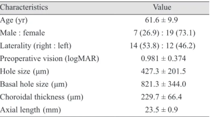
Table 1. Demographic characteristics and preoperative findings
Characteristics Value
Age (yr) 61.6 ± 9.9
Male : female 7 (26.9) : 19 (73.1)
Laterality (right : left) 14 (53.8) : 12 (46.2) Preoperative vision (logMAR) 0.981 ± 0.374
Hole size (μm) 427.3 ± 201.5
Basal hole size (μm) 821.3 ± 344.0
Choroidal thickness (μm) 229.7 ± 66.4
Axial length (mm) 23.5 ± 0.9
Values are presented as mean ± standard deviation or number (%).
logMAR = logarithm of the minimum angle of resolution.
Table 2. Preoperative factors affecting final visual acuity and changes in visual acuity
B coefficient p-value Final visual acuity
Sex 0.021 0.903
Age -0.162 0.835
Preoperative BCVA 0.173 0.371
Hole size -0.427 0.084
Basal hole size 0.471 0.006
Choroidal thickness -0.475 0.005
Axial length 0.062 0.707
Changes in visual acuity
Sex 0.000 0.999
Age 0.040 0.751
Preoperative BCVA 0.816 <0.001
Hole size -0.011 0.936
Basal hole size -0.269 0.057
Choroidal thickness 0.299 0.020
Axial length 0.006 0.961
BCVA = best-corrected visual acuity.
Fig. 2. Changes in best-corrected visual acuity (BCVA) after macular hole repair. logMAR = logarithm of the minimum angle of resolution. *p < 0.05, **p < 0.001.
BCVA (logMAR)
*
**
PreoperativeImmidiate 1 mom 3 mom 6 mom 9 mom 12 mom 3.5
3.0 2.5 2.0 1.5 1.0 0.5 0
Fig. 3. Analysis of correlation of choroidal thickness with (A) final best-corrected visual acuity (BCVA) and (B) changes in BCVA. logMAR = logarithm of the minimum angle of resolution.
Preoperaive subfoeval choroidal thickness
Preoperaive subfoeval choroidal thickness
12 mon BCVA ( logMAR)Changes in BCVA ( logMAR)
0 50 100 150 200 250 300 350 400 450
R2 = 0.2296
R2 = 0.2293 1.2
1 0.8 0.6 0.4 0.2 0
1.1 0.8 0.5 0.2 -0.1
-0.4 0 50 100 150 200 250 300 350 400 450
B A
To evaluate the factors affecting final BCVA, the vari- ables of sex, age, preoperative BCVA, MH size, basal hole diameter, choroidal thickness, and axial length were evalu- ated using multivariate linear regression analysis. This re- vealed that the final visual acuity was affected by basal hole diameter and choroidal thickness (R2 = 0.450, p = 0.001). A larger basal hole diameter was associated with a poor final BCVA (95% confidence interval [CI], 0.000 to 0.001). A greater preoperative choroidal thickness provided a protective effect, as patients with a thick choroid had a better final BCVA (95% CI, 0.000 to 0.001). When these factors were evaluated against changes in BCVA, preoper- ative BCVA and choroidal thickness were significantly as- sociated (R2 = 0.680, p < 0.001). Patients with poor preop- erative BCVA showed greater visual recovery (95% CI, 0.521 to 0.974). Choroidal thickness also had a positive re- lationship with visual acuity changes (95% CI, 0.000 to 0.003) (Table 2 and Fig. 3A, 3B).
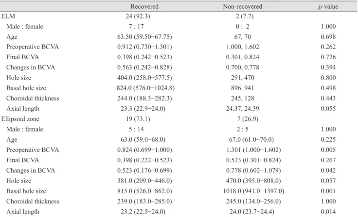
Twenty-four (92.3%) patients showed ELM recovery at a median of 1 month (1st to 3rd quartile, 1 to 4.50 months)
after surgery. No factors were significantly related to a dif- ference in ELM recovery. The ellipsoid zone recovered in 19 (73.1%) patients, by a median time of 3 months (1st to 3rd quartile, 1 to 8.0 months) after the operation. Patients with a recovered ellipsoid zone had better preoperative BCVA, smaller basal hole diameter, and shorter axial length. Although the final BCVA was better in the ellip- soid zone-recovered group, the difference was not statisti- cally significant (Table 3).
Discussion
In this study, we investigated the time to visual recovery after MH surgery and the related prognostic factors, in- cluding choroidal thickness. Previously, the closure rate after MH surgery was 58%, but advancements in surgical techniques and devices have led to an improvement in the closure rate to 89% to 100% [4,11,12]. In this study, we were able to achieve closure in 25 (96.2%) of 26 cases, us-
Table 3. Factors affecting ELM and ellipsoid zone recovery
Recovered Non-recovered p-value
ELM 24 (92.3) 2 (7.7)
Male : female 7 : 17 0 : 2 1.000
Age 63.50 (59.50−67.75) 67, 70 0.698
Preoperative BCVA 0.912 (0.730−1.301) 1.000, 1.602 0.262
Final BCVA 0.398 (0.242−0.523) 0.301, 0.824 0.726
Changes in BCVA 0.563 (0.242−0.828) 0.700, 0.778 0.394
Hole size 404.0 (258.0−577.5) 291, 470 0.800
Basal hole size 824.0 (576.0−1024.8) 896, 941 0.498
Choroidal thickness 244.0 (188.3−282.3) 245, 128 0.443
Axial length 23.3 (22.9−24.0) 24.37, 24.39 0.055
Ellipsoid zone 19 (73.1) 7 (26.9)
Male : female 5 : 14 2 : 5 1.000
Age 63.0 (59.0−68.0) 67.0 (61.0−70.0) 0.225
Preoperative BCVA 0.824 (0.699−1.000) 1.301 (1.000−1.602) 0.005
Final BCVA 0.398 (0.222−0.523) 0.523 (0.301−0.824) 0.267
Changes in BCVA 0.523 (0.176−0.699) 0.778 (0.602−1.079) 0.042
Hole size 381.0 (209.0−446.0) 470.0 (395.0−808.0) 0.057
Basal hole size 815.0 (526.0−862.0) 1018.0 (941.0−1397.0) 0.001
Choroidal thickness 239.0 (183.0−285.0) 245.0 (134.0−256.0) 1.000
Axial length 23.2 (22.5−24.0) 24.0 (23.7−24.4) 0.014
Values are presented as number (%), number, or median (25 percentile–75 percentile). Actual records of two patient is presented in non- recovered ELM group.
ELM = external limiting membrane; BCVA = best-corrected visual acuity.
ing a standard 23-gauge vitrectomy system. The single pa- tient with incomplete closure had a 395-µm sized MH, with a 1,397-µm basal hole diameter. He underwent a sec- ond operation 1 month after the first, during which the MH was successfully closed.
Shinoda et al. [13] reported visual recovery at 3 months after MH surgery in a 25-gauge vitrectomy group and at 9 months after surgery in a 20-gauge vitrectomy group. Less trauma to the ocular surface, a shorter operation time, and a reduced amount of irrigation fluid were considered to be reasons for the shorter time to visual recovery in the 25-gauge vitrectomy group. In this study, we performed 23-gauge vitrectomy, and vision stabilized by 6 months af- ter the operation. Leonard et al. [14] and Purtskhvanidze et al. [15] reported continuous visual improvement after MH surgery after following patients for up to 96 months. We observed continuous elevation in visual acuity for 6 months after the operation, and recovery of ELM and the ellipsoid zone was still observed at 35 and 38 months after surgery. However, changes in visual acuity were not statis- tically significant.
In 1986, Morgan and Schatz [16] hypothesized that the first step in the development of a MH is choroidal vascular change. They suggested that the majority of patients with an MH had cardiovascular disease, and decreased subfove- al choroidal circulation was noticed during fluorescein an- giography. Allen et al. [17] induced MHs in monkeys with an Nd-YAG laser, which resulted in a choriocapillaris per- fusion disorder. Histologically, the choriocapillaris was re- placed with fibroblasts and connective tissue. Other reports have also suggested that choroidal thickness is decreased in idiopathic MH patients compared to a control group when measured using OCT [8,9]. Aras et al. [18] have re- ported that patients with stage 4 and stage 1a MHs had re- duced subfoveal choroid circulation compared to controls, as determined using a scanning laser Doppler flowmeter. It is suspected that decreased choroidal circulation and a thinner choroid can cause macular hypoperfusion, and that deficits in several protective factors leave the macula vul- nerable to various types of damage [16,18]. In this study, choroidal thickness was not only related to the develop- ment of an MH, but final visual acuity and visual acuity change were also closely related. Choroidal thickness showed a protective effect on BCVA and on the degree of visual improvement obtained. We hypothesize that a thin- ner choroid can cause additional decreases in choroidal
circulation during compression by gas tamponade and can affect MH healing. If so, prolonged prone positioning of patients after surgery might have a hazardous effect on choroidal circulation. Further studies on the changes in choroidal circulation during the tamponade period are needed.
Choroidal thickness has been reported to decrease with age and with longer axial length [19,20]. It is also known that there is a diurnal variation of approximately 30 μm in choroidal thickness, and that the thickness is also affected by changes in blood pressure and drugs, such as sildenafil and steroids [21-23]. In this study, patients with an axial length exceeding 26.5 mm were excluded. However, fur- ther investigation of the relationship between choroidal thickness and visual prognosis in myopic MH patients is necessary. Choroidal thickness and choroidal circulation are not synonymous. Sogawa et al. [24] reported that cho- roidal blood flow was not correlated with choroidal thick- ness, as measured by laser Doppler, but their study sub- jects were young, healthy, and few in number.
ELM and ellipsoid zone recovery also affect visual re- covery after MH surgery [6,7]. We compared multiple fac- tors of the ELM and ellipsoid zone-recovered groups with those of the unrecovered group, but we found no factors affecting ELM recovery. Given that only two patients (7.7%) had unrecovered ELM, the small sample size might underlie the lack of statistically significant differences. In the case of ellipsoid zone recovery, patients with a larger basal hole diameter, poor initial visual acuity, and greater axial length had a poor recovery rate. The change in visual acuity was found to be greater in the group without recov- ery of the ellipsoid zone, but this was due to the differenc- es in preoperative visual acuity. Landa et al. [25] asserted that recovery of ELM is essential for ellipsoid zone recov- ery. We also analyzed OCT data and found that ellipsoid zone recovery was seen only after the ELM was restored.
There are several limitations to this study. First, we did not measure the choroidal circulation, and further investi- gation is needed to determine whether the choroidal circu- lation actually affects prognosis. If so, it will be necessary to confirm whether the postoperative course is different in cases where the choroidal circulation is increased. Second- ly, because this was a single center study of only Korean patients, the results might not be generalizable to different ethnicities. In addition, there are many patients who were not assessed for the duration of symptoms; thus, future
studies will need to consider various parameters, including duration of symptoms and choroidal thickness.
In conclusion, visual acuity stabilized after 6 months in MH patients who underwent 23-gauge vitrectomy. Final visual acuity was poor in patients with a larger basal hole diameter. Greater choroidal thickness was a protective prognostic factor for final BCVA. Greater visual improve- ment was seen in patients with a poor initial BCVA and thick subfoveal choroid. The ellipsoid zone showed poor recovery in patients with a large basal hole diameter and a greater axial length. Based on these results, we were able to identify the factors that influence visual recovery after MH surgery. This information will allow for better stratifi- cation of patients who are more likely to benefit from sur- gical MH repair.
Conflict of Interest
No potential conflict of interest relevant to this article was reported.
References
1. Gaudric A, Haouchine B, Massin P, et al. Macular hole formation: new data provided by optical coherence tomog- raphy. Arch Ophthalmol 1999;117:744-51.
2. Haritoglou C, Gandorfer A, Gass CA, et al. Indocyanine green-assisted peeling of the internal limiting membrane in macular hole surgery affects visual outcome: a clinicopath- ologic correlation. Am J Ophthalmol 2002;134:836-41.
3. Brooks HL Jr. Macular hole surgery with and without internal limiting membrane peeling. Ophthalmology 2000;107:1939-48.
4. Vaziri K, Schwartz SG, Kishor KS, et al. Rates of reoper- ation and retinal detachment after macular hole surgery.
Ophthalmology 2016;123:26-31.
5. Ullrich S, Haritoglou C, Gass C, et al. Macular hole size as a prognostic factor in macular hole surgery. Br J Ophthal- mol 2002;86:390-3.
6. Sano M, Shimoda Y, Hashimoto H, Kishi S. Restored pho- toreceptor outer segment and visual recovery after macular hole closure. Am J Ophthalmol 2009;147:313-8.
7. Itoh Y, Inoue M, Rii T, et al. Correlation between length of
foveal cone outer segment tips line defect and visual acuity after macular hole closure. Ophthalmology 2012;119:1438-46.
8. Reibaldi M, Boscia F, Avitabile T, et al. Enhanced depth imaging optical coherence tomography of the choroid in id- iopathic macular hole: a cross-sectional prospective study.
Am J Ophthalmol 2011;151:112-7.
9. Zeng J, Li J, Liu R, et al. Choroidal thickness in both eyes of patients with unilateral idiopathic macular hole. Oph- thalmology 2012;119:2328-33.
10. Boonarpha N, Zheng Y, Stangos AN, et al. Standardization of choroidal thickness measurements using enhanced depth imaging optical coherence tomography. Int J Ophthalmol 2015;8:484-91.
11. Kelly NE, Wendel RT. Vitreous surgery for idiopathic macular holes: results of a pilot study. Arch Ophthalmol 1991;109:654-9.
12. Briand S, Chalifoux E, Tourville E, et al. Prospective ran- domized trial: outcomes of SF6 versus C3F8 in macular hole surgery. Can J Ophthalmol 2015;50:95-100.
13. Shinoda H, Shinoda K, Satofuka S, et al. Visual recovery after vitrectomy for macular hole using 25-gauge instru- ments. Acta Ophthalmol 2008;86:151-5.
14. Leonard RE 2nd, Smiddy WE, Flynn HW Jr, Feuer W.
Long-term visual outcomes in patients with successful macular hole surgery. Ophthalmology 1997;104:1648-52.
15. Purtskhvanidze K, Treumer F, Junge O, et al. The long- term course of functional and anatomical recovery after macular hole surgery. Invest Ophthalmol Vis Sci 2013;54:4882-91.
16. Morgan CM, Schatz H. Involutional macular thinning: a pre-macular hole condition. Ophthalmology 1986;93:153-61.
17. Allen RD, Brown J Jr, Zwick H, et al. Laser-induced macu- lar holes demonstrate impaired choroidal perfusion. Retina 2004;24:92-7.
18. Aras C, Ocakoglu O, Akova N. Foveolar choroidal blood flow in idiopathic macular hole. Int Ophthalmol 2004;25:225-31.
19. Wei WB, Xu L, Jonas JB, et al. Subfoveal choroidal thick- ness: the Beijing Eye Study. Ophthalmology 2013;120:175- 80.
20. Tan CS, Ouyang Y, Ruiz H, Sadda SR. Diurnal variation of choroidal thickness in normal, healthy subjects measured by spectral domain optical coherence tomography. Invest Ophthalmol Vis Sci 2012;53:261-6.
21. Chakraborty R, Read SA, Collins MJ. Diurnal variations in axial length, choroidal thickness, intraocular pressure, and ocular biometrics. Invest Ophthalmol Vis Sci 2011;52:5121-9.
22. Sonoda S, Sakamoto T, Yamashita T, et al. Effect of intra- vitreal triamcinolone acetonide or bevacizumab on choroi- dal thickness in eyes with diabetic macular edema. Invest Ophthalmol Vis Sci 2014;55:3979-85.
23. Vance SK, Imamura Y, Freund KB. The effects of silde- nafil citrate on choroidal thickness as determined by enhanced depth imaging optical coherence tomography.
Retina 2011;31:332-5.
24. Sogawa K, Nagaoka T, Takahashi A, et al. Relationship between choroidal thickness and choroidal circulation in healthy young subjects. Am J Ophthalmol 2012;153:1129-32.
25. Landa G, Gentile RC, Garcia PM, et al. External limiting membrane and visual outcome in macular hole repair:
spectral domain OCT analysis. Eye (Lond) 2012;26:61-9.