• 검색 결과가 없습니다.

Normal Ranges and Physiological Changes of Midwall Fractional Shortening in Healthy Korean Population

N/A
N/A
Protected

Academic year: 2022

Share "Normal Ranges and Physiological Changes of Midwall Fractional Shortening in Healthy Korean Population"

Copied!
6
0
0

로드 중.... (전체 텍스트 보기)

전체 글

(1)

587

Open Access

Normal Ranges and Physiological Changes

of Midwall Fractional Shortening in Healthy Korean Population

Kyungil Park, MD, Sung-A Chang, MD, Hyung-Kwan Kim, MD, Hyo-Eun Park, MD, Sang-Hoon Na, MD, Yong-Jin Kim, MD, Dae-Won Sohn, MD, Byung-Hee Oh, MD and Young-Bae Park, MD

Division of Cardiology, Department of Internal Medicine, Seoul National University College of Medicine, Seoul, Korea ABSTRACT

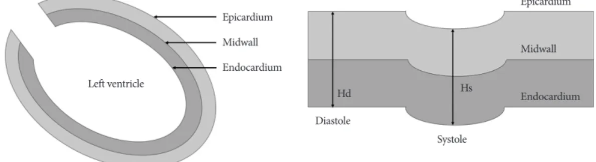
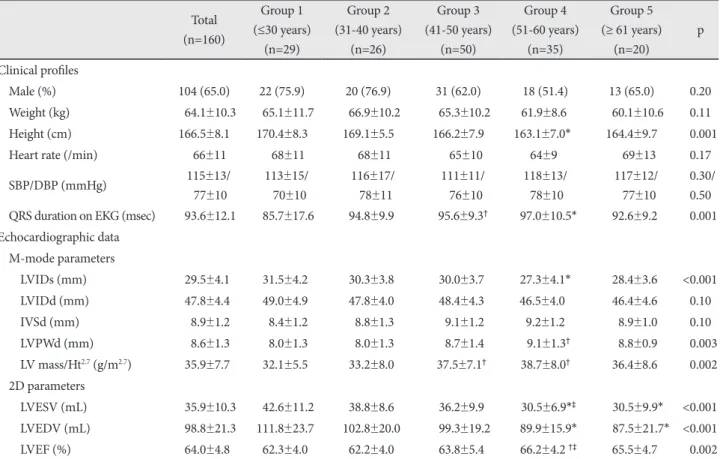
Background and Objectives: Left ventricular (LV) midwall fractional shortening (FSmw) reflects systolic function more ac- curately than LV endocardial fractional shortening (eFS) in patients with increased LV wall thickness. Although the normal reference ranges of LV-FSmw have been suggested in Western population studies, its reference values and age-related physi- ological changes in Eastern populations remain unknown. Subjects and Methods: Conventional echocardiographic param- eters, LV-FSmw, and stress-corrected LV-FSmw were assessed in 160 healthy and clinically normal subjects with a mean age of 45 (range, 11-72 years; 104 males, 56 women), all of whom were confirmed to be free of disease, based on laboratory investiga- tions, clinical and physical examination findings and computed tomographic coronary angiographic examinations. Results:

LV-FSmw was higher in women compared to men. However, the differences were without statistical significance (18.2±1.5%

for male gender and 19.4±2.5% for female gender, p=0.07). In contrast to LV-eFS that progressively increased with age (p=0.001), LV-FSmw and stress-corrected LV-FSmw was not influenced by changes in age (p=0.88 and 0.29, respectively).

The results remained unchanged when analyses were performed adjusting for gender. Conclusion: The results of this study provide normal reference values for LV-FSmw and stress-corrected LV-FSmw and their natural physiological changes with ad- vancing age. These measures can be used as reference standards for research on LV systolic function in the setting of pressure or volume overload. (Korean Circ J 2010;40:587-592)

KEY WORDS: Echocardiography; Left ventricular function; Systole.

Received: March 25, 2010 Revision Received: May 24, 2010 Accepted: June 1, 2010

Correspondence: Hyung-Kwan Kim, MD, Division of Cardiology, De- partment of Internal Medicine, Seoul National University College of Medi- cine, 101 Daehak-ro, Jongno-gu, Seoul 110-744, Korea

Tel: 82-2-2072-0243, Fax: 82-2-2072-3757 E-mail: [email protected]

cc

This is an Open Access article distributed under the terms of the Cre- ative Commons Attribution Non-Commercial License (http://creativecom- mons.org/licenses/by-nc/3.0) which permits unrestricted non-commer- cial use, distribution, and reproduction in any medium, provided the origi- nal work is properly cited.

Introduction

The assessment of left ventricular (LV) systolic function is of vital importance because it is closely associated with fu- ture patient outcomes.

1)2)

Evaluation of LV systolic function is usually achieved by measuring the ejection fraction (EF) and fractional shortening (FS). Albeit useful, the EF and FS obtained by conventional approach are derived from endo-

cardial movement. As such, they cannot reliably reflect the

“actual” systolic function in the setting of increased LV wall th-

ickness.

3)

In fact, LV systolic function measured by conven-

tional EF or endocardial FS (eFS) in patients with LV hyper-

trophy is estimated to be almost always normal or supernor-

mal in spite of the adverse prognosis of patients with LV hy-

pertrophy.

4)

This paradoxical trend may be explained by the

followings: first, the inner layer of the LV that includes the en-

docardium moves inward, further than the outer layer of the

LV. Second, the inner layer of the “thickened” LV tends to show

more inward movement during systole, compared to the in-

ner layer of the LV with “normal” thickness.

5)

Therefore, it has

been recommended that the LV midwall FS (LV-FSmw) be

used in place of conventional eFS in patients with increased

LV wall thickness for the assessment of LV systolic perform-

ance.

6)

From a prognostic point of view, LV-FSmw has prov-

en its value as a predictor of cardiac death and cardiovascular

morbidity in patients with arterial hypertension.

6)7)

Any gen-

eral use of LV systolic functional parameters should be pre-

(2)

ceded by determining their normal reference ranges. As the number and proportion of older individuals progressively in- crease in the general population, knowledge of the normal re- ference ranges and the physiological changes associated with aging will become important. Although the normal reference ranges for LV-FSmw have previously been established by st- udies in the Western population,

8)9)

the reference values and physiological changes with age have yet been explored in the Asian populations.

The objective of this study was to determine the normal reference values and physiologic changes of LV-FSmw, based on 160 clinically normal Koreans and utilize the outcomes to establish reference standards for the assessment of LV-FSmw in patients with pressure or volume overload in future studies.

Subjects and Methods

Study population

Volunteers who visited the Health Care Center at our hospi- tal for general routine check-up without laboratory abnor- malities, including electrocardiographic and echocardiogra- phic findings, were considered candidates for this study. Pa- tients with a history of cardiovascular or systemic diseases were excluded. For inclusion, subjects less than 40 years of age had to undergo a modified Bruce protocol-based treadmill test and those beyond 40 years of age required a treadmill test and a computed tomographic coronary angiography (CTCA) in or- der to exclude subclinical coronary artery disease. Volunteers who had stenosis of >25% in any coronary artery on the CTCA and who did not reach at least 85% of age-predicted maximal heart rate during the treadmill test were systematically exclud- ed. Subjects who were ≥50 years of age were screened by ca- rotid Doppler examination and patients who had stenoses gr- eater than 50% of the lumen diameter in any carotid arteries were excluded. The study protocol was approved by the insti- tutional review board of our hospital and written informed con- sent was obtained from all participants before study enrollment.

Echocardiographic analysis

Echocardiographies were performed by one cardiologist using commercially available equipment (GE Vivid 7, GE me-

dical systems, Milwaukee, WI, USA) with subjects in the left lateral decubitus position. LV cavity size, LV mass, LV relative wall thickness (RWT) and FSmw were determined using tis- sue harmonic imaging with M-mode measurements accord- ing to the guidelines suggested by the American Society of Echo- cardiography.

10)

Interventricular septal wall thickness (IVST), LV internal diameter (LVID) and posterior wall thickness (PWT) were measured at end-diastole and at end-systole. LV mass was calculated with the Devereux formula.

11)

To estab- lish the relationship between LV mass and linear height (Ht), LV mass was adjusted for height by dividing the value by the Ht

2.7

.

12)

LV-RWT was calculated using (LV-IVSTd+LV-PWTd)/

LVIDd. The LV-eFS was calculated using the conventional for- mula: e.g., LV-eFS (%)={(LVIDd-LVIDs)/LVIDd}×100, where s denotes end-systole and d, end-diastole. LV-EF was derived from modified Simpson’s method.

Left ventricular midwall fractional shortening calculation

Calculation of LV-FSmw was performed with the assump- tion of a cylindrical shaped LV, including consideration of epi- cardial migration of midwall circumferential fibers during systole.

13)

The model that assumed LV geometry to be cylindri- cal shaped considered LV as the union of two concentric cylin- drical shells, with equal thickness at end-diastole (Fig. 1). This model is based on the fact that LV wall volume is constant dur- ing the cardiac cycle. Using this model, the LV-FSmw can be calculated with the following formula: LV-FSmw={(LVIDd+

LV-IVSTd/2+LV-PWTd/2)-(LVIDs+Hs/2)}/LVIDd+LV- IVSTd/2+LV-PWTd/2)

Stress-corrected left ventricular midwall fractional shortening calculation

In order to eliminate the effects of LV afterload on LV- FSmw, stress-corrected LV-FSmw was calculated by normal- izing LV-FSmw to LV circumferential end-systolic wall stress (LV-cESS) in each subject.

14)

LV-cESS was calculated from the following formula: LV-cESS={[SBP×(LVIDs/2)

2

]×[1+(L VIDs/2+LV-PWTs)

2

/(LVIDs/2+LV-PWTs/2)

2

]}/{(LVIDs/

2+LV-PWTs)

2

-(LVIDs/2)

2

}, where SBP is the brachial cuff systolic blood pressure (BP).

Epicardium Midwall Endocardium

Epicardium

Midwall

Endocardium

Systole Diastole

Hd Hs

Left ventricle

Fig. 1. Schematic representation of two shell geometry model of midwall. Hd: diastolic thickness of shell, Hs: systolic thickness of shell.

(3)

Statistical analysis

All measurements were analyzed by one observer using St- atistical Package for the Social Sciences (SPSS) statistical soft- ware, version 14.0 (SPSS, Inc., Chicago, IL, USA). Data are pre- sented as the mean±SD for continuous variables and number (percentages) for categorical variables. To minimize the im- pact of confounding factors due to the increased proportion of women in the older age group, partial correlation and anal- ysis of covariance taking gender into account were performed.

A p<0.05 was considered statistically significant.

Results

Clinical and conventional echocardiographic data The study population was comprised of 160 subjects {104 males, 56 womens, age 44.9±2.7 years (range, 11-72 years)}.

Clinical and conventional echocardiographic results across age deciles are summarized in Table 1.

Comparison of left ventricular-endocardial fractional shortening and left ventricular midwall fractional shortening without gender breakdown

Table 2 shows the comparison between eFS and FSmw, with differentiation by age deciles in conjunction with data for the

entire cohort. LV mass/Ht

2.7

and LV-RWT showed significant increases with age (p for trend=0.002 and 0.001, respectively).

LV-eFS and LV-EF also displayed a gradual increase with age (p for trend=0.001 and 0.002, respectively), whereas LV-FSmw and SC LV-FSmw remained constant, regardless of age (p for trend=0.88 and 0.29, respectively).

Comparison of left ventricular-endocardial fractional shortening, left ventricular midwall fractional

shortening and stress-corrected left ventricular midwall fractional shortening according to gender

Echocardiographic findings of endocardial versus midwall mechanics by gender are shown in Table 3 and 4. Although the analyses were limited by small sample size, LV-RWT and LV-eFS showed significant increase, in parallel with age among men (p for trend=0.001 and 0.006, respectively) and women (p for trend=0.030 and 0.014, respectively). With advancing age, LV-FSmw tended to decrease, without statistical significance in men (p for trend=0.30). Despite a slightly higher LV-FSmw value in women compared to men (18.2±1.5% for male vs.

19.4±2.5% for female), this difference did not reach statisti- cal significance (p=0.07). In the women, LV-FSmw showed no trend in the relationship with age (p for trend=0.99), as shown in Table 4. SC LV-FSmw value also showed no trend with age Table 1. Clinical and conventional echocardiographic data

Total (n=160)

Group 1 (≤30 years)

(n=29)

Group 2 (31-40 years)

(n=26)

Group 3 (41-50 years)

(n=50)

Group 4 (51-60 years)

(n=35)

Group 5 (≥ 61 years)

(n=20)

p

Clinical profiles

Male (%) 104 (65.0) 22 (75.9) 20 (76.9) 31 (62.0) 18 (51.4) 13 (65.0) <0.20

Weight (kg) 0064.1±10.3 0065.1±11.7 0066.9±10.2 0065.3±10.2 061.9±8.60. 0060.1±10.6 <0.11 Height (cm) 166.5±8.1 170.4±8.3 169.1±5.5 166.2±7.9 163.1±7.0*0 164.4±9.7 <0.001

Heart rate (/min) 066±11 0068±11 0068±11 00.65±10 00.64±9000 00.69±13 <0.17

SBP/DBP (mmHg) 0.115±13/

0077±10

0.113±15/

00.70±10

0.116±17/

00.78±11

0.111±11/

00.76±10

0.118±13/0.

00.78±1000

0.117±12/

00.77±10

<0.30/

<0.50 QRS duration on EKG (msec) 0093.6±12.1 0085.7±17.6 094.8±9.9 0.95.6±9.3

097.0±10.5* 092.6±9.2 <0.001 Echocardiographic data

M-mode parameters

LVIDs (mm) 029.5±4.1 031.5±4.2 030.3±3.8 030.0±3.7 27.3±4.1* 028.4±3.6 <0.001 LVIDd (mm) 047.8±4.4 049.0±4.9 047.8±4.0 048.4±4.3 46.5±4.00 046.4±4.6 <0.10

IVSd (mm) 008.9±1.2 008.4±1.2 008.8±1.3 009.1±1.2 9.2±1.2 008.9±1.0 <0.10

LVPWd (mm) 008.6±1.3 008.0±1.3 008.0±1.3 008.7±1.4 09.1±1.3

008.8±0.9 <0.003 LV mass/Ht

2.7

(g/m

2.7

) 035.9±7.7 032.1±5.5 033.2±8.0 037.5±7.1

38.7±8.0

036.4±8.6 <0.002 2D parameters

LVESV (mL) 0035.9±10.3 0042.6±11.2 038.8±8.6 036.2±9.9 030.5±6.9*

0030.5±9.9* <0.001 LVEDV (mL) 0098.8±21.3 0111.8±23.7 0102.8±20.0 0099.3±19.2 089.9±15.9* 00087.5±21.7* <0.001 LVEF (%) 064.0±4.8 062.3±4.0 062.2±4.0 063.8±5.4 066.2±4.2

†‡

065.5±4.7 <0.002

*p<0.01 versus Group 1,

p<0.05 versus Group 1,

p<0.05 versus Group 2. S(D)BP: systolic (diastolic) blood pressure, EKG: electrocardiogram,

LVIDs(d): left ventricular systolic (diastolic) dimension, IVSd: diastolic interventricular septal thickness, LVPWd: diastolic left ventricular poste-

rior wall thickness, LVES(D)V: left ventricular end-systolic (diastolic) volume, LVEF: left ventricular ejection fraction

(4)

(p for trend= 0.29 for male and 0.50 for female).

Discussion

This is the first report on the normal reference values of LV- FSmw and the natural, physiological changes with age in a large number of healthy Korean individuals who underwent echocardiography using the harmonic imaging technique. In contrast to LV-eFS that showed a progressive increase in par-

allel with age, LV-FSmw displayed no significant changes in relation to aging in all subjects. This remained the case even when the analyses were performed to adjust for gender. Over- all, the LV-FSmw values obtained in this study were compa- rable to that of previous studies.

9)15)

However, the trend for LV- FSmw change is different from that of previous studies, in which LV-FSmw decreased slightly with advancing age in normal subjects.

9)15)

This discrepancy can be explained by differences in the characteristics of the study population. Specifically, the Table 2. Comparison of LV-eFS and LV-FSmw in the entire study cohort

Total (n=160)

Group 1 (≤30 years)

(n=29)

Group 2 (31-40 years)

(n=26)

Group 3 (41-50 years)

(n=50)

Group 4 (51-60 years)

(n=35)

Group 5 (≥61 years)

(n=20)

p for trend LV mass/Ht

2.7

(g/m

2.7

) 035.9±7.7 032.1±5.5 033.2±8.0 037.5±7.1

038.7±8.0

036.4±8.6 0.001 LV-RWT (cm) 00.37±0.06 00.34±0.06 00.35±0.05 00.37±0.06 00.39±0.05

00.39±0.06

0.001

LV-eFS (%) 038.3±4.8 035.9±3.1 036.9±4.6 038.1±4.0 041.4±5.5*

§

038.8±5.2 0.001

LV-FSmw (%) 018.6±2.0 018.6±1.6 018.5±1.8 018.6±2.0 018.9±2.4 018.3±2.2 0.880

cESS (kdyne/cm

2

) 123.5±27.9 138.7±31.9 133.7±36.3 121.6±27.9 117.0±24.5 123.9±20.5 0.101 SC LV-FSmw

(cm

2

/kdyne ∙ 10

-2

) 015.9±4.3 014.5±3.3 014.7±4.7 016.2±4.4 017.1±4.8 014.8±2.7 0.618

*p<0.01 versus Group 1,

p<0.05 versus Group 1,

§

p<0.01 versus Group 2. LV: left ventricle, RWT: relative wall thickness, eFS: endocardial frac- tional shortening, FSmw: mid wall fractional shortening, cESS: circumferential end-systolic wall stress, SC LV-FSmw: stress-corrected mid wall fractional shortening

Table 3. Comparison of LV-eFS and LV-FSmw in male subjects Total

(n=104)

Group 1 (≤30 years)

(n=22)

Group 2 (31-40 years)

(n=20)

Group 3 (41-50 years)

(n=31)

Group 4 (51-60 years)

(n=18)

Group 5 (≥61 years)

(n=13)

p for trend LV mass/Ht

2.7

(g/m

2.7

) 036.7±6.9 033.3±5.4 034.2±7.3 039.3±6.3

040.3±6.8

034.7±6.7 0.024 LV-RWT (cm) 00.37±0.06 00.34±0.06 00.35±0.05 00.38±0.06

00.40±0.04

†‡

00.38±0.05 0.001

LV-eFS (%) 037.3±4.2 035.4±3.2 036.4±4.4 037.5±3.7 040.2±5.1* 037.2±3.8 0.006

LV-FSmw (%) 018.2±1.5 018.5±1.4 018.3±1.3 017.9±1.6 018.1±1.6 018.0±1.8 0.296

cESS (kdyne/cm

2

) 127.9±28.1 148.5±23.6 139.5±30.3 123.9±28.4 119.0±28.9 132.2±19.2 0.123 SC LV-FSmw

(cm

2

/kdyne ∙ 10

-2

) 014.9±3.7 013.6±2.8 013.7±3.2 015.4±3.9 016.2±4.3 013.7±2.1 0.497

*p<0.01 versus Group 1,

p<0.05 versus Group 1,

p<0.05 versus Group 2. LV: left ventricle, RWT: relative wall thickness, eFS: endocardial frac- tional shortening, FSmw: mid wall fractional shortening, cESS: circumferential end-systolic wall stress, SC LV-FSmw: stress-corrected mid wall fractional shortening

Table 4. Comparison of LV-eFS and LV-FSmw in female subjects Total

(n=56)

Group 1 (≤30 years)

(n=7)

Group 2 (31-40 years)

(n=6)

Group 3 (41-50 years)

(n=19)

Group 4 (51-60 years)

(n=17)

Group 5 (≥61 years)

(n=7)

p for trend LV mass index (g/m

2

) 034.6±8.9 28.3±4.3 029.9±9.8 034.5±7.6 037.0±8.9 039.4±11.2 0.003

LV-RWT (cm) 00.37±0.06 0.34±0.06 00.35±0.07 00.35±0.06 00.38±0.06 00.40±0.08 0.030

LV-eFS (%) 040.3±5.2 37.5±2.4 038.3±5.4 039.2±4.4 042.8±5.7 041.6±6.4 0.014

LV-FSmw (%) 019.4±2.5 19.2±2.1 019.1±3.0 019.6±2.2 019.6±2.8 018.7±2.8 0.991

cESS (kdyne/cm

2

) 111.0±27.9 89.7±21.3 122.3±47.9 117.0±27.4 113.2±14.4 110.4±15.6 0.726 SC LV-FSmw

(cm

2

/kdyne ∙ 10

-2

) 018.8±6.8 18.8±2.5 016.8±6.8 017.9±5.0 017.2±2.2 016.7±2.8 0.680

LV: left ventricle, RWT: relative wall thickness, eFS: endocardial fractional shortening, FSmw: mid wall fractional shortening, cESS: circum-

ferential end-systolic wall stress, SC LV-FSmw: stress-corrected mid wall fractional shortening

(5)

subjects in the present study exhibited no increment in BP with age, whereas BP of the subjects enrolled in the study by Slot- winter et al.

9)

displayed stepwise BP increment in parallel with age, which can lead to a significant discrepancy between the two studies. In addition, silent myocardial diseases may ac- count for some of the modest age-related decline in LV-FSmw observed in previous studies,

15)

which was not the case for our study due to systematic exclusion of subjects with subclinical coronary artery disease based on treadmill test and CTCA. We believe that this attempt is of crucial value, given that coro- nary artery disease is now the most common cause of subcli- nical myocardial dysfunction in Korea. In terms of conven- tional echocardiographic parameters, LVIDs, LVIDd, LV- IVSd, and LV-PWTd values in patients less than 50 years are comparable to that obtained in previous studies, albeit there exist discrepancies in subjects aged more than 50 years.

16)

How- ever, the differences are very small and can be considered wi- thin a variability range that may be observed in routine echo- cardiographic examinations.

Echocardiography is a non-invasive and readily available dia- gnostic modality that performed in patients with known or suspected cardiovascular disease. Thanks to its versatility, the indications for echocardiography have progressively expand- ed. The assessment of LV systolic function is one of the most important clinical applications of echocardiography. Tradi- tionally, echocardiographic evaluation of LV systolic func- tion has been predicated using indexes acquired by measure- ments at the endocardial level. Many previous studies have shown that assessment of the LV systolic function based on endocardial movement affected the choice and timing of car- diac surgery and long-term survival.

17)18)

However, LV systolic function obtained on the basis of endocardial movement is of limited utility in patients with LV hypertrophy, given the re- ciprocal change of LV-EF in relation to LV wall thickness.

19)20)

In other words, the presence of ventricular hypertrophy can lead to over-estimation of LV-EF, the most frequently used in- dex for estimating LV systolic performance, despite decline in myocardial function, as demonstrated by tagging magne- tic resonance imaging.

21)

This apparent discrepancy could be explained, at least in part, by “cross-fiber shortening”, where endocardial shortening tends to be greater than normal in patients with LV hypertrophy.

22)

Because subendocardial fibers are longitudinally arranged, circumferential and radial short- ening observed at the endocardial level cannot be directly generated by shortening of subendocardial fibers per se.

To overcome the limitation of evaluating LV systolic func- tion in patients with thickened LV walls, direct estimation of LV-FSmw was introduced and found to be physiologically more appropriate than LV-eFS in estimating LV systolic per- formance when LV hypertrophy was present.

3)6)

Furthermore, LV-FSmw was shown to be an independent risk factor to predict cardiovascular events in patients with hypertension.

23-26)

As the number and proportion of older individuals in the population progressively increases, the prevalence of hyper- tensive patients is expected to increase steadily and thus the utility of LV-FSmw could be augmented. For widespread use of LV-FSmw in the clinical arena, knowledge of its normal re- ference range and physiologic changes associated with aging become more and more pivotal.

This study, for the first time, demonstrated the normal re- ference ranges of LV-FSmw and its physiological changes with age in the Asian population. Cardiovascular disease states can only be characterized in the aging populations with reference to age-adjusted normal values. However, in terms of defining normal reference ranges, the sole effect of aging on natural re- modeling of the heart is difficult to evaluate, because of sub- clinical diseases not infrequently found in clinical practice in- cidentally. Because all individuals in the present study were confirmed to be clinically normal based on laboratory tests, clinical and physical examination findings, CTCA and ca- rotid Doppler studies, we believe that our study is free of in- advertent enrollment of patients with subclinical disease.

It is interesting to note that in the current study, LV-FSmw obtained in male subjects showed slightly higher values com- pared to the females, although the difference did not reach sta- tistical significance (19.4±2.5% vs. 18.2±1.5%, p=0.068). In addition, while LV-eFS increased in a stepwise manner with age, LV-FSmw did not significantly vary by the age deciles, suggesting that age per se does not necessarily needed to be considered when interpreting LV-FSmw, which is useful in the clinical application of LV-FSmw when comparison is made between groups with different ages. This result is also sup- ported by the fact that no changes were observed in stress-cor- rected LV-FSmw with age, which was performed to eliminate the influence of LV afterload on LV-FSmw.

There were several limitations to this study. First, our data was derived from a single center and they may not be directly applicable to general populations. Normal echocardiographic range of any specific parameter population should be derived from a random sample of the general population. In this re- spect, our population may not reflect the general population.

However, we selected healthy subjects among the population who visited our health care center for general check-up at their discretion. Therefore, we believe that the present study popu- lation can be, to some extent, a representative sample of the Koreans.

In conclusion, the results of this study provide normative

data for LV-FSmw and its physiological changes with age in

a relatively large number of normal Asian subjects. LV-FSmw

remained stable with age, which was in contrast to LV-eFS

that showed progressive increase in parallel with age. The in-

formation obtained in the present study can be applicable to

future studies, and provides reference standards with which

LV systolic dysfunction can be defined more accurately.

(6)

REFERENCES

1) Quiñones MA, Greenberg BH, Kopelen HA, et al. Echocardiograph- ic predictors of clinical outcome in patients with left ventricular dys- function enrolled in the SOLVD registry and trials: significance of left ventricular hypertrophy. Studies of Left Ventricular Dysfunction. J Am Coll Cardiol 2000;35:1237-44.

2) Sohn JW, Shin DG, Kim DH, et al. Non-invasive parameters, includ- ing a low left ventricular ejection fraction, for predicting sudden cardi- ac death in Korean post myocardial infarction patients. Korean Circ J 2006;36:431-6.

3) Shimizu G, Hirota Y, Kita Y, Kawamura K, Saito T, Gaasch WH. Left ventricular midwall mechanics in systemic arterial hypertension: myo- cardial function is depressed in pressure-overload hypertrophy. Circu- lation 1991;83:1676-84.

4) Levy D, Garrison RJ, Savage DD, Kannel WB, Castelli WP. Prognos- tic implications of echocardiographically determined left ventricular mass in the Framingham Heart Study. N Engl J Med 1990;322:1561-6.

5) Mayet J, Ariff B, Wasan B, et al. Midwall myocardial shortening in athletic left ventricular hypertrophy. Int J Cardiol 2002;86:233-8.

6) de Simone G, Devereux RB, Roman MJ, et al. Assessment of left ven- tricular function by the midwall fractional shortening/end systolic st- ress relation in human hypertension. J Am Coll Cardiol 1994;23:

1444-51.

7) Aurigemma GP, Gaasch WH, McLaughlin M, McGinn R, Sweeney A, Meyer TE. Reduced left ventricular systolic pump performance and depressed myocardial contractile function in patients > 65 years of age with normal ejection fraction and a high relative wall thickness. Am J Cardiol 1995;76:702-5.

8) Calabrò R, Pisacane C, Pacileo G, Russo MG. Left ventricular mid- wall mechanics in healthy children and adolescents. J Am Soc Echo- cardiogr 1999;12:932-40.

9) Slotwiner DJ, Devereux RB, Schwartz JE, et al. Relation of age to left ventricular function in clinically normal adults. Am J Cardiol 1998;

82:621-6.

10) Lang RM, Bierig M, Devereux RB, et al. Recommendations for cham- ber quantification: a report from the American society of echocardiog- raphy’s guideline and standards committee and the chamber quanti- fication writing group, developed in conjunction with the European as- sociation of echocardiography, a branch of the European Society of Car- diology. J Am Soc Echocardiogr 2005;18:1440-63.

11) Devereux RB, Reichek N. Echocardiographic determination of left ventricular mass in man: anatomic validation of the method. Circula- tion 1977;55:613-8.

12) de Simone G, Daniels SR, Devereux RB, et al. Left ventricular mass and body size in normotensive children and adults: assessment of allo- metric relations and impact of overweight. J Am Coll Cardiol 1992;

20:1251-60.

13) Shimizu G, Conrad CH, Gaasch WH. Phase-plane analysis of left ventricular chamber filling and midwall fiber lengthening in patients with left ventricular hypertrophy. Circulation 1987;75 1 Pt 2:I34-9.

14) Yip GW, Shang Q, Tam LS, et al. Disease chronicity and activity pre- dict subclinical left ventricular systolic dysfunction in patients with systemic lupus erythematosus. Heart 2009;95:980-7.

15) de Simone G, Kimball TR, Roman MJ, et al. Relation of left ventricu- lar chamber and midwall function to age in normal children, adoles- cents and adults. Ital Heart J 2000;1:295-300.

16) Park SW. Multicenter trial for estimation of normal values of echocar- diograhic indices in Korea. Korean Circ J 2000;30:373-82.

17) Choi SH, Ha JW, Chang BC, et al. Long-term outcome of clinically in- significant aortic valve disease in patients undergoing mitral valve sur- gery for rheumatic heart disease. Korean Circ J 2001;31:1034-41.

18) Corti R, Binggeli C, Turina M, Jenni R, Luscher TF, Turina J. Predic- tors of long-term survival after valve replacement for chronic aortic re- gurgitation: is M-mode echocardiography sufficient? Eur Heart J 2001;22:866-73.

19) Wandt B, Bojö L, Tolagen K, Wranne B. Echocardiographic assess- ment of ejection fraction in left ventricular hypertrophy. Heart 1999;

82:192-8.

20) Seo JC, Lee MS, Chae CS, et al. Relation of hemodynamic load to left ventricular hypertrophy and performance in essential hypertension.

Korean Circ J 1993;23:380-9.

21) Palmon LC, Reichek N, Yeon SB, et al. Intramural myocardial short- ening in hypertensive left ventricular hypertrophy with normal pump function. Circulation 1994;89:122-31.

22) Rademakers FE, Rogers WJ, Guier WH, et al. Relation of regional cross-fiber shortening to wall thickening in the intact heart: three-di- mensional strain analysis by NMR tagging. Circulation 1994;89:

1174-82.

23) de Simone G, Devereux RB, Koren MJ, Mensah GA, Casale PN, Laragh JH. Midwall left ventricular mechanics: an independent pre- dictor of cardiovascular risk in arterial hypertension. Circulation 1996;

93:259-65.

24) Devereux RB, de Simone G, Pickering TG, Schwartz JE, Roman MJ.

Relation of left ventricular midwall function to cardiovascular risk factors and arterial structure and function. Hypertension 1998;31:

929-36.

25) Aurigemma GP, Gottdiener JS, Shemanski L, Gardin J, Kitzman D.

Predictive value of systolic and diastolic function for incident conges- tive heart failure in the elderly: the Cardiovascular Health Study. J Am Coll Cardiol 2001;37:1042-8.

26) Schussheim AE, Diamond JA, Jhang JS, Phillips RA. Midwall frac-

tional shortening is an independent predictor of left ventricular dia-

stolic dysfunction in asymptomatic patients with systemic hyperten-

sion. Am J Cardiol 1998;82:1056-9.

수치

Fig. 1. Schematic representation of two shell geometry model of midwall. Hd: diastolic thickness of shell, Hs: systolic thickness of shell.
Table 2 shows the comparison between eFS and FSmw, with  differentiation by age deciles in conjunction with data for the
Table 3. Comparison of LV-eFS and LV-FSmw in male subjects Total (n=104) Group 1 (≤30 years) (n=22) Group 2 (31-40 years)(n=20) Group 3 (41-50 years)(n=31) Group 4 (51-60 years)(n=18) Group 5 (≥61 years)(n=13) p for trend LV mass/Ht 2.7  (g/m 2.7 ) 036.7±6

참조

관련 문서

Method : : : : We evaluate clinical features of 714 Korean vitiligo patients who lived in the Gwangju chonnam province(315 males and 399 females).. Results

The purpose of this study is to investigate changes in listening, reading achievement, and learning attitudes of Korean elementary students who enrolled in

However, with the changes in the domestic and international environment after the reform and opening up, the new patriotism emerged in recognition of the

Measurement of proximal contact tightness between the left first molar and second molar(dental implant) in the mandible... Clinical dental anatomy, histology,

High complication rate in locking plate fixation of lower periprosthetic distal femur fractures in patients with total knee arthroplasties.. Management and

• Theory can extent to molten polymers and concentrated solutions.. The single-molecule bead spring models.. a)

Ha(2020), “The Effect of Forest Healing Program Experience on Psychological and Physiological Changes of Office Workers Exposed to Task Stress.” Chonnam

Although visceral fat adiposity has known to be associated with clinical, pathologic, and oncologic outcomes in patients with colorectal cancer (CRC), the