INTRODUCTION
Bowel ischemia caused by mesenteric occlusion would
Mesenteric Vascular Occlusion: Comparison of Ancillary CT Findings between Arterial and Venous Occlusions and Independent CT Findings Suggesting Life-Threatening Events
Yon-Cheong Wong, MD
1, Cheng-Hsien Wu, MD
1, Li-Jen Wang, MD
1, Huan-Wu Chen, MD
1, Being-Chuan Lin, MD
2, Chen-Chih Huang, MD
11Division of Emergency and Critical Care Radiology, Department of Medical Imaging and Intervention, Chang Gung Memorial Hospital, Chang Gung University, Taoyuan 333, Taiwan; 2Division of Trauma and Emergency, Department of Surgery, Chang Gung Memorial Hospital, Chang Gung University, Taoyuan 333, Taiwan
Objective: To compare the ancillary CT findings between superior mesenteric artery thromboembolism (SMAT) and superior mesenteric vein thrombosis (SMVT), and to determine the independent CT findings of life-threatening mesenteric occlusion.
Materials and Methods: Our study was approved by the institution review board. We included 43 patients (21 SMAT and 22 SMVT between 1999 and 2008) of their median age of 60.0 years, and retrospectively analyzed their CT scans. Medical records were reviewed for demographics, management, surgical pathology diagnosis, and outcome. We compared CT findings between SMAT and SMVT groups. Multivariate analysis was conducted to determine the independent CT findings of life- threatening mesenteric occlusion.
Results: Of 43 patients, 24 had life-threatening mesenteric occlusion. Death related to mesenteric occlusion was 32.6%. A thick bowel wall (p < 0.001), mesenteric edema (p < 0.001), and ascites (p = 0.009) were more frequently associated with SMVT, whereas diminished bowel enhancement (p = 0.003) and paralytic ileus (p = 0.039) were more frequent in SMAT.
Diminished bowel enhancement (OR = 20; p = 0.007) and paralytic ileus (OR = 16; p = 0.033) were independent findings suggesting life-threatening mesenteric occlusion.
Conclusion: The ancillary CT findings occur with different frequencies in SMAT and SMVT. However, the independent findings indicating life-threatening mesenteric occlusion are diminished bowel wall enhancement and paralytic ileus.
Index terms: CT; Superior mesenteric artery thromboembolism; Superior mesenteric vein thrombosis; Life-threatening mesenteric occlusion; Independent finding
Received April 12, 2012; accepted after revision May 29, 2012.
Corresponding author: Yon-Cheong Wong, MD, Division of Emergency and Critical Care Radiology, Department of Medical Imaging and Intervention, Chang Gung Memorial Hospital, Chang Gung University 5, Fu-Hsing Street, Gueishan, Taoyuan 333, Taiwan.
• Tel: 886-3-3281200 ext 3786 • Fax: 886-3-3970074
• E-mail: [email protected]
This is an Open Access article distributed under the terms of the Creative Commons Attribution Non-Commercial License (http://creativecommons.org/licenses/by-nc/3.0) which permits unrestricted non-commercial use, distribution, and reproduction in any medium, provided the original work is properly cited.
Korean J Radiol 2013;14(1):38-44
have a grave prognosis if not timely diagnosed and treated (1, 2). However, the diagnosis of mesenteric occlusion could be elusive based on physical examination because a wide spectrum of clinical presentations may occur ranging from nonspecific abdominal pain in the abdomen (3). The severity of mesenteric occlusion depends on whether the occlusion results in transient mucosal changes or a more severe transmural wall necrosis.
Catheter angiography or the state-of-the-art CT- angiography is the standard reference for diagnosis of mesenteric occlusion in selected patients (4-6). However, because of the limited availability of high-end multiple- row detector CT (MDCT) scanners and the lack of clinical suspicion prior to the CT scan, the more readily available
pISSN 1229-6929 · eISSN 2005-8330
causes (n = 1). Causes for SMVT were more variable and consist of abdominal infection (n = 6), hypercoagulation (n
= 3), liver disease (n = 3), renal disease (n = 2), cancer (n = 1), recent surgery (n = 1), others (n = 3), and unconfirmed causes (n = 3).
Clinical Data
The medical charts were independently reviewed by a co-investigator who did not know the CT findings for demographic data, management choice, surgical pathology diagnosis, and outcome (dead or alive). If bowel gangrene or hemorrhagic necrosis was recorded in surgical pathology diagnosis, then the mesenteric occlusion was defined as life-threatening. Life-threatening mesenteric occlusion was also assigned to patients who had not undergone surgery but died within 24 hours after CT examination was performed (11).
CT Techniques
All CT examinations were performed in the emergency room with a 4-detector MDCT scanner (LightSpeed QX/i Scanner, General Electric Medical Systems, Milwaukee, WI, USA). The routine protocol for an emergency abdominal CT examination in this study consisted of a non-enhanced and a contrast-enhanced series. All patients were scanned from the basal lung to pubic symphysis. The images were obtained with a high quality mode at 11.25 mm/rotation, 120 kVp, 200-250 mA, 5 mm slice thicknesses and 5 mm intervals. No oral contrast medium was ingested for gastrointestinal tract opacification. A uniphasic injection of 100-120 mL of intravenous iodinated contrast material Omnipaque 350 (Iohexol, 350 mgI/mL, GE Healthcare, Belfast, Ireland) was administered at a rate of 2-3 mL/
second for the contrast-enhanced series. The portal venous phase contrast-enhanced series was obtained at 70- 90 seconds after the initiation of the contrast material injection.
CT Data
Two abdominal radiologists with more than 10-years of experience were asked to review all CT examinations retrospectively on picture archiving communication systems for bowel and mesenteric abnormalities. They were aware of mesenteric occlusion but did not know the final condition of the bowel ischemia. They were free to use different window settings and discuss findings. A consensus interpretation obtained after discussion was regular CT examination has been used as a screening
modality in emergency rooms (7-9). This regular contrast- enhanced MDCT can clearly delineate the mesenteric vessels and ancillary findings of mesenteric occlusion (9-11).
Many nonspecific MDCT findings of mesenteric occlusion such as a thick bowel wall, thin bowel wall, strong bowel wall enhancement, diminished bowel wall enhancement, intramural gas, mesenteric edema, and ascites have been reported (10, 12-15). However, findings such as a thick bowel wall and thin bowel wall, strong wall enhancement and diminished wall enhancement could be mutually contradictory and confusing (16).
Our first hypothesis is that the ancillary findings of superior mesenteric artery thromboembolism (SMAT) and superior mesenteric vein thrombosis (SMVT) on contrast- enhanced MDCT are different or occur at different
frequencies. Consequently, confusion would occur if we do not address the CT findings of SMAT and SMVT separately.
Second, regardless of SMAT or SMVT, life-threatening mesenteric occlusion would occur if the collateral
circulation for bowel perfusion is inadequate. Therefore, we conducted a retrospective study to compare the ancillary CT findings between SMAT and SMVT as well as between patients of life-threatening and non-life-threatening mesenteric occlusion.
MATERIALS AND METHODS
Subjects
This retrospective study was approved by our institution
review board and informed consent was waived. We searched
from our radiology information system for emergency CT
reports containing impression keywords of main mesenteric
occlusion (SMAT and SMVT) between January 1999 and
December 2008. We defined SMAT and SMVT as total
occlusion of the SMA trunk and SMV trunk, respectively,
by thromboembolism or thrombosis. A total of 68 CT
reports of mesenteric occlusion were identified. Eighteen
CT examinations were incomplete or of poor enhancement
quality and were thus excluded. Also, an additional seven
were excluded because they were not admitted or had been
transferred to another hospital. The remaining 43 patients
(21 SMAT, 22 SMVT), which comprised of 17 women and 26
men, with a median age of 60.0 years (interquartile range
46.0, 78.0 years) formed the study group. The 21 SMATs
were caused by atrial fibrillation (n = 13), other heart
diseases (n = 3), atherosclerosis (n = 4), and unconfirmed
recorded. Small bowel walls greater than 3 mm in thickness were recorded as abnormal bowel wall thickening. Wall enhancement of the small bowel was compared with that of the duodenum and rectosigmoid colon. Diminished enhancement was recorded if a segment of the small bowel wall was hypodense compared to that of the duodenum or rectosigmoid colon based on qualitative assessment.
Enhancement of the duodenum and rectosigmoid colon was used as a standard reference for comparison because enhancement of the duodenum and rectosigmoid colon was mainly supplied by the gastroduodenal artery and hemorrhoidal arteries, respectively. Pneumatosis intestinalis was defined as small bowel intramural gas, and paralytic ileus as small bowel distention more than 3 cm in diameter without a transitional zone. An increase of mesenteric fat density was recorded as mesenteric edema. The presence of peritoneal fluid was recorded as ascites. Another radiologist was requested to measure the cross-sectional diameters of the main mesenteric vessels (SMV and SMA) at the level of occlusion and compute a ratio of SMV to SMA diameters (VA ratio).
Statistics
We compared the ancillary CT findings between patients of SMAT and SMVT to determine the difference of their occurring frequencies. Demographics and CT findings were compared with life-threatening mesenteric occlusion in a univariate analysis. The categorical variables were tested by the chi-square test or Fisher’s exact test if one of the cells had an expected count less than 5. The VA ratio and age were computed with the Mann-Whitney U-test and their respective optimal cutoff values were determined by the receiver operating characteristic (ROC) curve.
Logistic regression was used to determine the independent factors and adjusted odds ratios (ORs) of life-threatening mesenteric occlusion. A two-tailed p value of less than 0.05 was considered statistically significant.
RESULTS
Of the 43 patients, 24 had life-threatening mesenteric occlusion. Among these 24 patients, 22 were proven by surgery, and 2 critical patients were non-operable and died of sepsis and acidosis within twenty-four hours after CT examination. Of these two patients, who presented with atrial fibrillation and mesenteric occlusion, one had concurrent spleen infarct and the other had kidney infarct.
Overall, 16 patients died in this series. Among them, 14 patients died of mesenteric occlusion and bowel ischemia.
The death rate related to mesenteric occlusion was 32.6%
(14/43). Of the other 2 patients, 1 died of cancer, and the other died of fulminant hepatitis.
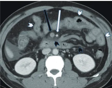
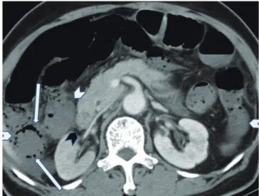
The CT findings (Fig. 1) of the thick bowel wall (p < 0.001), mesenteric edema (p < 0.001), and ascites (p = 0.009) were more frequently associated with SMVT (Table 1). In contrast, diminished bowel wall enhancement (p = 0.003) and paralytic ileus (p = 0.039) were more significantly found in SMAT (Fig. 2). Although pneumatosis intestinalis was more frequent in SMAT (Fig. 3), the difference did not reach a statistical significance (p = 0.185). The VA ratio was larger in the SMVT group than in the SMAT group (p < 0.001).
Table 2 shows that life-threatening mesenteric occlusion had no predilection in gender but was associated with the necessity of surgery (p < 0.001), death (p = 0.001), SMAT (p = 0.001), smaller VA ratio (p = 0.001) and older age (p = 0.019). The area under the ROC curve for age and VA ratio was 0.709 (p = 0.020) and 0.802 (p = 0.001), respectively.
The optimal cutoff values of age ≥ 50 years and VA ratio < 1.5 for life-threatening mesenteric occlusion were determined by ROC curves.
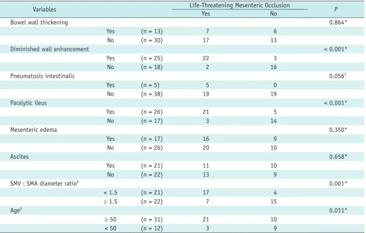
As detailed in univariate analysis (Tables 2, 3), the significant variables of life-threatening mesenteric occlusion with a p value < 0.100 were SMAT (p = 0.001),
Fig. 1. 46 year-old male of nephrotic syndrome. CT scans shows acute superior mesenteric vein thrombosis (black arrow) with bowel wall thickening (white arrowhead), mesenteric edema (black arrowhead) and ascites. Note the normal SMA enhancement (white arrow) and ratio of SMV to SMA diameters > 2. He did not receive surgery but was treated with anticoagulation therapy. Follow-up CT showed abundant collateral veins and no bowel necrosis. SMA = superior mesenteric artery, SMV = superior mesenteric vein
diminished bowel wall enhancement (p < 0.001), paralytic ileus (p < 0.001), pneumatosis intestinalis (p = 0.056), VA ratio < 1.5 (p = 0.001), and age ≥ 50 years (p = 0.011). By controlling above variables using logistic regression (Table
4), diminished bowel wall enhancement (OR = 20; p = 0.007) and paralytic ileus (OR = 16; p = 0.033) were independent factors of life-threatening mesenteric occlusion. This model could correctly predict 95.8% of the life-threatening
Fig. 3. Although the SMA orifice is normally enhanced, CT scans caudal to this level (not shown) reveal acute superior mesenteric artery occlusion. Note diminished enhancement of thin bowel walls (white arrowheads) as compared to normal duodenum (black arrowhead) and pneumatosis intestinalis (white arrows). Resection of small bowel and right colon showed coagulative necrosis. This 86 year-old female patient died of sepsis 4 days later. SMA = superior mesenteric artery Fig. 2. 73 year-old female of atrial fibrillation and paralytic
ileus. CT scan shows acute superior mesenteric artery occlusion (white arrow) with diminished enhancement of thin bowel walls (white arrowheads) as compared to normal duodenum (black arrowhead). Note normal SMV enhancement (black arrow) and ratio of SMV to SMA diameters of about 1. Extensive bowel gangrene was found at exploratory laparotomy. Bowel resection was not possible and she died of sepsis and acidosis. SMA = superior mesenteric artery, SMV = superior mesenteric vein
Table 1. Comparison of Ancillary CT Findings between SMAT and SMVT Groups
CT Findings SMAT SMVT P
Bowel wall thickening < 0.001*
Yes (n = 13) 1 12
No (n = 30) 20 10
Diminished wall enhancement 0.003*
Yes (n = 25) 17 8
No (n = 18) 4 14
Pneumatosis intestinalis 0.185†
Yes (n = 5) 4 1
No (n = 38) 17 21
Paralytic ileus 0.039*
Yes (n = 26) 16 10
No (n = 17) 5 12
Mesenteric edema < 0.001*
Yes (n = 17) 1 16
No (n = 26) 20 6
Ascites 0.009*
Yes (n = 21) 6 15
No (n = 22) 15 7
SMV : SMA diameter ratio (n = 43) 1.0 (0.94, 1.15) 2.0 (1.54, 2.32) < 0.001§ Note.— *chi-square test, †Fisher’s exact test, §Mann-Whitney U-test. SMAT = superior mesenteric artery thromboembolism, SMVT = superior mesenteric vein thrombosis, SMV = superior mesenteric vein, SMA = superior mesenteric artery
mesenteric occlusion in this study group.
DISCUSSION
Many abnormal CT findings of mesenteric occlusion have been reported (10, 12-17). Investigators have been attempting to explain these findings by using different physiology states of mesenteric ischemia (14, 18, 19). We were unable to correctly retrieve the exact time of disease onset and therefore could not correlate the CT findings with the changes in pathophysiology states. However, we have confirmed our hypothesis that the abnormal ancillary CT findings occur at different frequencies between patients of SMAT and SMVT.
The mesentery is richly innervated by the autonomic nervous system that coordinates the contractile activity of the bowel (19, 20). As seen in the SMAT patients of this series, the ischemic nervous system would paralyze the coordinated bowel peristalsis and result in paralytic ileus (14, 21). Arterial occlusion could also interrupt bowel perfusion and cause diminished wall enhancement of the affected bowels (16, 19-21). The poor inner- layer enhancement indicates mucosal ischemia, whereas a diminished transmural enhancement is consistent with a whole-layer wall ischemia that may lead to a life- threatening bowel infarct (22). The ischemic mucosa of SMAT may allow dissection of intraluminal gas into the mural layer as pneumatosis intestinalis (19). Moreover, it could be exacerbated by translocation of gas-forming
bacilli and the increase of intraluminal pressure as a result of paralytic ileus (19). Although there was no statistical significance, pneumatosis intestinalis was more frequently associated with SMAT than SMVT.
In contrast, SMVT would result in an increase of intravascular volume and elevation of hydrostatic pressure if the collateral draining veins are inadequate (19, 20).
The blood cells, plasma and the administered contrast medium are forced through the endothelium fenestrations into submucosal and mucosal spaces (19, 20) that result in concentric bowel wall thickening if the mucosal and serosal enhancement is still preserved. If the venous pressure is not alleviated, it could gradually compromise the in-flow of arterioles and capillaries (15). The concentric bowel wall would then become a thick wall with diminished enhancement. Hemorrhagic necrosis of the affected bowel may subsequently occur (23). In this series, bowel necrosis was less common in SMVT than in SMAT. The increase of mesenteric fat density in SMVT patients is caused by mesenteric edema as a result of the elevated hydrostatic pressure (19, 20). Intravascular fluid could also be forced out into the mesenteric space and other peritoneal spaces as ascites (19, 20).
We have confirmed the observation of Kurimoto et al.
(24) that a small VA ratio is a helpful screening criterion for arterial mesenteric occlusion. The SMV diameter is usually larger than the adjacent SMA diameter. However, upon mesenteric occlusion, the diameter of the occluded segment would become larger than its otherwise non-occlusive
Table 2. Comparison of Clinical Characteristics between Life-Threatening and Non-Life-Threatening Mesenteric Occlusion GroupsVariables Life-Threatening Mesenteric Occlusion
Yes No P
Management < 0.001*
Surgery (n = 22) 22 0
No surgery (n = 21) 2 19
Outcome 0.001*
Death (n = 16) 14 2
Alive (n = 27) 10 17
Gender 0.748*
Man (n = 26) 14 12
Woman (n = 17) 10 7
Occlusion type 0.001*
SMAT (n = 21) 17 4
SMVT (n = 19) 4 15
Age (n = 43) 73.0 (51.5, 80.0) 53.0 (41.0, 70.0) < 0.019†
SMV : SMA diameter ratio (n = 43) 1.0 (0.98, 1.50) 2.0 (1.50, 2.04) 0.001†
Note.— *chi-square test, †Mann-Whitney U-test. SMAT = superior mesenteric artery thromboembolism, SMVT = superior mesenteric vein thrombosis, SMV = superior mesenteric vein, SMA = superior mesenteric artery
status (24). In our series, the VA ratio in SMAT was reduced to 1 : 1 due to engorgement of the occluded SMA. In contrast, the VA ratio of SMVT was increased to 2 : 1 due to SMV engorgement.
Although life-threatening mesenteric occlusion was associated with elderly patients, small VA ratio, SMAT, diminished wall enhancement, pneumatosis intestinalis and paralytic ileus in univariate analysis, the significant independent factors were diminished wall enhancement and paralytic ileus. Patients exhibiting mesenteric occlusion (regardless of SMAT or SMVT) with diminished bowel wall enhancement were twenty times more likely than otherwise to proceed to a life-threatening condition. Likewise, patients of mesenteric occlusion with paralytic ileus were
sixteen times more likely to proceed to a life-threatening condition. Death is still significantly associated with life-threatening mesenteric occlusion despite aggressive surgical intervention to remove the nonviable bowel and restore mesenteric blood flow. Death related to mesenteric occlusion in this study was 32.6%, consistent with the high mortality rates previously reported (1, 2).
Although small VA ratio (< 1.5) and pneumatosis intestinalis were more frequently associated with life- threatening mesenteric occlusion, this study of the small sample was not able to prove that they were significant independent predictors. Unlike CT-angiography, the regular contrast-enhanced CT has a limitation in detecting embolic occlusion of peripheral arterial branches leading
Table 4. Ancillary CT Findings with Adjusted Odds Ratios and 95% Confidence Interval for Life-Threatening Mesenteric OcclusionVariable Adjusted Odds Ratios and 95% Confidence Interval P
Diminished wall enhancement 20.426 (2.279, 183.104) 0.007
Paralytic ileus 15.716 (1.249, 197.714) 0.033
SMV : SMA diameter < 1.5 9.226 (0.777, 109.562) 0.078
Note.— SMV = superior mesenteric vein, SMA = superior mesenteric artery
Table 3. Comparison of Ancillary CT Findings and Optimal Cutoff Values of SMV : SMA Diameter Ratio and Age between Life- Threatening and Non-Life-Threatening Mesenteric Occlusion Groups
Variables Life-Threatening Mesenteric Occlusion
Yes No P
Bowel wall thickening 0.864*
Yes (n = 13) 7 6
No (n = 30) 17 13
Diminished wall enhancement < 0.001*
Yes (n = 25) 22 3
No (n = 18) 2 16
Pneumatosis intestinalis 0.056†
Yes (n = 5) 5 0
No (n = 38) 19 19
Paralytic ileus < 0.001*
Yes (n = 26) 21 5
No (n = 17) 3 14
Mesenteric edema 0.350*
Yes (n = 17) 16 9
No (n = 26) 20 10
Ascites 0.658*
Yes (n = 21) 11 10
No (n = 22) 13 9
SMV : SMA diameter ratio§ 0.001*
< 1.5 (n = 21) 17 4
≥ 1.5 (n = 22) 7 15
Age§ 0.011*
≥ 50 (n = 31) 21 10
< 50 (n = 12) 3 9
Note.— *chi-square test, †Fisher’s exact test, §Optimal cutoff values determined by receiver operating characteristic curves. SMV = superior mesenteric vein, SMA = superior mesenteric artery
to a false negative diagnosis and underestimation of acute SMAT. Selection bias was unavoidable because we retrieved records of main trunk mesenteric occlusion from the radiology information system and excluded those with poor CT quality or those without hospital admission.
Regarding CT interpretation, we recorded the consensus of two experienced radiologists and did not analyze if there was any discrepancy in interpretation between experienced radiologists and junior radiologists. Another limitation of this study is that two patients of the life-threatening group did not have a pathology diagnosis and died within twenty- four hours after CT examination. They were un-operable at the time of presentation. Furthermore, autopsy is seldom performed in our hospital unless it is court ordered.
Conclusion
Abnormal ancillary CT findings occur at different frequencies in arterial or venous mesenteric occlusion.
Diminished bowel wall enhancement, paralytic ileus and pneumatosis intestinalis are more frequent in SMAT. In contrast, bowel wall thickening, mesenteric edema, and ascites are more frequent in SMVT. The independent CT findings of life-threatening mesenteric occlusion are diminished bowel wall enhancement and paralytic ileus.
REFERENCES
1. Schoots IG, Koffeman GI, Legemate DA, Levi M, van Gulik TM.
Systematic review of survival after acute mesenteric ischaemia according to disease aetiology. Br J Surg 2004;91:17-27 2. Yasuhara H. Acute mesenteric ischemia: the challenge of
gastroenterology. Surg Today 2005;35:185-195
3. Martinez JP, Hogan GJ. Mesenteric ischemia. Emerg Med Clin North Am 2004;22:909-928
4. Bakal CW, Sprayregen S, Wolf EL. Radiology in intestinal ischemia. Angiographic diagnosis and management. Surg Clin North Am 1992;72:125-141
5. Klein HM, Lensing R, Klosterhalfen B, Töns C, Günther RW.
Diagnostic imaging of mesenteric infarction. Radiology 1995;197:79-82
6. Kim JH, Ha HK, Sohn MJ, Shin BS, Lee YS, Chung SY, et al.
Usefulness of MR imaging for diseases of the small intestine:
comparison with CT. Korean J Radiol 2000;1:43-50 7. Leschka S, Alkadhi H, Wildermuth S, Marincek B. Multi-
detector computed tomography of acute abdomen. Eur Radiol 2005;15:2435-2447
8. Federle MP. CT of the acute (emergency) abdomen. Eur Radiol 2005;15 Suppl 4:D100-D104
9. Horton KM, Fishman EK. Multi-detector row CT of mesenteric ischemia: can it be done? Radiographics 2001;21:1463-1473
10. Rosow DE, Sahani D, Strobel O, Kalva S, Mino-Kenudson M, Holalkere NS, et al. Imaging of acute mesenteric ischemia using multidetector CT and CT angiography in a porcine model. J Gastrointest Surg 2005;9:1262-1274; discussion 1274-1275
11. Kirkpatrick ID, Kroeker MA, Greenberg HM. Biphasic CT with mesenteric CT angiography in the evaluation of acute mesenteric ischemia: initial experience. Radiology 2003;229:91-98
12. Wiesner W, Khurana B, Ji H, Ros PR. CT of acute bowel ischemia. Radiology 2003;226:635-650
13. Segatto E, Mortelé KJ, Ji H, Wiesner W, Ros PR. Acute small bowel ischemia: CT imaging findings. Semin Ultrasound CT MR 2003;24:364-376
14. Romano S, Romano L, Grassi R. Multidetector row computed tomography findings from ischemia to infarction of the large bowel. Eur J Radiol 2007;61:433-441
15. Tang ZH, Qiang JW, Feng XY, Li RK, Sun RX, Ye XG. Acute mesenteric ischemia induced by ligation of porcine superior mesenteric vein: multidetector CT evaluations. Acad Radiol 2010;17:1146-1152
16. Rha SE, Ha HK, Lee SH, Kim JH, Kim JK, Kim JH, et al. CT and MR imaging findings of bowel ischemia from various primary causes. Radiographics 2000;20:29-42
17. Moschetta M, Stabile Ianora AA, Pedote P, Scardapane A, Angelelli G. Prognostic value of multidetector computed tomography in bowel infarction. Radiol Med 2009;114:780- 791
18. Chang RW, Chang JB, Longo WE. Update in management of mesenteric ischemia. World J Gastroenterol 2006;12:3243- 3247
19. Gore RM, Yaghmai V, Thakrar KH, Berlin JW, Mehta UK, Newmark GM, et al. Imaging in intestinal ischemic disorders.
Radiol Clin North Am 2008;46:845-875, v
20. Romano S, Niola R, Maglione F, Romano L. Small bowel vascular disorders from arterial etiology and impaired venous drainage. Radiol Clin North Am 2008;46:891-908, vi
21. Romano S, Lassandro F, Scaglione M, Romano L, Rotondo A, Grassi R. Ischemia and infarction of the small bowel and colon: spectrum of imaging findings. Abdom Imaging 2006;31:277-292
22. Chou CK, Wu RH, Mak CW, Lin MP. Clinical significance of poor CT enhancement of the thickened small-bowel wall in patients with acute abdominal pain. AJR Am J Roentgenol 2006;186:491-498
23. Cenedese A, Monneuse O, Gruner L, Tissot E, Mennesson N, Barth X. Initial management of extensive mesenteric venous thrombosis: retrospective study of nine cases. World J Surg 2009;33:2203-2208
24. Kurimoto Y, Morishita K, Fukada J, Kawaharada N, Komatsu K, Yama N, et al. A simple but useful method of screening for mesenteric ischemia secondary to acute aortic dissection.
Surgery 2004;136:42-46