http://e-nrp.org
The effects of Momordica charantia on obesity and lipid profiles of mice fed a high-fat diet
Jun Wang and Ho Kyung Ryu
§Department of Food Science and Nutrition, Pusan National University, Busandaehak-ro, 63beon-gil, Geumjeong-gu, Busan 609-735, Korea
BACKGROUND/OBJECTIVES: The present study was conducted to investigate the effects of dried Momordica charantia aqueous extracts (MCA) and ethanol extracts (MCE) on obesity and lipid profiles in mice fed a high-fat diet.
MATERIALS/METHODS: Forty two ICR mice were randomly divided into six groups. The normal group was fed a basal diet, and other groups were fed a 45% high-fat diet (HFD) for 7 weeks. The normal and HFD groups were also orally administered distilled water each day for 7 weeks. The remaining groups received Momordica charantia extract (0.5 or 1.0 g/kg/day MCA, and 0.5 or 1.0 g/kg/day MCE). In order to measure the anti-obesity and lipid profile improvement effects, body and visceral tissue weight, lipid profiles, plasma insulin levels, hepatic malondialdehyde (MDA) levels and superoxide dismutase (SOD) activity were measured.
RESULTS: Both MCA and MCE significantly decreased body and visceral tissue weight relative to those of the HFD group (P
< 0.05). Additionally high doses of MCE and MCA significantly reduced the plasmatic insulin levels compared to the HFD groups (P < 0.05) to concentrations comparable to those found in the normal group. MCA and MCE supplementation also significantly modulated the lipid profiles in plasma, liver, and feces compared to mice fed the HFD (P < 0.05). Furthermore MCA and MCE significantly increased hepatic SOD activity, and reduced MDA generation in the liver of the HFD mice (P < 0.05).
CONCLUSIONS: Results from the present study suggest that Momordica charantia extracts have anti-obesity effects and the ability to modulate lipid prolife of mice fed a HFD by suppressing body weight gain, visceral tissue weight, plasma and hepatic lipid concentrations, and lipid peroxidation along with increasing lipid metabolism.
Nutrition Research and Practice 2015;9(5):489-495; doi:10.4162/nrp.2015.9.5.489; pISSN 1976-1457 eISSN 2005-6168
Keywords: Momordica charantia, ICR mouse, obesity, lipid profiles, weight
INTRODUCTION
6)In 1997, obesity was declared a major public health problem and global epidemic by the World Health Organization (WHO).
The rate of obesity has globally increased dramatically during the last three decades. Over 200 million men and nearly 300 million women were obese in 2008 according to the estimates made by the WHO [1]. The proportions of obese people have increased in Asia [2]. In South Korea, marked environmental and lifestyle changes have occurred during the recent decades. Due to increased alcohol intake and decreased physical activity, the number of obese patients has significantly risen in South Korea.
Additionally the prevalence of obesity-related diseases has also increased remarkably [3]. In China, lifestyle has also changed a lot since reforms have been made and the country opened up. An increasingly common high-fat, low-carbohydrate diet and general decline in physical activity intensity has led to the rapid growth of obesity rates among Chinese people. The prevalence of overweight and obese Chinese was 23.2%, close to a quarter of the total population. In China, obesity has become an important epidemic disease. To keep maintain health and
increase the quality of life, many people are looking for the ways to treat obesity and promote weight loss [4].
Obesity is an underlying risk factor for cardiovascular disease [5]. Patients suffering from cardiovascular disease usually have increased blood cholesterol and low density lipoprotein choles- terol levels, as well as elevated lipid peroxidation activity [6].
Furthermore obesity is a major risk factor for dyslipidemia [7].
Thus, obesity is closely associated with lipid profiles. Hyperlipi- demia is considered to be major risk factors for atherosclerosis, myocardial infarction, heart attacks, stoke, and cerebrovascular diseases [6]. Lipid peroxidation can damage to cell membranes, lipoproteins and other lipid-containing structures. This process can further influence the normal function of cells. Many human diseases, such as cancer, vascular sclerosis, and aging are associated with lipid peroxidation [8].
Momordica charantia is a climber belonging to the family Cucurbitaceae and has not been widely used in South Korea.
However this plant has long been consumed as food and medicine in many countries, including China, India, Malaya, Thailand [9]. In Chinese and Ayurvedic traditional medicine, Momordica charantia has been typically used to treat hypogly-
§Corresponding Author: Ho Kyung Ryu, Tel. 82-51-510-7397, Fax. 82-55-350-5359, Email. hokryu @pusan.ac.kr Received: June 9, 2014, Revised: January 19, 2015, Accepted: March 18, 2015
This is an Open Access article distributed under the terms of the Creative Commons Attribution Non-Commercial License (http://creativecommons.org/licenses/by-nc/3.0/) which permits unrestricted non-commercial use, distribution, and reproduction in any medium, provided the original work is properly cited.
cemic and diabetes [10]. In previous studies, this plant was reported to have effect on diabetic complications as well as antibacterial, antiviral (including anti-HIV), abortifacient, antifertility, anti-ulcer, and antimalarial activities along with immunomo- dulatory activity and miscellaneous effects [11]. In mice fed a high-fat diet (HFD), freeze-dried Momordica charantia juice was found to possess a potential for reducing adiposity and extracts from this plant may reduce visceral obesity [12,13]. However, no study about the anti-obesity and lipid improvement effects of Momordica charantia extracts in mice fed a HFD has been conducted. In freeze-dried Momordica charantia juice, a portion of the dietary fiber could remain. Therefore, the adiposity reduction effect of Momordica charantia juice might be partly due to the presence of dietary fiber. For practical applications, fresh Momordica charantia is difficult to preserve for a long time and has bitter taste. In China, dried Momordica charantia has been widely used to make tea. Therefore, the aqueous extract and ethanol extract of dried Momordica charantia was evaluated in the present study. The aim of this investigation was to determine the effects of Momordica charantia extracts on obesity and lipid profiles of mice fed a HFD.
MATERIALS AND METHODS Extracts preparation
Dried Momordica charantia was purchased from Yangwon Food (Chungcheongnamdo, Korea). The plant were deseeded and ground to powder. The aqueous extract (MCA) was recovered by boiling 300g of Momordica charantia powder in 3L of water for 1 hour three times. The mixtures were filtered with filter paper (Whatman No.20). The filtrates were concentrated, lyophilized and freeze-dried. The ethanol extract (MCE) was prepared by mixing 300g of Momordica charantia powder with 3L of 70%
ethanol for 3 days. The extract was filtered and the residue underwent re-extraction under same conditions. The filtrates were combined, concentrated, lyophilized and freeze-dried. The average yield obtained for the MCA and MCE was about 30%
and 23%, respectively.
Chemicals
Phosphate buffered saline (PBS), HEPES buffer were purchased from Biosesang Inc. (Gyonggi-do, Korea). Chloroform was obtained from Junsei Chemical Co. (Tokyo, Japan). Methyl alcohol was purchased from Hayman Limited (Essex, England). All chemicals were of analytical grade.
Animals
Five-week-old male ICR mice were purchased from Central Lab Animal, Inc. (Seoul, Korea) and maintained at 23 ± 1°C with a 12-h light-dark cycle during the entire experimental procedure.
The ICR mouse is a general-purpose model used for studies in a wide range of fields including toxicity, pharmacology, drug efficacy, immunology, aging, and nutritional biochemical research [13]. After 1 week of acclimation, the animals were randomly divided into six groups of, seven mice each. The mice were housed in stainless steel cages, and all had free access to water and food. Animals in the normal group were fed a basal diet (Diet 12450B, Research Diets. Inc) with 10% fat and
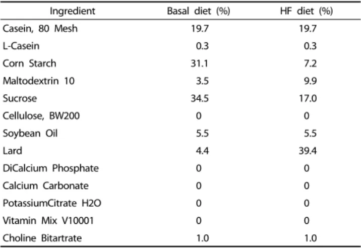
mice in the other groups were fed HFD with 45% fat (Diet 12451, Research Diets. Inc) for 7 weeks. Compositions of diets are shown in Table 1.
The normal and HFD control groups were orally administered distilled water each day for 7weeks. The other four groups received 0.5 g/kg/day MCA, 1.0 g/kg/day MCA, 0.5 g/kg/day MCE, or 1.0 g/kg/day MCE. LD50 for the MCE is 362.34 mg/100 g body weight. The dosages of Momordica charantia extracts used in the current study were far less than the LD50. Body weight and food intake were measured once a week. All animal experiment procedures were approved by Pusan National University-Institutional Animal Care and Use Committee (PNU-IACUC; approval number PNU-2013-0463).
Sample collection
At the end of the feeding period, the mice were sacrificed using ethyl ether after 12 hours of fasting. Blood samples were collected from the inferior vena cava in tubes containing sodium citrate as an anticoagulant. Plasma was recovered by centrifugation at 3,000 rpm for 20 minutes at 4°C. The plasma was frozen at -80°C until further analysis. The liver, kidney, spleen, testis, and testis fat were removed and weighed. The liver was divided into five parts and stored at -80°C for analysis.
Slices of liver were preserved in formalin for histological analysis.
Plasma aspartate aminotransferase (AST), Alanine aminotransferase (ALT), and insulin levels
AST and ALT levels in plasma were measured by using a commercially available kit (AM101-K; Asan Pharm., Korea). The plasma insulin levels were determined using an insulin mouse ELISA kit (KMC2281; Novex, USA).
Plasma lipid profiles
The plasma triglyceride (TG), plasma total cholesterol (TC) and high-density lipoprotein cholesterol (HDL-C) levels were measured using commercially available kits (AM157S-K, AM 202-K, and AM 203-K; Asan Pharm.). The low-density lipoprotein cholesterol (LDL-C) levels were calculated as follows:
LDL-C = TC - HDL-C Hepatic lipid profiles
To determine the hepatic TG and TC concentrations, frozen liver tissues were mixed with a volume of PBS buffer 10 times the weight of the tissue and homogenized. The liver homo- genate was centrifuged at 1600 x g for 10 minutes at 4°C and the supernatant was stored on ice for analysis. The concentra- tions of TG and TC in liver were measured using the same kits (AM157S-K and AM 202-K; Asan Pharm.) and identical methods as those performed for the plasma.
Lipid peroxide and antioxidant enzyme activity in liver
In order to determine hepatic malondialdehyde (MDA) concen-
trations, the liver homogenate was analyzed with a commer-
cially available kit (TBARS assay kit; Cayman Chemical Company,
USA). For the superoxide dismutase (SOD) assay, frozen liver
tissue was mixed with a volume of HEPES buffer 10 times that
of the tissue weight and homogenized. SOD activity in the
supernatant was measured with a corresponding commercial
Ingredient Basal diet (%) HF diet (%)
Casein, 80 Mesh 19.7 19.7
L-Casein 0.3 0.3
Corn Starch 31.1 7.2
Maltodextrin 10 3.5 9.9
Sucrose 34.5 17.0
Cellulose, BW200 0 0
Soybean Oil 5.5 5.5
Lard 4.4 39.4
DiCalcium Phosphate 0 0
Calcium Carbonate 0 0
PotassiumCitrate H2O 0 0
Vitamin Mix V10001 0 0
Choline Bitartrate 1.0 1.0
HF: high-fat
Table 1. Ingredient composition of diets
Group Normal HFD HFD + momordica charantia extracts
MCA0.5 MCA1 MCE0.5 MCE1
Initial body weight (g) 26.60 ± 0.86 26.52 ± 0.86 26.73 ± 0.97 26.00 ± 0.66 26.37 ± 0.82 26.50 ± 0.32
Final body weight (g) 36.83 ± 2.84ab 44.47 ± 2.36c 38.07 ± 2.07b 36.52 ± 3.82ab 35.98 ± 2.25ab 33.62 ± 2.52a
Food intake (g) 3.83 ± 0.38b 3.31 ± 0.22a 3.20 ± 0.27a 3.06 ± 0.26a 3.12 ± 0.31a 3.16 ± 0.15a
Liver weight (g/g B.W.) 3.30 ± 0.36a 3.83 ± 0.42b 3.37 ± 0.35a 3.35 ± 0.43a 3.28 ± 0.17a 3.25 ± 0.36a
Kidney weight (g/g B.W.) 1.35 ± 0.08 1.39 ± 0.32 1.37 ± 0.21 1.20 ± 0.19 1.36 ± 0.16 1.35 ± 0.31
Spleen weight (g/g B.W.) 0.25 ± 0.02ab 0.33 ± 0.11c 0.29 ± 0.11bc 0.27 ± 0.06bc 0.24 ± 0.04ab 0.22 ± 0.04a
Testicle weight (g/g B.W.) 0.53 ± 0.18 0.72 ± 0.07 0.67 ± 0.16 0.69 ± 0.16 0.66 ± 0.17 0.53 ± 0.07
Testicle fat weight (g/g B.W.) 2.07 ± 0.77ab 4.10 ± 0.99c 4.00 ± 1.13c 3.35 ± 1.04bc 2.90 ± 1.22abc 1.81 ± 1.34a Values are presented as mean ± SD (n = 6). The different letters in same line indicate statistically significant difference (P< 0.05) according to Duncan's multiple range test.
Tissue weight was expressed as tissue weight (g) × 100/final body weight. HFD: high-fat diet; MCA0.5: Momordica charantia aqueousextracts 0.5 g/kg/day; MCA1: Momordica charantia aqueousextracts 1 g/kg/day; MCE0.5: Momordica charantia ethanolextracts 0.5 g/kg/day; MCE1: Momordica charantia ethanolextracts 1 g/kg/day Table 2. Effect of Momordica charantia extracts on body weight, food intake, and tissue weight
kit (SOD assay kit; Cayman Chemical Company).
Fecal lipids profiles
To assess fecal TG and TC concentrations, fecal material was freeze-dried and mixed with volume of CHCl
3and MeOH solution (2:1, v:v) 10 times that of the fecal material weight.
After 4 hours of extraction, the mixture was centrifuged at 800 x g for 20 minutes at 4°C and the supernatant was recovered.
The fecal level of TG and TC were measured using the same kits (AM157S-K and AM 202-K, Asan Pharm.) and identical methods as those used for plasma and liver analyses.
Histological analysis
The liver samples preserved in formalin were embedded in paraffin wax and cut into section 4-μm thick. Next, the liver section was stained with hematoxylin and eosin (H&E; Sigma- Aldrich, St. Louis, MO, USA). Morphological features of the hepatocytes were observed using light microscopy at 400 × magnification [14].
Statistical analysis
All data are expressed as the mean ± standard error of the mean. A one way analysis of variance (ANOVA) followed by Duncan’s multiple range test were performed to identify signifi- cance differences among the groups. P-values < 0.05 were considered significant. All statistical analyses were performed with the SPSS program (ver.21).
RESULTS
Body weight, food intake, and visceral tissue weight
As shown in Table 2, the initial body weights of the mice in the six groups were statistically identical. After 7 weeks of oral administration, the final body weight of mice in the HFD group (44.47 ± 2.36 g) was 20.74% higher compared to animals in the normal group (36.83 ± 2.84 g, P < 0.05). Final body weights of the Momordica charantia-treated groups (MCA0.5, 38.07 ± 2.07 g; MCA1, 36.52 ± 3.82 g; MCE0.5, 35.98 ± 2.25 g;
and MCE1, 33.62 ± 2.52 g) were significantly lower (P < 0.05) than that of the HFD group. There was no difference in food intake between the HFD and Momordica charantia- treated mice. However, food intake of the normal group was signifi- cantly higher than of the other groups (P < 0.05).
The relative kidney weight and relative testicle weight did not differ significantly among the six groups. In contrast, mice in the HFD group had a higher relative liver weight, and relative spleen weight, relative testicle fat weight compared to the normal group (P < 0.05). The relative liver weights of the Momordica charantia-treated groups (MCA0.5, 3.37 ± 0.35 100 g/g; MCA1, 3.35 ± 0.43 100 g/g; MCE0.5, 3.28 ± 0.17 100 g/g;
and MCE1, 3.25 ± 0.36 100 g/g) were significantly lower (P <
0.05) than that of the HFD groups (3.83 ± 0.42 100 g/g). Relative spleen weights of the MCE-treated mice (MCE0.5, 0.24 ± 0.04 100 g/g and MCE1, 0.22 ± 0.04 100 g/g) were lower (P < 0.05) than that of the HFD group (0.33 ± 0.11 100 g/g). Additionally, mice in the MCE1 group had a lower relative testicle fat weight (1.81 ± 1.34 100 g/g) compared to that of the HFD group (4.10
± 0.99 100 g/g; P < 0.05).
Plasma AST and ALT levels
Both AST and ALT levels are commonly used to identify the
liver damage [15]. Plasma AST and ALT levels measured in the
current study are shown in Table 3. Plasma AST and ALT levels
of mice only fed the HFD were found to be significantly higher
than that of mice in all normal groups. Plasma AST levels of
the Momordica charantia-treated groups (MCA0.5, 32.40 ± 2.93
Karmen units/mL; MCA1, 25.38 ± 1.89; MCE0.5, 32.26 ± 0.72 Karmen
units/mL; and MCE1, 25.53 ± 4.24 Karmen units/mL) were
Group Normal HFD HFD + Momordica charantia extracts
MCA0.5 MCA1 MCE0.5 MCE1
AST (Karmen unit/ml) 20.97 ± 0.63a 36.17 ± 3.85d 32.40 ± 2.93c 25.38 ± 1.89b 32.26 ± 0.72c 25.53 ± 4.24b ALT (Karmen unit/ml) 17.39 ± 2.85a 23.61 ± 2.04b 22.96 ± 1.28b 16.60 ± 2.00a 20.36 ± 4.94b 16.75 ± 3.67a Values are presented as mean ± SD (n = 6). The different letters in same line indicate statistically significant difference (P< 0.05) according to Duncan's multiple range test.
HFD: high-fat diet; MCA0.5: Momordica charantia aqueousextracts 0.5 g/kg/day; MCA1: Momordica charantia aqueousextracts 1 g/kg/day; MCE0.5: Momordica charantia ethanol extracts 0.5 g/kg/day; MCE1: Momordica charantia ethanolextracts 1 g/kg/day; AST: aspartate aminotransferase; ALT: Alanine aminotransferase
Table 3. Effect of Momordica charantia extracts on liver function
Group Normal HFD HFD + momordica charantia extracts
MCA0.5 MCA1 MCE0.5 MCE1
In plasma
TG (mg/dl) 103.38 ± 21.65a 197.02 ± 15.43c 145.84 ± 18.37b 143.96 ± 20.16b 108.84 ± 22.47a 105.41 ± 27.75a TC (mg/dl) 153.65 ± 15.02a 207.03 ± 11.44c 198.31 ± 25.36c 175.80 ± 19.38b 191.85 ± 26.89bc 154.31 ± 17.06a HDL-C (mg/dl) 114.12 ± 13.61bc 95.32 ± 7.96a 107.41 ± 9.14ab 102.47 ± 9.82ab 124.93 ± 13.81c 125.70 ± 14.95c LDL-C (mg/dl) 50.46 ± 4.18b 96.31 ± 12.23d 76.18 ± 13.20c 68.36 ± 11.04c 70.77 ± 10.10c 33.76 ± 2.69a In liver
TG (mg/g liver) 9.41 ± 0.49ab 13.27 ± 1.61d 11.42 ± 0.28c 8.69 ± 0.44a 9.50 ± 0.41b 9.16 ± 0.53ab TC (mg/g liver) 6.45 ± 2.85a 9.07 ± 1.90b 6.72 ± 1.94a 6.12 ± 1.24a 8.51 ± 2.16b 6.85 ± 1.36a In feces
TG (mg/g feces) 9.27 ± 0.99a 10.25 ± 2.17a 9.78 ± 1.34a 9.96 ± 1.28a 9.82 ± 0.85a 12.81 ± 2.77b
TC (mg/g feces) 7.87 ± 1.34 7.52 ± 1.12 7.23 ± 1.20 6.81 ± 0.90 7.30 ± 1.08 7.23 ± 0.07
Values are presented as mean ± SD (n = 6). The different letters in same line indicate statistically significant difference (P< 0.05) according to Duncan's multiple range test.
HFD: high-fat diet; MCA0.5: momordica charantia aqueousextracts 0.5 g/kg/day; MCA1: momordica charantia aqueousextracts 1 g/kg/day; MCE0.5: momordica charantia ethanolextracts 0.5 g/kg/day; MCE1: momordica charantia ethanolextracts 1 g/kg/day; TG: total triglyceride; TC: total cholesterol; HDL-C: high density lipoprotein cholesterol;
LDL-C: low density lipoprotein cholesterol
Table 4. Effect of momordica charantia extracts on plasma, hepatic and fecal lipid levels
significantly lower (P < 0.05) compared to that of the HFD group (36.17 ± 3.85 Karmen units/mL; P < 0.05). Additionally ALT activity in mice fed a high dose of MCA (16.60 ± 2.00 Karmen units/mL) or MCE (16.75 ± 3.67 Karmen units/mL) was also significantly lower (P < 0.05) than that of the HFD group (23.61
± 2.04 Karmen units/mL).
Plasma insulin levels
Obesity is associated with an increase risk of developing insulin resistance. To compensate for this resistance, more insulin is produced. Therefore, insulin levels were measured. The level of plasma insulin for mice in the HFD group was significantly higher compared to the normal group (P < 0.05). The insulin levels of mice fed a high dose MCE or MCA were lower than that of the HFD group (P < 0.05) and comparable to the level found in the normal group (Fig. 1).
Plasma, hepatic, and fecal lipid levels
In order to study the effects of Momordica charantia extracts on lipid profiles, plasma, hepatic, and fecal lipid levels were measured. The levels of plasma TC, TG, and LDL-C in the HFD group were significantly higher than those in the normal group.
Plasma TG levels were significantly higher in the HFD group (197.02 ± 15.43 mg/dL) than that of the normal control group (103.38 ± 21.65 mg/dL; P < 0.05). Furthermore, plasma TG levels of the Momordica charantia-treated groups (MCA0.5, 145.84 ± 18.37 mg/dL; MCA1, 143.96 ± 20.16 mg/dL; MCE0.5, 108.84 ± 22.47 mg/dL; and MCE1, 105.41 ± 27.75 mg/dL) were significantly lower compared to the HFD group (103.38 ± 21.65 mg/dL; P
< 0.05). Mice fed a high dose of MCA (175.80 ± 19.38 mg/dL)
or MCE (154.31 ± 17.06 mg/dL) had a lower plasma TC level compared to the HFD group (207.03 ± 11.44 mg/dL; P < 0.05).
Treatment with a high dose of MCE (33.76 ± 2.69 mg/dL) significantly lowered plasma TG concentrations by 185.2%
compared to the HFD group (96.31 ± 12.23 mg/dL). The HDL-C levels of mice in the HFD group (95.32 ± 7.96 mg/dL) were significantly lower than those of the normal group (114.12 ±
13.61 mg/dL, P < 0.05). In addition, MCE-treated mice had a higher HDL-C level (MCE0.5, 124.93 ± 13.81 mg/dL and MCE1, 125.70 ± 14.95 mg/dL) compared to the HFD group (P < 0.05).
The levels of hepatic TC and TG for the HFD group were significantly higher than those for the normal group. All of the Momordica charantia-treated animals had lower TG levels (MCA0.5, 11.42 ± 0.28 mg/dL; MCA1, 8.69 ± 0.44 mg/dL; MCE0.5, 9.50 ± 0.41 mg/dL and MCE1, 9.16 ± 0.53 mg/dL) than that of the HFD group (13.27 ± 1.61 mg/dL; P < 0.05). The hepatic TC levels of the MCA- and high dose MCE-treated group (MCA0.5, 6.72 ± 1.94 mg/dL; MCA1, 6.12 ± 1.24 mg/dL; and MCE1, 6.85
± 1.36 mg/dL) were significantly lower relative to the HFD group (13.27 ± 1.61 mg/dL; P < 0.05).
There were no significant differences in fecal TG levels between the normal and HFD groups. However, the fecal TG levels of the in MCE1 group were significantly lower than those of the other groups (P < 0.05). No significant differences in fecal TC levels were observed among the groups (Table 4).
Lipid peroxide and antioxidant enzyme activity in the liver
Lipid peroxidation is a well-established mechanism of cellular
injury in both plants and animals and is used as an indicator
of oxidative stress in cells and tissues [16]. MDA is a naturally
a
b
ab
a
ab a
0.00 0.20 0.40 0.60 0.80 1.00 1.20 1.40 1.60
normal HFD MCA0.5 MCA1 MCE0.5 MCE1
Plasma insulin (ng/ml)
Fig. 1. Effect of momordica charantia extracts on plasma insulin. Values are presented as mean ± SD (n = 6). a-bDifferent letters over the bars indicate that the mean values are significantly different (P< 0.05) according to Duncan's multiple range test. HFD:
high-fat diet; MCA0.5: high-fat diet +momordica charantia aqueousextracts 0.5 g/kg/day;
MCA1: high-fat diet +momordica charantia aqueousextracts 1 g/kg/day; MCE0.5: high-fat diet +momordica charantia ethanolextracts 0.5 g/kg/day; MCE1: momordica charantia ethanolextracts 1 g/kg/day;
a
b
ab
ab ab
a
0.00 5.00 10.00 15.00 20.00 25.00 30.00 35.00 40.00 45.00
Normal HFD MCA0.5 MCA1 MCE0.5 MCE1
MDA content(μM)
Fig. 2. Effect of momordica charantia extracts on MDA content. Each value is expressed as mean ± SD (n = 6). a-bDifferent letters over the bars indicate that the mean values are significantly different (P< 0.05) according to Duncan's multiple range test. HFD:
high-fat diet; MCA0.5: high-fat diet +momordica charantia aqueousextracts 0.5 g/kg/day;
MCA1: high-fat diet +momordica charantia aqueousextracts 1 g/kg/day; MCE0.5: high-fat diet +momordica charantia ethanol extracts 0.5 g/kg/day; MCE1: high-fat diet + momordica charantia ethanolextracts 1 g/kg/day; MDA: Malondialdehyde
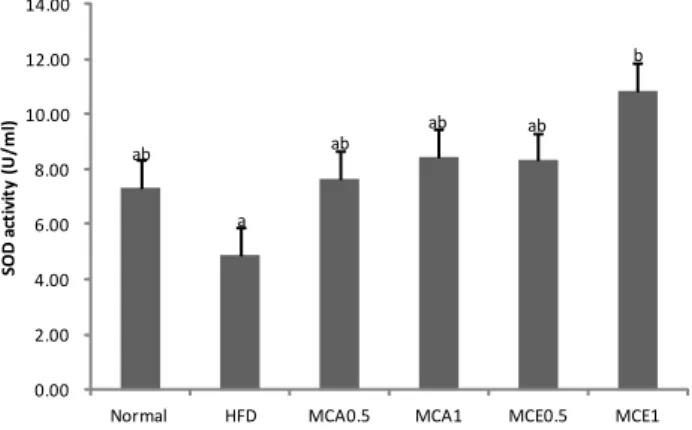
ab
a
ab
ab ab
b
0.00 2.00 4.00 6.00 8.00 10.00 12.00 14.00
Normal HFD MCA0.5 MCA1 MCE0.5 MCE1
SOD activity (U/ml)
Fig. 3. Effect of momordica charantia extracts on SOD activity. Each value is expressed as mean ± SD (n = 6). a-bDifferent letters over the bars indicate that the mean values are significantly different (P< 0.05) according to Duncan's multiple range test. HFD:
high-fat diet; MCA0.5: high-fat diet +momordica charantia aqueousextracts 0.5 g/kg/day;
MCA1: high-fat diet +momordica charantia aqueousextracts 1 g/kg/day; MCE0.5: high-fat diet +momordica charantia ethanol extracts 0.5 g/kg/day; MCE1: high-fat diet + momordica charantia ethanolextracts 1 g/kg/day; SOD: superoxide dismutase
Fig. 4. Effects of momordica charantia extracts on histopathologic changes in mice fed a high-fat diet. Representative H&E stained mouse liver samples from (A):
Normal group (B): HFD group (C): MCA0.5 group (D): MCA1 group (E): MCE0.5 group (F): MCE1 group. H&E: hematoxylin and eosin; HFD: high-fat diet; MCA0.5: high-fat diet
+momordica charantia aqueousextracts 0.5 g/kg/day; MCA1: high-fat diet +momordica charantia aqueousextracts 1 g/kg/day; MCE0.5: high-fat diet +momordica charantia ethanolextracts 0.5 g/kg/day; MCE1: high-fat diet +momordica charantia ethanolextracts 1 g/kg/day; magnification: 400 ×
occurring product of lipid peroxidation [17]. SOD is an enzyme that catalyzes the dismutation of the superoxide anion into molecular oxygen and hydrogen peroxide. SOD is crucial for the prevention of diseases linked to oxidative stress [18]. In order to determine the effects of Momordica charantia on liver lipid peroxidation in mice, hepatic MDA and SOD levels were examined. MDA levels in the liver are shown in Fig. 2. MDA concentration in the HFD group (36.30 ± 2.11 μM) was signi- ficantly higher than that in the normal group (24.10 ± 4.44 μM, P < 0.05). The MDA level in mice treated with a high dose of MCE was significantly lowered by 41.9% compared to the HFD group (P < 0.05). As shown in Fig. 3, there was no significant difference in SOD levels when comparing the normal and HFD groups. However mice given a high dose of MCE had a higher level of SOD activity compared to the other groups (P < 0.05).
Histopathological analysis of the hepatocytes
H&E-stained hepatic sections are shown in Fig. 4. Mice in the normal group had normal histology. No excess lipid was observed in these animals. Mice in the HFD group had severe macrovesicular steatosis and abundant lipid droplets accumu- lation inside the parenchyma cells. But only slight changes in dosage groups. Lipid deposition was reduced remarkably in hepatocytes of mice treated with Momordica charantia extracts compared to the HFD group. Liver histology of the extract- treated groups was similar to that of the normal group. Overall, histological analysis revealed that HFD consumption induced lipid accumulation in the hepatocytes that could be effectively prevented by treatment with Momordica charantia extracts.
DISCUSSION
Momordica charantia contains bioactive compounds that include glycosides, saponins, alkaloids, fixed oils, triterpenes, proteins and steroids. Several phytochemicals such as momor- charins, momordenol, momordolol, charantin, charine, cryptox- anthin, cucurbitacins, cycloartenols, diosgenin, elaeostearic acids, erythrodiol, galacturonic acids, gentisic acid, goyaglycosides, , multiflorenol, have been isolated [11].
The aim of the current study was to determine the effects
of dried Momordica charantia extracts on obesity and lipid profiles of mice fed a HFD. We found that the extracts effectively suppressed body weight gain of mice that consumed a HFD.
Chen et al. previously demonstrated that Momordica charantia juice reduced weight gain without affecting energy intake or apparent fat absorption [12]. In the present investigation, Momordica charantia-s-extracts treated mice also had a lower relative liver weight, spleen weight, and testicle fat weight than those of the HFD group. Furthermore, Momordica charantia was also found to suppress the HFD-induced increase in liver weight in a study by Shih et al. [19]. Momordica charantia treatment of 3T3-L1 preadipocytes cells resulted in a decreased lipid accumulation. And peroxisome proliferator-activated receptor-γ (PPARγ) expression was significantly down-regulated with the treatment [18]. So Momordica charantia extracts reduced the body weight gain possibly by reducing the expression of PPARγ and reduce the lipid accumulation [20].
We also found that insulin levels of mice treated with high doses of MCE or MCA were lower than those of the HFD group and comparable to the levels of the normal group. The hypoglycemic potential of Momordica charantia in normal and diabetic rats or humans with type 2 diabetes has been previously reported [21-23]. This plant may also increase insulin-positive cell numbers in the pancreas [24] and improve insulin resistance [23,25].
In our study, supplementation with Momordica charantia extracts significantly lowered the plasma TG, TC, and LDL-C levels along with hepatic TG and TC concentrations in mice fed a HFD. Treatment with the extracts also elevated plasma HDL-C levels and fecal TG concentration in animals given the HFD.
These findings indicate that Momordica charantia extracts could improve lipid profiles and lipid metabolism. Similar results have been found in previous studies, Momordica charantia extracts were also discovered to reduce serum cholesterol along with hepatic TC and TG in normal rats [26] while increasing HDL-C in streptozotocin-treated diabetic rats [10]. In the present investigation, mice treated with the Momordica charantia extracts had lower MDA contents and higher SOD activity than those in the HFD group, indicating that the extracts can significantly suppress lipid peroxidation. Momordica charantia extracts possess potent antioxidant and free radical scavenging activities. These antioxidant activities could have contributed, at least partly, to the effect of Momordica charantia extracts on suppressing lipid peroxidation [27].
There was no significant difference between MCA or MCE treated groups in general. But the plasma TG level in MCE treated groups was significantly lower compared to MCA treated groups. And the plasma TC and LDL-C level in MCE1 group was lower than that in the MCA treated groups. The plasma MCA treated mice also had significantly higher HDL-C level compared to MCE treated groups. So the MCE treated groups perhaps had better effect on the plasma lipid profiles of mice fed a high-fat diet.
In conclusion, we demonstrated that Momordica charantia extracts have potent in vitro antioxidant activity. Furthermore, the extracts have anti-obesity effects and can improve profiles of lipid improvement effect in mice fed a HFD by suppressing body weight , reducing plasma and hepatic lipid concentrations,
and inhibiting lipid peroxidation while increasing lipid metabolism.
REFERENCES
1. Haththotuwa RN, Wijeyaratne CN, Senarath U. Worldwide epidemic of obesity. In: Mahmood T, Arulkumaran S, editors. Obesity: A Ticking Time Bomb for Reproductive Health. London: Elsevier; 2013.
p.4-10.
2. Yoon KH, Lee JH, Kim JW, Cho JH, Choi YH, Ko SH, Zimmet P, Son HY. Epidemic obesity and type 2 diabetes in Asia. Lancet 2006;368:1681-8.
3. Oh SW. Obesity and metabolic syndrome in Korea. Diabetes Metab J 2011;35:561-6.
4. Wu YF, Ma GS, Hu YH, Li YP, Li X, Cui ZH, Chen CM, Kong LZ.
The current prevalence status of body overweight and obesity in China: data from the China National Nutrition and Health Survey.
Zhonghua Yu Fang Yi Xue Za Zhi 2005;39:316-20.
5. Grundy SM. Obesity, metabolic syndrome, and cardiovascular disease. J Clin Endocrinol Metab 2004;89:2595-600.
6. Parikh NH, Parikh PK, Kothari C. Indigenous plant medicines for health care: treatment of Diabetes mellitus and hyperlipidemia.
Chin J Nat Med 2014;12:335-44.
7. Liao CC, Su TC, Chien KL, Wang JK, Chiang CC, Lin CC, Lin RS, Lee YT, Sung FC. Elevated blood pressure, obesity, and hyperlipidemia.
J Pediatr 2009;155:79-83, 83.e1.
8. Halliwell B, Chirico S. Lipid peroxidation: its mechanism, measure- ment, and significance. Am J Clin Nutr 1993;57:715S-724S.
9. Abd El Sattar El Batran S, El-Gengaihi SE, El Shabrawy OA. Some toxicological studies of Momordica charantia L. on albino rats in normal and alloxan diabetic rats. J Ethnopharmacol 2006;108:
236-42.
10. Ahmed I, Lakhani MS, Gillett M, John A, Raza H. Hypotriglyceridemic and hypocholesterolemic effects of anti-diabetic Momordica charantia (karela) fruit extract in streptozotocin-induced diabetic rats. Diabetes Res Clin Pract 2001;51:155-61.
11. Grover JK, Yadav SP. Pharmacological actions and potential uses of Momordica charantia: a review. J Ethnopharmacol 2004;93:
123-32.
12. Chen Q, Chan LL, Li ET. Bitter melon (Momordica charantia) reduces adiposity, lowers serum insulin and normalizes glucose tolerance in rats fed a high fat diet. J Nutr 2003;133:1088-93.
13. Park SH, Ko SK, Chung SH. Euonymus alatus prevents the hyperglycemia and hyperlipidemia induced by high-fat diet in ICR mice. J Ethnopharmacol 2005;102:326-35.
14. Xu SP, Mao XY, Cheng X, Chen B. Ameliorating effects of casein glycomacropeptide on obesity induced by high-fat diet in male Sprague-Dawley rats. Food Chem Toxicol 2013;56:1-7.
15. M'Kada H, Munteanu M, Perazzo H, Ngo Y, Ramanujam N, Imbert-Bismut F, Ratziu V, Bonnefont-Rousselot D, Souberbielle B, Schuppe-Koistinen I, Poynard T; DILI group of the SAFE-T consortium.
What are the best reference values for a normal serum alanine transaminase activity (ALT)? Impact on the presumed prevalence of drug induced liver injury (DILI). Regul Toxicol Pharmacol 2011;
60:290-5.
16. Yagi K. Simple assay for the level of total lipid peroxides in serum or plasma. Methods Mol Biol 1998;108:101-6.
17. Armstrong D, Browne R. The analysis of free radicals, lipid peroxides, antioxidant enzymes and compounds related to oxidative stress as
applied to the clinical chemistry laboratory. In: Armstrong D, editor.
Free Radicals in Diagnostic Medicine: A Systems Approach to Laboratory Technology, Clinical Correlations, and Antioxidant Therapy.
New York (NY): Plenum Press; 1994. p.43-58.
18. Malmstrom BG, Andreasson LE, Reinhammar B. Copper-containing oxidases and superoxide dismutase. In: Boyer PD, editor. The Enzymes. New York (NY): Academic Press; 1975. p.533.
19. Shih CC, Lin CH, Lin WL. Effects of Momordica charantia on insulin resistance and visceral obesity in mice on high-fat diet. Diabetes Res Clin Pract 2008;81:134-43.
20. Popovich DG, Li L, Zhang W. Bitter melon (Momordica charantia) triterpenoid extract reduces preadipocyte viability, lipid accumu- lation and adiponectin expression in 3T3-L1 cells. Food Chem Toxicol 2010;48:1619-26.
21. Srivastava Y, Venkatakrishna-Bhatt H, Verma Y. Effect of Momordica charantia Linn. pomous aqueous extract on cataractogenesis in murrin alloxan diabetics. Pharmacol Res Commun 1988;20:201-9.
22. Akhtar MS, Athar MA, Yaqub M. Effect of Momordica charantia on blood glucose level of normal and alloxan-diabetic rabbits. Planta
Med 1981;42:205-12.
23. Srivastava Y, Venkatakrishna-Bhatt H, Verma Y, Venkaiah K, Raval BH. Antidiabetic and adaptogenic properties of Momordica charantia extract: an experimental and clinical evaluation. Phytother Res 1993;7:285-9.
24. Ahmed I, Adeghate E, Sharma AK, Pallot DJ, Singh J. Effects of Momordica charantia fruit juice on islet morphology in the pancreas of the streptozotocin-diabetic rat. Diabetes Res Clin Pract 1998;40:
145-51.
25. Leatherdale BA, Panesar RK, Singh G, Atkins TW, Bailey CJ, Bignell AH. Improvement in glucose tolerance due to Momordica charantia (karela). Br Med J (Clin Res Ed) 1981;282:1823-4.
26. Jayasooriya AP, Sakono M, Yukizaki C, Kawano M, Yamamoto K, Fukuda N. Effects of Momordica charantia powder on serum glucose levels and various lipid parameters in rats fed with cholesterol-free and cholesterol-enriched diets. J Ethnopharmacol 2000;72:331-6.
27. Wu SJ, Ng LT. Antioxidant and free radical scavenging activities of wild bitter melon (Momordica charantia Linn. var. abbreviata Ser.) in Taiwan. Lebenson Wiss Technol 2008;41:323-30.