ABSTRACT
Purpose: Obesity is a serious public health issue for the modern society and is considered a chronic health hazard. There are many surgical and pharmacological approaches to treat obesity. However, various potentially hazardous side effects remain the biggest challenge.
Therefore, diets based on foods derived from natural products have gained increasing attention compared to anti-obesity drugs. Recently, research on edible insects as a food source has been a topic of considerable interest in the scientific communities. This study examined the anti- obesity effects of ingesting an edible insect by feeding a high-fat diet (HFD)-induced obese mouse models with a diet containing Tenebrio molitor larvae powder (TMLP).
Methods: Six-week-old female C57BL/6J mice were divided into 4 groups according to
treatment: 100% normal diet (ND), 100% HFD (HFD), HFD 99% + TMLP 1% (TMLP), and HFD 97% + TMLP 3% (TMLP 3%). TMLP was added to the HFD for 6 weeks for the latter two groups.
Results: Compared to the HFD group, mice in the TMLP group showed weight loss, and micro-computed tomographic imaging revealed that the volume of the adipose tissue in the abdominal area also showed significant reduction. After an autopsy, the fat weight was found to be significantly reduced in the TMLP group compared to the HFD group. In addition, the degree of fat cell deposition in the liver tissue and the size of the adipocytes significantly decreased in the TMLP group. Reverse transcription polymerase chain reaction analysis for the mRNA expression of adipogenesis-related genes namely CCAAT-enhancer-binding proteins (C/EBP-β, C/EBP-δ), and fatty acid-binding protein 4 (FABP4) showed that the expression levels of these genes were significantly reduced in the TMLP group compared to the HFD group. Serum leptin level also decreased significantly in the TMLP group in the comparison with the HFD group. In addition, total cholesterol, triglyceride, and glucose levels in mouse serum also decreased in the TMLP group.
Conclusion: Taken together, our results showed that TMLP effectively inhibited adipocyte growth and reduced body weight in obese mice.
Keywords: obesity, Tenebrio, larva, food, insecta
Research Article
Received: Jun 9, 2021 Revised: Jul 26, 2021 Accepted: Aug 9, 2021 Correspondence to Bong Joo Lee
Department of Veterinary Infectious Diseases, College of Veterinary Medicine, Chonnam National University, 77 Yongbong-ro, Buk-gu, Gwangju 61186, Korea.
Tel: +82-62-530-2850 E-mail: [email protected]
© 2021 The Korean Nutrition Society This is an Open Access article distributed under the terms of the Creative Commons Attribution Non-Commercial License (http://
creativecommons.org/licenses/by-nc/3.0/) which permits unrestricted non-commercial use, distribution, and reproduction in any medium, provided the original work is properly cited.
ORCID iDs Bo Mi Park
https://orcid.org/0000-0003-3979-9510 Hyung Ju Lim
https://orcid.org/0000-0001-7904-1435 Bong Joo Lee
https://orcid.org/0000-0002-4756-7376 Conflict of Interest
There are no financial or other issues that might lead to conflict of interest.
Bo Mi Park 1, Hyung Ju Lim 2, and Bong Joo Lee 1
1 Department of Veterinary Infectious Diseases, College of Veterinary Medicine, Chonnam National University, Gwangju 61186, Korea
2Jeollanamdo Veterinary Service Laboratory, Suncheon 57923, Korea
Anti-obesity effects of Tenebrio
molitor larvae powder in high-fat diet-
induced obese mice
INTRODUCTION
Obesity is a chronic health hazard growing rapidly worldwide, causing significant human health risks. Obesity increases the incidence of comorbidities such as stroke, atherosclerosis, myocardial infarction, cardiovascular disease, diabetes, hypertension, and hyperlipidemia [1].
Moreover, obesity is associated with an accumulation of excess energy in the triglyceride-type fatty tissues caused by the intake of food providing an amount of energy that exceeds energy consumption. The surplus energy activates pre-adipocytes in the adipose tissue, increasing the number and size of adipocytes [2]. Therefore, inhibiting the activity of pre-adipocytes and reducing the size and number of adipocytes may be an effective treatment for obesity. To date, various treatment regimens have been proposed for obesity management, including exercise, gastrectomy, and prescription weight-loss drugs [3]. Most known anti-obesity drugs play a role in appetite suppression and nutrient absorption, which is often accompanied by varying degrees of hazardous side effects [4-7]. Therefore, due to the undesirable and detrimental effects of anti-obesity drugs, it is of vital importance to ramp up research on identifying natural products that offer safe therapeutic effects to tackle the obesity epidemic.
To date, many studies have explored the use of insects as food to prevent obesity. Feeding Protaetia brevitarsis larvae extract to a high-fat diet (HFD)-induced obesity mouse model resulted with decreases in weight, liver fat cells, and fat-related levels in the serum [8]. A study on the effect of administering Allomyrina dichotoma larvae powder to HFD mice found that the volume of fat tissue decreased significantly due to the upregulation of the fat metabolism genes [9]. Insects, as a substitute for animal-based meat, has recently become a developing research focus due to its naturally rich properties in protein, fats, minerals and vitamins [10]. Tenebrio molitor larvae (mealworm) is an edible insect that is widely used as pet food. Insect breeding offers the advantage of rapid growth, very low feed cost as well as tremendous cost savings on space for breeding and handling compared to animal farming [11]. The fatty acid components of mealworms are rich in oleic and palmitic acids, known as immune-enhancing components; therefore, more studies are imperative to develop mealworms as functional health foods [12]. In terms of nutrition, freeze-dried mealworms contain 33% fat and 51% crude protein, and are rich in minerals, vitamins, amino acids, and fatty acids. Several T. molitor studies have shown anti-obesity effects. A recent study has shown that administering a diet containing the larvae of T. molitor to the obese Zucker rat model resulted in a decrease of fatty acid synthesis in the liver, as well as in the plasma triglyceride and cholesterol levels. Results also showed that ingestion of T. molitor larvae may alleviate hyperlipidemia [13]. Other study, the T. molitor larvae were powdered, fermented, ethanol-extracted thereafter fed to mice or applied to 3T3-L1 adipocytes to regulate
adipogenesis-related pathways and regulate the protein involved in glucose, lipid, amino acid metabolism [14,15], or reduce endoplasmic reticulum (ER) stress and food intake [15,16]. In addition to this study, there have been other recent research activities using T. molitor showing anti-obese effects and the potential to develop into treatment modalities with further investigations warranted.
In this study, we investigated the effectiveness of administering T. molitor larvae powder (TMLP) to HFD-induced obese mice for obesity prevention. To determine the anti-obesity effects, micro-computed tomographic (CT) images of the abdominal area were examined to determine the volume of the adipose tissue and the size of the fat cells in the liver and adipose tissues. Additionally, serum levels of adiponectin and leptin, total cholesterol, triglyceride and glucose levels, as well as adipogenesis-related gene expression were examined.
METHODS
Animals and experimental diets
Six-week-old female mice (C57BL/6J) were purchased from Damule Science (Daejeon, Korea).
TMLP was obtained from Greeinsect Co. (Gwangju, Korea). All diets used in this study were prepared by adding TMLP to the HFD (Damule Science) that had a 45% fat content. The mice were then randomly divided into 4 groups (n = 5 each) according to treatment: 100% normal diet 100% (ND group), 100% HFD (HFD group), HFD 99% + TMLP 1% (TMLP 1% group), and HFD 97% + TMLP 3% (TMLP 3% group). Each group was fed the designated experimental meal for 6 weeks. The mice were kept in an SPF facility at constant temperature of 21 ± 2°C and humidity of 50 ± 5% under a 12-hour light:dark cycle. All protocols related to animal handling and experiments were according to the Guide for the Care and Use of Laboratory Animals of the National Institutes of Health and approved by the Chonnam National University Animal Experimental Ethics Committee (CNU IACUC-YB-2018-32). During the treatment period, mice were checked for weight changes every week. After six-week, mice were anesthetized with avertin and autopsied. Subcutaneous and visceral adipose tissues were dissected and weighed.
Whole blood samples (from the heart) were collected and centrifuged at 2,000 × g for 15 minutes at 4°C, and the separated serum samples were stored at −80°C until use.
In vivo micro-CT for evaluation of the area and volume of the abdominal adipose tissue
Micro-CT analysis for evaluating the area and volume of mouse visceral and subcutaneous adipose tissue was performed using a Quantum GX μCT imaging system (PerkinElmer, Hopkinton, MA, USA). During scanning, mice were immobilized by administering a light dose of anesthesia (2% isoflurane/O2 gas).
Histopathological examination of liver and adipose tissue
At the end of the experiment, the liver and adipose tissues (subcutaneous and visceral fat) were removed from the mice and weighed. Liver and adipose tissues were fixed in 10% neutral buffered formalin, processed for paraffin embedding, sectioned, and then stained with hematoxylin and eosin (H&E). To evaluate the degree of hepatocellular fat change induced by HFD, a histological rating of the nonalcoholic hepatitis clinical study network scoring system was used, which was represented by the non-alcoholic fatty liver disease activity score, NAS. Hepatic fat changes were scored according to the percentage of hepatocytes showing the changes: 0 ≤ 5%, 1 = 5%–33%, 2 ≥ 33%–66%, and 3 ≥ 66% [17].
To quantify the degree of histological liver lesions, 10 representative images were taken from H&E-stained liver sections, and 2 pathologists used blinded evaluation techniques to calculate the score of steatosis. To quantify the size of the adipocytes in the adipose tissues, a total of 10 representative images were taken from H&E-stained adipose tissues and then converted to pixels using ImageJ software (https://imagej.nih.gov/ij/). The average size of 10 cells in 10 representative images of each group was calculated using ImageJ software.
RNA extraction and quantitative reverse transcription polymerase chain reaction (RT-PCR)
Total RNA from visceral adipose tissue was extracted using RNeasy Mini kit (Qiagen, Hilden, Germany) and quantified using a NanoDrop ND-1000 device (Thermo Fisher Scientific, Wilmington, DE, USA). Extracted RNA was used as the template for the synthesis of single- stranded cDNA using SuperScript™ III RT kit (Invitrogen, Waltham, MA, USA). Quantitative RT-PCR was performed on an Applied Biosystems 7500 Real-Time PCR system (Applied
Biosystems, Forster, CA, USA). Primer sequences are shown in Table 1. The most common housekeeping gene, glyceraldehyde-3-phosphate dehydrogenase (GAPDH), was used as an internal control. RT-PCR reaction mixtures were prepared using Power SYBR Green PCR master mix (Thermo Fisher Scientific). The relative expression levels of the target genes were determined using the comparative Ct method (threshold cycle number at the cross-point between the amplification plot and threshold) with GAPDH as an internal control. mRNA expression levels of the target genes were normalized to those of GAPDH.
Enzyme-linked immunosorbent assay (ELISA)
The serum concentrations of adiponectin and leptin were measured using a mouse adiponectin ELISA kit (EMD Millipore Corporation, Burlington, MA, USA), and a mouse Quantikine ELISA kit (R&D Systems, Minneapolis, MN, USA), respectively. Measurements were performed according to the manufacturer's instructions. Absorbance was quantified using a microplate spectrophotometer (Thermo Fisher Scientific, Wilmington, DE, USA).
The limits of detection for adiponectin and leptin were 166 ng/mL and 4,000 pg/mL, respectively. The concentrations of total cholesterol, triglycerides, and glucose in the mouse serum were measured using ELISA kits (total cholesterol, mouse triglyceride, and mouse glucose ELISA kits; Mybiosource, San Diego, CA, USA). ELISA was performed according to the manufacturer's instructions. Total concentrations for blood cholesterol, triglycerides, and glucose were measured at optical density 450 nm and calculated by referring to the standard curve.
Statistical analysis
Each data value is presented as the mean ± SD. Statistical analysis for the data was performed using one-way analysis of variance followed by Dunnett's multiple comparison test between groups. Data were considered statistically significant if the probability value was 0.05 or less (p ≤ 0.05). All data were analyzed using GraphPad Prism 5.00 (GraphPad Software Inc., San Diego, CA, USA).
RESULTS
Body weight gain
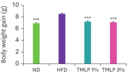
After the end of the 6-week experimental period, the weight gain values by groups were 8.49 g (HFD), 7.15 g (TMLP 1%), 7.02 g (TMLP 3%), and 7 g (ND). Weight gain of the mice in the TMLP groups was significantly lower than that of the mice in the HFD group (Fig. 1).
In vivo quantification of adipocytes using micro-CT
To measure the area and volume of mouse subcutaneous and visceral fat, micro-CT images were analyzed using the Analyze Direct (Overland Park, KS, USA) and return on investment Table 1. Primer sequences for mouse adipogenesis-related genes and GAPDH
GENE F primer (5′-3′) R primer (5′-3′)
GAPDH CTGGAGAAACCTGCCAAGTA AGTGGGAGTTGCTGTTGAAG
Pparg CAAAACACCAGTGTGAATTA ACCATGGTAATTTCTTGTGA
Cebpb CACAGCGACGACTGCAAGATCC CTTGAACAAGTTCCGCAGGGTG
Cebpd CAGGCTTGGACGGCTAAGTA TCCAAGTTGGCTGTCACCTC
Fabp4 ATCCCCAACATGCCCATTCG TTCAATGCGAACTTCAGTCC
GAPDH, glyceraldehyde-3-phosphate dehydrogenase; Pparg, peroxisome proliferator-activated receptor gamma;
Cebpb, CCAAT-enhancer-binding protein-β; Cebpd, CCAAT-enhancer-binding protein-δ; Fabp4, fatty acid-binding protein 4.
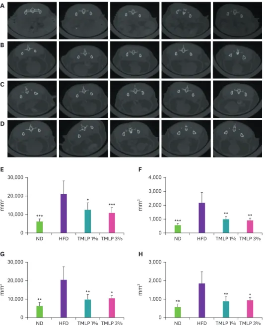
(ROI) calculators (Fig. 2). HFD group showed a significant increase in the area and volume of both the subcutaneous and visceral fat compared to the ND group. However, the accumulation of subcutaneous and visceral fat in the TMLP groups was reduced compared to that in the HFD group. Area measurement for subcutaneous fat revealed that TMLP 1% and TMLP 3% groups showed significant reductions of 42 and 49%, respectively, compared with the HFD group (Fig. 2E). The volume of subcutaneous fat also decreased by 55 and 60% in TMLP 1% and 3%
groups, respectively, compared with the volume in the HFD group (Fig. 2F). Upon comparing the area size of visceral fat, TMLP 1% and 3% groups showed a decrease of 52 and 50%, respectively, compared with the value observed in the HFD group (Fig. 2G). Additionally, the volume of the visceral fat decreased by 53 and 50% in TMLP 1% and 3% groups, respectively, compared with the volume observed in the HFD group (Fig. 2H).
Weight of the adipose tissues
At the end of the experimental period, the weights of the visceral and subcutaneous adipose tissues were measured. The weight of the subcutaneous adipose tissue in TMLP 1% and TMLP 3% groups decreased by 0.6 g (43%) and 0.8 g (55%), respectively, compared to that in the HFD group (Fig. 3A). The weight of the visceral adipose tissue in TMLP 1% and TMLP 3%
groups was also reduced by 0.9 g, compared to that in the HFD group (Fig. 3B).
Histopathological analysis of liver
Analysis of adipocyte accumulation in the hepatocytes was based on histopathological findings and quantification methods. Observed histopathological changes in the liver showed that HFD group mice showed numerous lipid droplets throughout the liver lobe, but not the ND group mice. However, liver tissues of the mice in the TMLP groups showed reduced HFD-induced adipocyte accumulation in a dose-dependent manner (Fig. 4A).
Quantification of hepatocellular fat change was performed using the steatosis class system [17] and results showed that compared with the mice in the HFD group, the mice in the TMLP groups exhibited significant inhibitory effects denoted by the accumulation of adipocytes in liver tissues. The steatosis scores by groups were 1 (ND), 1 (TMLP 1%), 1.6 (TMLP 3%), and 2.6 (HFD) (Fig. 4B).
Adipocyte size in subcutaneous and visceral adipose tissues
Histopathological examination of the subcutaneous adipocyte tissue showed a significant increase in adipocyte size in the HFD group compared with the ND group. However, in TMLP 1% and TMLP 3% groups, adipocyte sizes were reduced in a dose-dependent manner compared with the HFD group (Fig. 5A and B). The visceral adipocyte tissue also showed a significant increase in adipocyte size in the HFD group, compared with ND group. However,
10
6 8
4 2 ain (Body weight gg) 0
ND HFD TMLP 1% TMLP 3%
*** ***
***
Fig. 1. Body weight gain in mice after 6-week of administering different TMLP treatments. Values are presented as the mean ± SD and compared to the HFD group by using Dunnett's multiple comparison test.
ND, 100% normal diet; HFD, 100% high-fat diet; TMLP, high-fat diet 99% + Tenebrio molitor larvae powder 1%;
TMLP 3%, high-fat diet 97% + Tenebrio molitor larvae powder 3%.
***p < 0.001.
TMLP groups showed a dose-dependent decrease in adipocyte size, compared with the HFD group (Fig. 5C and D). Microscopy-based quantification showed that adipocyte size decreased significantly in TMLP groups compared with the HFD group (Fig. 5B and D).
Expression of adipogenesis-related genes
Genes related to adipogenesis in the RNA extracted from visceral fat were quantified and compared by using RT-PCR. The mRNA expression levels of CCAAT-enhancer-binding
D C B A
* 30,000
20,000 10,000 0
mm2
ND HFD TMLP 1% TMLP 3%
***
***
E
**
4,000 3,000 2,000 1,000 0
mm3
ND HFD TMLP 1% TMLP 3%
*** **
F
**
30,000 20,000 10,000 0
mm2
ND HFD TMLP 1% TMLP 3%
** *
G
**
3,000 2,000 1,000 0
mm3
ND HFD TMLP 1% TMLP 3%
** *
H
Fig. 2. Micro-CT images of adipose tissue and comparison of fat area and volume in the abdomen of C57BL6 mice fed with different TMLP-enriched diets. The abdominal fat images among (A) ND, (B) HFD (C) TMLP 1%, (D) TMLP 3% groups were compared (bones in light white, internal organs are bright gray, and fat tissue is dark gray).
The calculated results of (E) subcutaneous fat area, (F) subcutaneous fat volume, (G) visceral fat area and (H) visceral fat volume are plotted. Data represent the mean ± SD compared to the HFD group by using Dunnett's multiple comparison test.
Micro-CT, micro-computed tomographic; TMLP, Tenebrio molitor larvae powder; ND, 100% normal diet; HFD, 100% high-fat diet; TMLP, high-fat diet 99% + Tenebrio molitor larvae powder 1%; TMLP 3%, high-fat diet 97% + Tenebrio molitor larvae powder 3%.
*p < 0.05, **p < 0.01, and ***p < 0.001.
protein (C/EBP)-β (Cebpb) and C/EBP-δ (Cebpd) increased significantly in the HFD group, compared with the ND group, but were significantly decreased in the TMLP 1% and TMLP 3% groups, compared with the HFD group (p < 0.05, respectively) (Fig. 6A and B). mRNA expression of fatty acid-binding protein 4 (Fabp4) gene was significantly higher in the HFD group than in the ND group, but showed a significant decrease in the TMLP 1% and 3%
groups, compared with the HFD group (p < 0.01 and p < 0.05, respectively) (Fig. 6C). Despite the increase in the mRNA expression levels of peroxisome proliferator-activated receptor gamma (PPAR-γ; Pparg) gene in all experimental groups, compared with the ND group, the difference was not significant, and similarly, the TMLP groups showed lower expression compared to the HFD group but the difference was not statistically significant (Fig. 6D).
Status of serum metabolites
The concentrations of leptin and adiponectin in mouse serum were measured using ELISA.
Leptin levels were significantly increased in the HFD group compared with the ND group, whereas the TMLP 1% and TMLP 3% groups showed a decrease by approximately 44% and 55%, respectively, compared with the HFD group (Fig. 7A). However, adiponectin levels tended to increase significantly in the HFD group, compared with the ND group, but no
2.5 2.0 1.5 1.0 0.5
g)Subcutaneous fat ( 0
ND HFD TMLP 1% TMLP 3%
*
***
A
* 2.5
2.0 1.5 1.0 0.5 0
Visceral fat (g)
ND HFD TMLP 1% TMLP 3%
*
**
B
Fig. 3. Weight of subcutaneous and visceral fat. (A) subcutaneous fat and (B) visceral fat. Data represents the mean ± SD and compared to the HFD group by using Dunnett's multiple comparison test.
ND, 100% normal diet; HFD, 100% high-fat diet; TMLP, high-fat diet 99% + Tenebrio molitor larvae powder 1%;
TMLP 3%, high-fat diet 97% + Tenebrio molitor larvae powder 3%.
*p < 0.05, **p < 0.01, and ***p < 0.001.
3 2 1 0
Steatosis grade
ND HFD TMLP 1% TMLP 3%
**
**
ND HFD B
TMLP 1% TMLP 3%
A
Fig. 4. Effect of Tenebrio molitor larvae powder on the inhibition of hepatic steatosis in the mice. (A) Comparison of histological hepatocyte fatty changes. A representative set of results are shown. Hematoxylin and eosin stain (bar = 50 µm). (B) The severity of steatosis in each experimental group was calculated and statistically analyzed.
Values are shown as the means ± SD. Significant differences of steatosis compared to HFD group by using Dunnett's multiple comparison test.
ND, 100% normal diet; HFD, 100% high-fat diet; TMLP, high-fat diet 99% + Tenebrio molitor larvae powder 1%;
TMLP 3%, high-fat diet 97% + Tenebrio molitor larvae powder 3%.
**p < 0.01.
ND HFD
TMLP 1% TMLP 3%
A ND HFD
TMLP 1% TMLP 3%
B
15 10 5
0 ND HFD TMLP 1% TMLP 3%
***
**
3Pixcels (× 10) ***
C
Pixcels (× 103)
15 10 5
0 ND HFD TMLP 1% TMLP 3%
***
***
***
D
Fig. 5. Inhibitory effect of Tenebrio molitor larvae powder on adipocyte sizes in gonadal (A, C) and subcutaneous (B, D) adipose tissue. Hematoxylin and eosin stain (bar = 100 µm) of adipocyte were compared in the gonadal and subcutaneous adipose tissues, respectively (A and B). A representative set of results is shown. The average size of adipocytes in each experimental group was calculated and statistically analyzed (C and D). Values are shown as the means ± SD.
Significant differences of adipocyte sizes compared to HFD group by using Dunnett's multiple comparison test.
ND, 100% normal diet; HFD, 100% high-fat diet; TMLP, high-fat diet 99% + Tenebrio molitor larvae powder 1%; TMLP 3%, high-fat diet 97% + Tenebrio molitor larvae powder 3%.
**p < 0.01 and ***p < 0.001.
2.0 1.5 1.0 0.5
0 ND HFD TMLP 1% TMLP 3%
* *
*
Relative Cebpb level
A
1.5 1.0 0.5
0 ND HFD TMLP 1% TMLP 3%
* *
Relative Cebpd level
B
2.0 1.5 1.0 0.5
0 ND HFD TMLP 1% TMLP 3%
** *
*
Relative Fabp4 level
C
2.0 1.5 1.0 0.5
0 ND HFD TMLP 1% TMLP 3%
Relative Pparg level
D
Fig. 6. Expression of mRNA in the adipocyte differentiation-related genes in visceral adipose tissues of the mice. (A) Cebpb, (B) Cebpd, (C) Fabp4, and (D) Pparg. Data represents the mean ± SD and compared to HFD group by using Dunnett's multiple comparison test. Expression level of each mRNA was normalized to the level of GAPDH expression.
Cebpb, CCAAT-enhancer-binding protein-β; Cebpd, CCAAT-enhancer-binding protein-δ; Fabp4, fatty acid-binding protein 4; Pparg, peroxisome proliferator- activated receptor gamma; ND, 100% normal diet; HFD, 100% high-fat diet; TMLP, high-fat diet 99% + Tenebrio molitor larvae powder 1%; TMLP 3%, high-fat diet 97% + Tenebrio molitor larvae powder 3%.
*p < 0.05 and **p < 0.01.
significant differences were observed in the TMLP 1% and TMLP 3% groups, compared with the HFD group (Fig. 7B). Total cholesterol, triglyceride, and glucose concentrations in the serum of mice were measured. Total cholesterol in HFD group (p < 0.001) was significantly increased compared to the ND group and was lower in the TMLP 1% group compared to the HFD group, but without any significant difference, except for the TMLP 3% group (p
< 0.05) (Fig. 7C). Triglyceride levels in the HFD group were significantly increased (p <
0.001) compared with those in the ND group, and were significantly reduced in the TMLP 1% and TMLP 3% groups compared with those in the HFD group (p < 0.001 and p < 0.01, respectively) (Fig. 7D). Glucose concentrations were significantly higher in the HFD group than in the ND group, and significantly reduced in both TMLP 1% and TMLP 3% groups compared to the HFD group (p < 0.001) (Fig. 7E).
DISCUSSION
Obesity is caused by excessive energy accumulation through excessive food intake. As such, energy intake and expenditure in the body continue to be imbalanced, causing chronic metabolic diseases such as diabetes, hyperlipidemia, dyslipidemia, and cardiovascular
2,500 2,000 1,500 1,000 500
0 ND HFD TMLP 1% TMLP 3%
* **
***
pg/mL
A
20 15 10 5
0 ND HFD TMLP 1% TMLP 3%
*
ng/mL
B
300 200 100
0 ND HFD TMLP 1% TMLP 3%
* /dLmg ***
C
100 80 60 40 20
0 ND HFD TMLP 1% TMLP 3%
*** *** **
mg/dL
D
250 200 150 100 50
0 ND HFD TMLP 1% TMLP 3%
*** ***
/dLmg **
E
Fig. 7. The serum levels of metabolites in mice. (A) leptin, (B) adiponectin, (C) total cholesterol, (D) triglycerides, and (E) glucose. Data represents the mean ± SD and compared to HFD group by using Dunnett's multiple comparison test.
ND, 100% normal diet; HFD, 100% high-fat diet; TMLP, high-fat diet 99% + Tenebrio molitor larvae powder 1%;
TMLP 3%, high-fat diet 97% + Tenebrio molitor larvae powder 3%.
*p < 0.05, **p < 0.01, and ***p < 0.001.
diseases. Obesity can be chronically damaging to health, and many different types of surgery and pharmacological approaches have been studied to treat obesity. However, with the emergence of large and small side effects on developed drugs and surgical methods, finding candidate substances for obesity treatment through a new approach has become a major issue in modern research [1-3,18].
In this study, we investigated the anti-obesity effect of TMLP, which has gained increasing attention as a healthy food due to its abundance of proteins, minerals, amino acids, and fatty acids [19]. To investigate the anti-obesity effects of TMLP, an HFD-induced obese mouse model fed with a diet containing TMLP was used. After six weeks, weight gain of mice was significantly decreased in the TMLP-fed group compared to the HFD group. Accumulation of adipocytes in the liver tissue also decreased in the TMLP group mice compared to the mice in the HFD group, and steatosis score [20] was significantly reduced in the TMLP 1% group compared with the HFD group, while the TMLP 3% group showed a level similar to that of the ND group. In addition, the mean area of adipocytes in the adipose tissue was significantly decreased in the TMLP groups compared with the HFD group. Adipocytes in obese animals increase in size to store the excess energy [21]. Fat cells that reach their maximum size can activate macrophages, causing inflammation and increased insulin resistance. Therefore, chronic obesity leads to chronic inflammation, the activation of specific tumors, and type 2 diabetes. Therefore, reducing the size of fat cells is an important factor in mitigating obesity- induced diseases [22]. Increased adipose tissue volume in the subcutaneous and visceral tissues of mice is an important indicator of obesity. Micro-CT scans showed a decrease in the adipose tissue area and volume in the TMLP group mice compared with the mice in the HFD group. In addition, subcutaneous and visceral adipose tissue weights were significantly reduced in the TMLP group compared with the HFD group.
In this study, the expression of adipocyte differentiation genes was investigated. The mRNA expression levels of Cebpb, Cebpd, and Fabp4 genes in adipose tissue in the TMLP groups were significantly lower than those in the HFD group. The Cebpb, Cebpd, the Pparg, and Fabp4 genes are important for adipocyte differentiation [23]. These genes were found to show increased expression in the case of obesity and function as lipid chaperone proteins in the adipose tissue [24]. In the HFD-fed group, the reduction in fat volume and adipocyte size showed a dose-dependent relation with the expression of adipocyte differentiation genes.
Adipocytes not only store energy but also secrete endocrine factors such as adipokines, cytokines, and growth factors [25,26]. Leptin and adiponectin secreted by adipocytes are adipokines, a type of cytokine [27]. The concentrations of leptin and adiponectin in the blood are closely related to obesity and body weight change [28]. Leptin is an adipokine that controls food and is involved in maintaining weight. In obese individuals, the level of leptin in the blood increases proportionally [29,30]. Adiponectin is an adipokine that contributes to fatty acid, glucose, and carbohydrate metabolism, and improves insulin resistance.
Therefore, it is thought to be a substance that can treat obesity and diabetes [22]. In this study, serum leptin levels were significantly lower in the TMLP groups than in the HFD group. Therefore, reduced leptin levels may have affected the reduced fat accumulation in the adipose tissue. Other studies have shown that HFD-induced obese mice have increased levels of blood sugar and total cholesterol and triglycerides in the blood [31].
A study by Huang et al. [32] showed significant reduction in food intake and adipose tissue accumulation in chitin fed obesity rats which prevented weight gain and significantly reduced
serum lipid levels such as total cholesterol, triglycerides and low-density cholesterol. Other studies have shown that unsaturated fatty acids (UFAs) and polyunsaturated fatty acids (PUFAs) can also contribute to obesity regulation, contributing to the production of lipid intermediates that are particularly involved in food intake, energy detection and food-related disorders [33,34]. T. molitor larvae contains more than 50% protein. Their exoskeletons also contain plenty of chitin. It also has a high composition of UFAs which accounts for nearly 80% of its total fatty acid content and has essential PUFAs such as omega 3 and 6 [35]. Thus, in this study TMLP was administered to obese mice which showed abdominal fat reduction in both volume and weight as well as a decrease in adipogenesis genes and metabolites.
However, it is yet to be answered as to which components of the TMLP has the lipid-lowering effect. The high content of chitin, UFAs and PUFAs of the TMLP administered to mice are thought to have anti-obesity effects. Thus, further research is needed to investigate which components of T. molitor larvae are effective against obesity and its mechanism of action.
SUMMARY
Our findings suggested that the high protein, chitin, UFAs and PUFAs in TMLP had an anti-obesity effect in obesity mice. Compared to the HFD group, TMLP-fed mice showed decreases in weight, subcutaneous and visceral fat volumes, adipocyte accumulation in the liver, adipocyte size, leptin concentration, adipogenesis-related gene levels, as well as serum levels for total cholesterol, triglyceride, and glucose. Therefore, T. molitor larvae as an edible insect, is a natural protein-rich nutrient source and has low health risk, and may be a safer alternative to reduce fat cells and control obesity compared to weight-loss drugs.
REFERENCES
1. Pi-Sunyer FX. The obesity epidemic: pathophysiology and consequences of obesity. Obes Res 2002; 10 Suppl 2: 97S-104S.
PUBMED | CROSSREF
2. Spiegelman BM, Flier JS. Adipogenesis and obesity: rounding out the big picture. Cell 1996; 87(3): 377-389.
PUBMED | CROSSREF
3. Kissane NA, Pratt JS. Medical and surgical treatment of obesity. Best Pract Res Clin Anaesthesiol 2011;
25(1): 11-25.
PUBMED | CROSSREF
4. Heck AM, Yanovski JA, Calis KA. Orlistat, a new lipase inhibitor for the management of obesity.
Pharmacotherapy 2000; 20(3): 270-279.
PUBMED | CROSSREF
5. Palacios-Martinez D, Garcia-Alvarez JC, Montero-Santamaria N, Villar-Ruiz OP, Ruiz-Garcia A, Diaz- Alonso RA. Macrocytic anemia and thrombocytopenia induced by orlistat. Int J Endocrinol Metab 2013;
11(4): e6721.
PUBMED | CROSSREF
6. Krentz AJ, Fujioka K, Hompesch M. Evolution of pharmacological obesity treatments: focus on adverse side-effect profiles. Diabetes Obes Metab 2016; 18(6): 558-570.
PUBMED | CROSSREF
7. Eroglu E, Gemici G, Bayrak F, Kalkan AK, Degertekin M. Acute myocardial infarction in a 24 year-old man possibly associated with sibutramine use. Int J Cardiol 2009; 137(2): e43-e45.
PUBMED | CROSSREF
8. Jeong EJ, Jegal J, Ahn J, Kim J, Yang MH. Anti-obesity effect of Dioscorea oppositifolia extract in high-fat diet- induced obese mice and its chemical characterization. Biol Pharm Bull 2016; 39(3): 409-414.
PUBMED | CROSSREF
9. Yoon YI, Chung MY, Hwang JS, Han MS, Goo TW, Yun EY. Allomyrina dichotoma (Arthropoda: Insecta) larvae confer resistance to obesity in mice fed a high-fat diet. Nutrients 2015; 7(3): 1978-1991.
PUBMED | CROSSREF
10. Yi L, Lakemond CM, Sagis LM, Eisner-Schadler V, van Huis A, van Boekel MA. Extraction and characterisation of protein fractions from five insect species. Food Chem 2013; 141(4): 3341-3348.
PUBMED | CROSSREF
11. Han SR, Lee BS, Jung KJ, Yu HJ, Yun EY, Hwang JS, et al. Safety assessment of freeze-dried powdered Tenebrio molitor larvae (yellow mealworm) as novel food source: evaluation of 90-day toxicity in Sprague- Dawley rats. Regul Toxicol Pharmacol 2016; 77: 206-212.
PUBMED | CROSSREF
12. Zhao X, Vázquez-Gutiérrez JL, Johansson DP, Landberg R, Langton M. Yellow mealworm protein for food purposes - extraction and functional properties. PLoS One 2016; 11(2): e0147791.
PUBMED | CROSSREF
13. Sung YY, Kim SH, Yoo BW, Kim HK. The nutritional composition and anti-obesity effects of an herbal mixed extract containing Allium fistulosum and Viola mandshurica in high-fat-diet-induced obese mice. BMC Complement Altern Med 2015; 15(1): 370.
PUBMED | CROSSREF
14. Seo M, Goo TW, Chung MY, Baek M, Hwang JS, Kim MA, et al. Tenebrio molitor larvae inhibit adipogenesis through AMPK and MAPKs signaling in 3T3-L1 adipocytes and obesity in high-fat diet-induced obese mice. Int J Mol Sci 2017; 18(3): 518.
PUBMED | CROSSREF
15. Ham JR, Choi RY, Lee Y, Lee MK. Effects of edible insect Tenebrio molitor larva fermentation extract as a substitute protein on hepatosteatogenesis and proteomic changes in obese mice induced by high-fat diet.
Int J Mol Sci 2021; 22(7): 3615.
PUBMED | CROSSREF
16. Seo M, Kim J, Moon SS, Hwang JS, Kim MA. Intraventricular administration of Tenebrio molitor larvae extract regulates food intake and body weight in mice with high-fat diet-induced obesity. Nutr Res 2017;
44: 18-26.
PUBMED | CROSSREF
17. Kleiner DE, Brunt EM, Van Natta M, Behling C, Contos MJ, Cummings OW, et al. Design and validation of a histological scoring system for nonalcoholic fatty liver disease. Hepatology 2005; 41(6): 1313-1321.
PUBMED | CROSSREF
18. Flegal KM, Graubard BI, Williamson DF, Gail MH. Cause-specific excess deaths associated with underweight, overweight, and obesity. JAMA 2007; 298(17): 2028-2037.
PUBMED | CROSSREF
19. Nowak V, Persijn D, Rittenschober D, Charrondiere UR. Review of food composition data for edible insects. Food Chem 2016; 193: 39-46.
PUBMED | CROSSREF
20. Brunt EM. Nonalcoholic steatohepatitis. Semin Liver Dis 2004; 24(1): 3-20.
PUBMED | CROSSREF
21. Kim KY, Lee HN, Kim YJ, Park T. Garcinia cambogia extract ameliorates visceral adiposity in C57BL/6J mice fed on a high-fat diet. Biosci Biotechnol Biochem 2008; 72(7): 1772-1780.
PUBMED | CROSSREF
22. Weyer C, Foley JE, Bogardus C, Tataranni PA, Pratley RE. Enlarged subcutaneous abdominal adipocyte size, but not obesity itself, predicts type II diabetes independent of insulin resistance. Diabetologia 2000;
43(12): 1498-1506.
PUBMED | CROSSREF
23. Darlington GJ, Ross SE, MacDougald OA. The role of C/EBP genes in adipocyte differentiation. J Biol Chem 1998; 273(46): 30057-30060.
PUBMED | CROSSREF
24. Prentice KJ, Saksi J, Hotamisligil GS. Adipokine FABP4 integrates energy stores and counterregulatory metabolic responses. J Lipid Res 2019; 60(4): 734-740.
PUBMED | CROSSREF
25. Flier JS. The adipocyte: storage depot or node on the energy information superhighway? Cell 1995; 80(1):
15-18.
PUBMED | CROSSREF
26. Mohamed-Ali V, Pinkney JH, Coppack SW. Adipose tissue as an endocrine and paracrine organ. Int J Obes Relat Metab Disord 1998; 22(12): 1145-1158.
PUBMED | CROSSREF
27. Fasshauer M, Kralisch S, Klier M, Lossner U, Bluher M, Klein J, et al. Adiponectin gene expression and secretion is inhibited by interleukin-6 in 3T3-L1 adipocytes. Biochem Biophys Res Commun 2003; 301(4):
1045-1050.
PUBMED | CROSSREF
28. Maffei M, Halaas J, Ravussin E, Pratley RE, Lee GH, Zhang Y, et al. Leptin levels in human and rodent:
measurement of plasma leptin and ob RNA in obese and weight-reduced subjects. Nat Med 1995; 1(11):
1155-1161.
PUBMED | CROSSREF
29. Friedman JM, Halaas JL. Leptin and the regulation of body weight in mammals. Nature 1998; 395(6704):
763-770.
PUBMED | CROSSREF
30. Leibowitz SF, Chang GQ, Dourmashkin JT, Yun R, Julien C, Pamy PP. Leptin secretion after a high-fat meal in normal-weight rats: strong predictor of long-term body fat accrual on a high-fat diet. Am J Physiol Endocrinol Metab 2006; 290(2): E258-E267.
PUBMED | CROSSREF
31. Li J, Wu H, Liu Y, Yang L. High fat diet induced obesity model using four strains of mice: Kunming, C57BL/6, BALB/c and ICR. Exp Anim 2020; 69(3): 326-335.
PUBMED | CROSSREF
32. Huang J, Wu Q, Lin Z, Liu S, Su Q, Pan Y. Therapeutic effects of chitin from Pleurotus eryngii on high-fat diet induced obesity in rats. Acta Sci Pol Technol Aliment 2020; 19(3): 279-289.
PUBMED | CROSSREF
33. de Oliveira PA, Kovacs C, Moreira P, Magnoni D, Saleh MH, Faintuch J. Unsaturated fatty acids improve atherosclerosis markers in obese and overweight non-diabetic elderly patients. Obes Surg 2017; 27(10):
2663-2671.
PUBMED | CROSSREF
34. D'Angelo S, Motti ML, Meccariello R. ω-3 and ω-6 polyunsaturated fatty acids, obesity and cancer.
Nutrients 2020; 12(9): 2751.
PUBMED | CROSSREF
35. Hong J, Han T, Kim YY. Mealworm (Tenebrio molitor larvae) as an alternative protein source for monogastric animal: a review. Animals (Basel) 2020; 10(11): 2068.
PUBMED | CROSSREF