Natural Product Sciences 24(2) : 115-118 (2018)
https://doi.org/10.20307/nps.2018.24.2.115
115
Ethyl Haematommate from Stereocaulon graminosum Schaer.:
Isolation and Crystal Structure
Friardi Ismed
1,*, Nurwahidatul Arifa
1, Erizal Zaini
1, Amri Bakhtiar
1, Daiki Umeda
2, Okky Dwichandra Putra
2,3, and Etsuo Yonemochi
2,*
1The Laboratory of Natural Resource of Sumatra and Faculty of Pharmacy, Andalas University, 26163 Padang, Indonesia
2School of Pharmacy and Pharmaceutical Sciences, Hoshi University, 2-4-41, Ebara, Shinagawa, Tokyo 142-8501, Japan
3Current Address: Pharmaceutical Technology and Development, AstraZeneca, Pepparedsleden 1, Mölndal S-431 83, Sweden
Abstract − Herein, we reported the phytochemical investigation of whole thallus Sumatran lichen, Stereocaulon graminosum Schaer, and isolated a mono aromatic compound, ethyl haematommate (1). The structure of compound 1 have been established based on spectroscopic data and confirmed by single crystal X-ray structure analysis.
Keywords − Stereocaulon, Lichen, Mono aromatic, Crystal structure
Introduction
The genus Stereocaulon includes fruticose lichens distributed worldwide, from polar to tropical zones. This genus is usually found on plateaus with low temperature and on rocky surfaces exposed to direct sunlight.
1To best of our knowledge, only a few phytochemical studies have been conducted on this genus. Previously, we isolated lobarin, a diphenyl ether compound, from S. halei and stictic acid derivatives from S. montagneanum.
2-3Following our continuous interest in the phytochemical composition of this genus, herein we focused on one of the Sumatran species from this genus, namely S. graminosum Schaer.
that was harvested from Mount Talang (altitude ~2.300 m above sea level), West Sumatra, Indonesia.
This paper aimed to describe the isolation and elucidate the structure of the monoaromatic compound ethyl haematommate (1) obtained from the ethyl acetate extract of S. graminosum. Compound 1 is known to be an intermediate product in depside biosynthesis.
4,5This com- pound has been reported to have antioxidant,
6antiglycation,
7and antimicrobial
8,9properties. The chemical structure of
compound 1 was established by spectroscopic analysis (UV, IR,
1H-NMR,
13C-NMR and HMBC). The structure was also confirmed by single crystal X-ray crystallographic analysis.
Experimental
General experimental procedures − Melting point was measured on a Fisher Melting Point apparatus. UV spectra were performed on a Shimadzu 1700 spectro- photometer. FTIR spectra were run on a PerkinElmer FTIR spectrometer.
1H- and
13C-NMR spectra were recorded at 500 and 125 MHz, respectively, on a Jeol 500 MHz NMR spectrometer using CDCl
3as solvents. Chro- matographic separation was performed using vacuum liquid chromatography on silica gel (Merck 35 - 70 μm).
TLC analytic (Merck silica gel 60F254) were eluted using two standard solvent systems
9: toluene/acetic acid (85:15) (C); toluene/EtOAc/formic acid (139:83:8) (G). Visualization of plates was carried out under UV light (254 and 365 nm) and using anisaldehyde H
2SO
4(v/v) reagent then heating.
Lichen Material − S. graminosum was collected on Mount Talang, Solok, West Sumatra (altitude ~2.300 m above sea level). The voucher specimens were identified by Harrie Sipman (Berlin Botanical Museum) and deposited at Biota Sumatran Laboratory, Andalas University, West Sumatra (Indonesia) under the reference number FS13.
*Author for correspondence
Friardi Ismed, The Laboratory of Natural Resource of Sumatra and Faculty of Pharmacy, Andalas University, 26163 Padang, Indonesia.
E-mail: [email protected]
Etsuo Yonemochi, School of Pharmacy and Pharmaceutical Sci- ences, Hoshi University, 2-4-41, Ebara, Shinagawa, Tokyo 142- 8501, Japan.
Tel: +81-3-5498-5048; E-mail: [email protected]
116 Natural Product Sciences
Extraction and isolation − Three kilograms of air- dried whole thalli of S. graminosum were macerated with n-hexane, ethyl acetate (EtOAc), acetone, and methanol successively. Each filtrate was concentrated in vacuo to obtain n-hexane, EtOAc, acetone, and methanol extracts.
Twenty-two grams of ethyl acetate extract was chromato- graphed by vacuum liquid chromatography on silica gel (35 - 70 μm, 350 g) with a solvent gradient consisting of n-hexane/EtOAc (100:0 → 0:100) to yield 10 subfractions.
The subfraction 1 - 2 (7 g) was selected for further chro- matography using a silica gel column (35 - 70 μm, 140 g) and eluted employing n-hexane/EtOAc (80:20 v/v). Eight fractions were subsequently obtained and 130 mg of compound 1 was purified by recrystallization in n-hexane/
EtOAc (1:1) (v/v).
Ethyl haematommate (1) − colorless block powder crystals (EtOAc), mp 106 - 107
oC; IR (KBr) ν
maxcm
−1: 2923, 1729, 1628, 1416, 1303, 1253; UV (CHCl
3) λ
maxnm (log ε): 246 (4.41), 289 (4.06), 349 (3.51);
1H-NMR (CDCl
3, 500 MHz) and
13C-NMR (CDCl
3, 125 MHz); see Table 1.
Single-crystal X-ray diffraction and refinement − Single-crystal X-ray diffraction patterns were recorded at 93 K in ω-scan mode (R-AXIS RAPID II, Rigaku, Japan) using a Cu K α X-ray source (50 kV, 100 mA) and a graphite monochromator. The integrated and scaled data were empirically corrected for absorption effects using
ABSCOR
10and the initial structure was solved with SIR 2014
11using the direct method and refined on F
o2with SHELXL 2014.
12All non-hydrogen atoms were anisotro- pically refined, and hydrogen atom positions were calculated geometrically and included in the calculation using the riding model. Hydrogen atoms attached to oxygen atoms were located using a differential Fourier map. All hydrogen atoms were freely refined, and molecular graphics were produced using Mercury 3.7 software.
13Result and Discussion
Compound 1 was obtained as colorless block powder crystals. The UV spectrum of compound 1 with absorption maxima at 246, 289, and 349 nm suggested that it has a monoaromatic type structure.
14The IR spectrum showed bands for hydroxyl phenolic, carbonyl, and aromatic groups at 2923, 1729, and 1628 cm
−1, respectively.
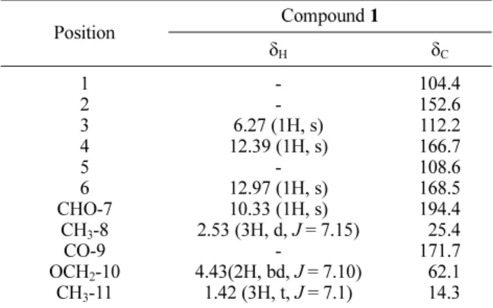
Table 1 shows the
1H-NMR and
13C-NMR spectra of compound 1. The
1H-NMR spectral data of 1 indicated the presence of signals for aromatic protons at δ
H6.27 (1H, s), two hydroxyl groups at δ
H12.39 (1H, s) and δ
H12.97 (1H, s), one carbonyl group at δ
H10.33 (1H, s), one ethoxy group at δ
H4.43 (bd, J = 7.10 Hz), and two methyl groups at δ
H2.53 (d, J = 7.15Hz) and 1.42 (t, J = 7.10 Hz). The
13C-NMR spectrum exhibited 11 carbon signals comprising two carbonyl carbon atom groups (aldehyde and ester) at δ
C194.1 and δ
C177.7, one aromatic carbon signal at δ
C168.5, 166.7, 152.6, 112.2, 108.6, 104.1), one ethoxyl at δ
C62.1, and two methyl carbon signals at δ
C25.4 and δ
C14.3.
In the HMBC spectrums (Fig. 2), the complete structure is described with the following connectivity: aromatic proton H-3 at δ
H6.27 with CH
3-8 ( δ
C25.4), C-1 ( δ
C104.1) and C-5 ( δ
C108.6), aldehyde CHO-7 at δ
H10.33 with C-4 ( δ
C166.7), ethoxy proton OCH
2-10 at δ
H4.43 with CO-9 and CH
3-11 ( δ
C177.7 and 14.3, respectively).
Therefore, the chemical structure of compound 1 was identified as ethyl haematommate according to the NMR data as well as its comparison to the literature.
9Single crystal X-ray crystallography is one of the most advanced methods for structural determination of many
Fig. 1. Chemical structure of compound 1 isolated from lichen S.graminosum.
Table 1. 1H- and 13C-NMR data of ethyl haematommate (1) in CDCl3 (δ in ppm, 500 MHz for 1H and 125 MHz for 13C)a
Position Compound 1
δH δC
1 2 3 4 5 6 CHO-7
CH3-8 CO-9 OCH2-10
CH3-11
- - 6.27 (1H, s) 12.39 (1H, s)
- 12.97 (1H, s) 10.33 (1H, s) 2.53 (3H, d, J = 7.15)
-
4.43(2H, bd, J = 7.10) 1.42 (3H, t, J = 7.1)
104.4 152.6 112.2 166.7 108.6 168.5 194.4 25.4 171.7 62.1 14.3
a J value are in parentheses and reported in Hz
Fig. 2. Key HMBC correlations of compound 1.
Vol. 24, No. 2, 2018 117
compounds including the natural products.
15-17To determine the crystal structure of compound 1 through singe crystal X-ray crystallography, the isolate was recrystallized.
Compound 1 was dissolved in n-hexane:EtOAc (1:1, v/v) solvent mixture and the solution was maintained under ambient conditions until colorless block-shaped single crystals were obtained. A suitable large single crystal (0.646 × 0.121 × 0.100 mm
3) was selected for single crystal X-ray diffraction. The analysis of single crystal X-ray crystallography revealed that compound 1 is crystallized in the orthorhombic crystal system and the P2
12
12
1space group. The crystallographic data and thermal ellipsoid drawing for compound 1 are shown in Table 2 and Fig. 3, respectively.
Compound 1 expectedly adopts a planar conformation.
All bond distances, angles, and dihedral angles appear within normal range except C8 −C2−C1 (124.8 (2)°) and C8 −C2−C3 (115.7 (2)°) compared with the mean value reported in Cambridge Structural Database (CSD, version 5.38, updated July 2017).
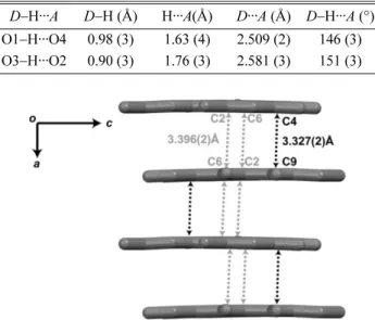
18The deviation in the bond angle may be a result of the strong intramolecular O1 − H1···O4 interaction. Interestingly, only the intramolecular
hydrogen bonds were observed in the crystal. The numerical details of the hydrogen bond in the crystal structure of compound 1 are listed in Table 3. The two hydroxyl-carbonyl intramolecular hydrogen bonds of O1 − H···O4 and O3 −H···O2 enclose (6) loops. The inter- molecular interactions between compound 1 molecules are mainly composed by π–π interaction in which the perpendicular distances between two rings are between 3.327(2) Å and 3.396(2) Å for C4···C9 and C2···C6, respectively. Together, this π–π interaction forms a layered architecture propagating in the (001) plane (Fig. 4).
In summary, we have succeeded to isolate compound 1 from S. graminosum and analyze its chemical structure by spectroscopy. In addition, the crystal structure of com-
S11 Table 2. Crystallographic data of compound 1
Chemical formula C11H12O5
Mr 224.21
Crystal system, space group Orthorhombic, P212121
Temperature (K) 93
a, b, c (Å) 6.7257 (4), 12.1568 (7), 12.3739 (8)
Tmin, Tmax 0.455, 0.905
No. of measured, independent, and observed [I > 2σ(I)] reflections 11356, 1848, 1490
Rint 0.079
(sin θ/λ)max (Å−1) 0.602
Tmin, Tmax 0.455, 0.905
R[F2> 2σ(F2)], wR(F2), S 0.043, 0.101, 0.96
No. of reflections 1848
No. of parameters 159
Fig. 3. Thermal ellipsoid structures of compound 1 with atom labelling drawn at a 50% probability level. The asymmetric unit contained one molecule of compound 1.
Table 3. Hydrogen-bond geometry of compound 1
D−H···A D−H (Å) H···A(Å) D···A (Å) D−H···A (°) O1−H···O4 0.98 (3) 1.63 (4) 2.509 (2) 146 (3) O3−H···O2 0.90 (3) 1.76 (3) 2.581 (3) 151 (3)
Fig. 4. The π–π interactions in compound 1 create a layered architecture propagating in the (001) plane. Hydrogen atoms are omitted for clarity.
118 Natural Product Sciences
pound 1 was determined for the first time by single crystal X-ray structure analysis. Our current study is underway to identify other isolates from S. graminosum and other species from the same genus. This paper is expected to provide an additional phytochemical contribu- tion of the poorly understood Stereocaulon genus.
Acknowledgments
We are grateful to Andalas University for the financial support (contract No. 14/UN.16.17/PP.HGB/LPPM/2017).
Dr Harrie J. M. Sipman, Botanischer Garten und Botanisches Museum Berlin-Dahlem, Freie Universitat Berlin, is acknowledged for the identification of the lichen. We also thank Dr. Ahmad Darmawan and Sofa Fajriah, Pusat Penelitian Kimia, LIPI, Serpong, for the collection of NMR data.
Supplementary Materials
The online version of this article contains supplementary materials: experimental procedures and characterizations of compound 1. CCDC 1585998 contains the supple- mentary crystallographic data presented in this paper.
These data can be obtained free of charge at www.ccdc.
cam.ac.uk/data_request/cif, by e-mailing data_request@
ccdc.cam.ac.uk, or by contacting The Cambridge Crystallo- graphic Data Centre, 12, Union Road, Cambridge CB2 1EZ, UK; fax: +44 1223 336033.
References
(1) Singh, R.; Ranjan, S.; Nayaka, S.; Pathre, U. V.; Shirke, P. A. Acta Physiol. Plant. 2013, 35, 1605-1615.
(2) Ismed, F.; Lohézic-Le Dévéhat, F.; Delalande, O.; Sinbandhit, S.;
Bakhtiar, A.; Boustie, J. Fitoterapia 2012, 83, 1693-1698.
(3) Ismed, F.; Lohézic-Le Dévéhat, F.; Rouaud, I.; Ferron, S.; Bakhtiar, A.; Boustie, J. Z. Naturforsch. C. 2017, 72, 55-62.
(4) Stocker-Wörgötter, E. Nat. Prod. Rep. 2008, 25, 188-200.
(5) Seshadri, T. R. Proc. Indian Acad. Sci. 1944, 20, 1-14.
(6) Marante, F. J. T.; Castellano, A. G.; Rosas, F. E.; Aguiar, J. Q.;
Barrera, J. B. J. Chem. Ecol, 2003, 29, 2049-2071.
(7) Choudhary, M. I.; Ali, M.; Wahab, A.; Khan, A.; Rasheed, S.;
Shyaula, S. L.; Rahman, A. Sci. China Chem. 2011, 54, 1926-1931.
(8) Manojlovic, N. T.; Vasiljevic, P. J.; Maskovic, P. Z.; Juskovic, M.;
Bogdanovic-Dusanovic, G. Evid. Based Complement. Alternat. Med.
2012, 2012, 452431.
(9) Huneck, S.; Yoshimura, I. Identification of lichen substances;
Springer: United States, 1996. pp 421-422.
(10) Calculated using ABSCOR. Empirical Absorption Correction Based on Fourier Series Approximation; Rigaku: Texas, 1994.
(11) Burla, M. C.; Caliandro, R.; Camalli, M.; Carrozzini, B.;
Cascarano, G. L.; De Caro, L.; Giacovazzo, C.; Polidori, G.; Spagna, R. J.
Appl. Cryst. 2005, 38, 381-388.
(12) Sheldrick, G. M. Acta Crystallogr. A 2008, 64, 112-122.
(13) Macrae, C. F.; Bruno, I. J.; Chisholm, J. A.; Edgington, P. R.;
McCabe, P.; Pidcock, E.; Rodriguez-Monge, L.; Taylor, R.; van de Streek, J.; Wood, P. A. J. Appl. Cryst. 2008, 41, 466-470.
(14) Rao, P. S.; Sarma, K. G.; Seshadri, T. R. Proc. Indian Acad. Sci.
1967, 66, 1-14.
(15) Putra, O. D.; Furuishi, T.; Yonemochi, E.; Terada, K.; Uekusa, H.
Cryst. Growth Des. 2016, 16, 3577-3581.
(16) Ismed, F.; Farhan, A.; Bakhtiar, A.; Zaini, E.; Nugraha, Y. P.;
Dwichandra Putra, O. Uekusa, H. Acta Crystallogr. E Crystallogr.
Commun. 2016, 72, 1587-1589.
(17) Putra, O. D.; Umeda, D.; Nugraha, Y. P.; Furuishi, T.; Nagase, H.;
Fukuzawa, K.; Uekusa, H.; Yonemochi, E. CrystEngComm. 2017, 19, 2614-2622.
(18) Groom, C. R.; Bruno, I. J.; Lightfoot, M. P.; Ward, S. C.; Acta Crystallogr. B Struct. Sci. Cryst. Eng. Mater. 2016, 72, 171-179.
Received December 13, 2017 Revised February 19, 2018 Accepted February 22, 2018