A RARE CASE OF PRIMARY OVARIAN LEIOMYOMA WITH ATYPICAL MEIGS’ SYNDROME
Seung-Yeon Choi, MD, Jin-Sun Park, MD, Jeong-Won Lee, MD, PhD, Byoung-Gie Kim, MD, PhD, Duk-Soo Bae, MD, PhD
Department of Obstetrics and Gynecology, Samsung Medical Center, Sungkyunkwan University School of Medicine, Seoul, Korea
Primary ovarian leiomyoma is a rare benign ovarian tumor. Ovarian leiomyoma accompanied with atypical Meigs’ syndrome is extremely rare. A 13-year-old woman had underlying primary lymphedema and visited our clinic due to associated vulvar edema.
During a gynecologic examination, we detected a right ovarian tumor with large amounts of ascites on transrectal ultrasonography.
The tumor size increased from 4 to 7 cm during 6 months follow-up. After laparoscopic right ovarian tumorectomy, the final pathology of the tumor was primary ovarian leiomyoma. Ovarian leiomyomas are not typically suspected before surgery due to their extreme rarity and because they are easily misdiagnosed as fi broma during frozen biopsy due to similarities between both diseases. Here we introduce the case of a primary ovarian leiomyoma accompanied with atypical Meigs’ syndrome.
Keywords:
Ovarian leiomyoma; Meigs syndrome; Rare diseases; Ovary
Received: 2011.12.5. Revised: 2012.1.24. Accepted: 2012.2.27.
Corresponding author: Jeong-Won Lee, MD
Department of Obstetrics and Gynecology, Samsung Medical Center, Sungkyunkwan University School of Medicine, 81 Irwon-ro, Gangnam-gu, Seoul 135-710, Korea
Tel: +82-2-3410-1382 Fax: +82-2-3410-0630 E-mail: [email protected]
Th is is an Open Access article distributed under the terms of the Creative Commons Attribution Non-Commercial License (http://creativecommons.org/licenses/
by-nc/3.0/) which permits unrestricted non-commercial use, distribution, and reproduction in any medium, provided the original work is properly cited.
Copyright © 2012. Korean Society of Obstetrics and Gynecology http://dx.doi.org/10.5468/KJOG.2012.55.4.285
pISSN 2233-5188 · eISSN 2233-5196
Ovarian leiomyomas, either primary or parasitic in origin, account for about 0.5% to 1% of all benign ovarian tumors [1]. In previ- ous reported cases, most ovarian leiomyomas were unilateral, asymptomatic, of relatively small size and occurred in premeno- pausal women. Primary ovarian leiomyomas originate from ovar- ian tissues, including intraovarian blood vessels, smooth muscle fi bers, or similar tissues within the ovarian stroma and tunica al- buginea [2,3]. In contrast, parasitic ovarian leiomyomas originate from extraovarian tissues [4]. Due to their rarity, about 70 cases of primary ovarian leiomyomas have been reported worldwide [1].
Primary ovarian leiomyoma with Meigs’ syndrome is extreme rare case and previously reported only 4 cases [5]. The purpose of this report is to present a patient with a primary ovarian leiomyoma with atypical Meigs’ syndrome.
Case Report
A 13-year-old, para 0-0-0-0, virgin woman was referred to our clinic for evaluation of right vulvar edema. She had right leg ede- ma due to underlying primary lymphedema and lymphaticovenular anastomosis and no improvement in leg edema. She complained of right vulvar area edema and was referred to our clinic due to
the vulvar edema. We examined the external genitalia and de- tected right majora and minora edematous changes. Then, we per- formed routine transrectal ultrasonography. We detected a 4 cm ovarian tumor on transrectal sonography incidentally. She had no abdomino-pelvic discomfort and we decided to observe at regular intervals.
Six months later, the ovarian tumor had increased to about 7 cm
and we decided to perform a laparoscopic operation. The sono-
graphic images showed a 7.59 × 4.66 cm heterogeneous echo-
genic solid mass of the right ovary. The tumor was smooth mass
with round contour and was suspected to be a benign solid tumor.
We detected fairly large amounts of fl uid collection in posterior cul de sac, but did not think abnormal fl uid collections.
We performed abdomino-pelvic computed tomography (CT) preop- eratively and the abdomino-pelvic CT showed an 8 cm enhancing solid mass with central low-attenuation from the right ovary. Poor delineation of the right margin of the mass was observed (Fig. 1).
There were moderate amounts of ascites in pelvic cavity and it was thought as hemoperitoneum because of the high attenuation.
And there were small amounts of omental infi ltration which was considered as peritoneal seeding. Also there were multiple small lymph nodes along the mesenteric root. Finally, radiologist recom- mended differentiation of malignant germ cell tumor and juvenile granulosa cell tumor. In addition, subcutaneous soft tissue swell-
Fig. 1. Poor delineation of the right ovarian mass measuring about 8cm in length on computed tomography.
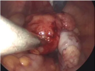
Fig. 2. Gross picture of 5cm sized solid tumors originating from right ovary. The ovarian tumor and the surrounding whitish fragile tissues are separated from right tube and is limited to the right ovary.
Fig. 3. (A) Spindle-shaped cells with wavy nuclei and extensive hyalinization was seen in the right ovarian tumor (H&E, ×100). (B) On the high powered magnititude, no signifi cant nuclear atypia or pleomorphism was seen (H&E, ×200).
A B
ing on right side lower abdomen and pelvis was detected and was thought as involvement of lymphedema.
Laboratory fi ndings such as complete blood cell count, urinalysis, chest X-ray and electrocardiography were normal. Because clini-
cian was not consider the ovarian tumor as malignancies, tumor markers were not checked.
On laparoscopy, 5 cm sized solid tumors were observed and the tumors were surrounded by fragmented whitish tissues (Fig. 2).
The tumor was sent to frozen biopsy and suspected to be a fi bro- ma or leiomyoma arising from the ovary. The ovary mass looked like two divided masses: a round solid mass and surrounding whit- ish fragile tissues. The total size of the ovarian mass was about 5 cm. The vulvar mass looked like a mass arising vulvar subcutane- ous layer and was suspected to be a lymphangioma, considering the patient’s underlying primary lymphedema. The uterus and left ovary were grossly normal and bilateral tubes were in hydrosal- pinxes. No adhesion between ovary and salpinx was seen. We only did a right ovary tumorectomy on laparoscopy considering the patient’s age and further fertility. After a laparoscopic right ovary tumorectomy, we excised the right 4 cm vulvar mass which had been occured due to vulvar edema. The vulvar mass was easily removed without hematoma or other complications.
On microscopy, a round ovarian mass and surrounding whitish friable tissues were noted. Sections from the round solid mass re- vealed a tumor, composed of interlacing bundles of fusiform cells.
Spindle-shaped cells had wavy nuclei and extensive hyalinization was seen in the tumor (Fig. 3A). On a high power view of the tu- mor, there was no signifi cant nuclear atypia or pleomorphism (Fig.
3B). The fragile whitish mass was also compatible with leiomyoma.
Immunohistochemically, the tumor cells were strongly positive for smooth muscle actin (SMA) (Fig. 4). The tumor cells were strongly positive for reticulin and the tumor was confi rmed as leiomyoma (Fig. 5) [6-8].
The patient had normal postoperative care and was discharged 2 days postoperatively without any other complications.
Discussion
A primary leiomyoma of the ovary is a very rare case and ap- proximately 70 cases are reported in the literature since Sangalli fi rst described this tumor in 1862. Even though the cases were re- ported that primary ovary leiomyoma occurred in postmenopausal women, most primary ovarian leiomyomas were reported in pre- menopausal women as seen in our case [2]. The size of primary ovarian leiomyoma was usually <3 cm, the size of the tumor was relatively large in our case.
Because of the rarity of the condition, primary ovarian leiomyomas are not diagnosed initially by ultrasonography or radiography.
Fig. 4. Immunohistochemically, the tumor cells were strongly positive for smooth muscle actin (smooth muscle actin, ×200).
Fig. 5. The tumor cells were strongly positive for reticulin and the tumor was confi rmed as leiomyoma (reticulin, ×200).
Ultrasound has been well documented as the best diagnostic modalities for pelvic organ masses, but in ovarian leiomyomas, it is diffi cult to distinguish ovarian leiomyomas from other ovarian tumors. Even on abdominopelvic CT, it is difficult to distinguish ovarian leiomyomas from other ovarian masses, and in some cases, the ovarian mass is reported as malignancy [8]. In addition, there were some cases misdiagnosed as an ovarian fi broma even though it will be revealed as an ovarian myoma on fi nal pathology as our case showed.
Immunohistochemistry was stated to be useful in establishing the diagnosis in this case, since the results of frozen section was confusing between fi broma with leiomyoma. The diffuse positive staining for desmin and SMA are characteristic of a leiomyoma, whereas the negative staining for inhibin makes the diagnosis of a sex cord/stromal tumor unlikely. Because only focal stromal stain- ing of SMA was observed in fi broma, we did reticulin immunohis- tochemistry staining for differentiation of leiomyoma and fi broma instead of inhibin staining [2].
We experienced an ovarian leiomyoma associated with ascites but not hydrothorax which is called Meigs’ syndrome; an benign ovarian tumor associated with ascites and/or hydrothorax. Ap- proximately 1% of benign ovarian tumors are associated with Meigs’ syndrome. Atypical Meigs’ syndrome is characterized by an ovarian tumor and ascites, but no pleural effusion, as in our case.
Meigs’ syndrome caused by ovarian leiomyoma is extremely rare.
We found only one reported case in English and another in Japa- nese, which presented with both ascites and pleural effusion and two additional cases of ovarian leiomyoma presenting only ascites without pleural effusion as atypical Meigs’ syndrome [5,9]. As a rule, resolution of ascites and pleural effusions occurs after re- moval of the ovarian lesion. We followed up the patient 6 months later and confi rmed the resolution of ascites wirh on transrectal ultrasonograpy.
Concentrations of CA-125 can be raised in Meigs’ syndrome [9].
Although the reason is unknown, benign solid ovarian tumors might secrete unknown factors that mediate the accumulation of fl uid. Therefore preoperative diagnosis of the ovarian tumor was thought as ovarian malignancy in Meigs’ syndrome and the surgi- cal treatment can reach abdominal total hysterectomy plus bilat- eral salpingooophorectomy. We did not conduct the preoperative serum CA-125, because the clinician presumed that the ovarian tumor would be benign.
All cases of ovarian leiomyomas demonstrated an excellent prog- nosis without recurrence despite the active mitosis observed in the
tumor [6]. Therefore the surgeon should make an effort to perform less invasive surgery, particularly in young women, to preserve fer- tility [10].
In conclusion, it is difficult to diagnose ovarian leiomyomas ac- curately due to their rarity and diagnostic difficulties. However, because primary ovarian leiomyomas occur in young women, the nature of the tumor is typically benign, prognosis is excellent and recurrence is rare, surgeons should consider ovary-preserving sur- gery as a fi rst choice in surgical management.
References
1. van Esch EM, van Wijngaarden SE, Schaafsma HE, Smeets MJ, Rhemrev JP. The diagnostic and therapeutic approach of a pri- mary bilateral leiomyoma of the ovaries: a case report and a literature review. Arch Gynecol Obstet 2011;283:1369-71.
2. Güney M, Ozsoy M, Oral B, Mungan T, Kapucuo ğlu N. Unilat- eral primary ovarian leiomyoma in adolescent: a case report.
Arch Gynecol Obstet 2007;275:507-10.
3. Koo YJ, Cho YJ, Kim JY, Lee JE, Kim ML, Kim JM, et al. Ovarian leiomyoma as a potential cause of compromised fertility. Fertil Steril 2011;95:1120.e11-4.
4. Abdel-Gadir A, Francis ND, Oyawoye OO, Chander BP. Second- ary amenorrhoea with high inhibin B level caused by parasitic ovarian leiomyoma. Gynecol Endocrinol 2010;26:93-5.
5. Kurai M, Shiozawa T, Noguchi H, Konishi I. Leiomyoma of the ovary presenting with Meigs’ syndrome. J Obstet Gynaecol Res 2005;31:257-62.
6. Bucella D, Limbosch JF, Buxant F, Simon P, Fayt I, Anaf V, et al.
Recurrence of mitotically active cellular fi broma of the ovary.
Obstet Gynecol Int 2009;2009:803062.
7. Tomas D, Lenicek T, Tuckar N, Puljiz Z, Ledinsky M, Kruslin B.
Primary ovarian leiomyoma associated with endometriotic cyst presenting with symptoms of acute appendicitis: a case report. Diagn Pathol 2009;4:25.
8. Paladini D, Testa A, Van Holsbeke C, Mancari R, Timmerman D, Valentin L. Imaging in gynecological disease (5): clinical and ultrasound characteristics in fi broma and fi brothecoma of the ovary. Ultrasound Obstet Gynecol 2009;34:188-95.
9. Shiau CS, Chang MY, Hsieh CC, Hsieh TT, Chiang CH. Meigs’
syndrome in a young woman with a normal serum CA-125 level. Chang Gung Med J 2005;28:587-91.
10. Son CE, Choi JS, Lee JH, Jeon SW, Hong JH, Bae JW. Laparo-
scopic surgical management and clinical characteristics of ovarian fi bromas. JSLS 2011;15:16-20.
비정형적 메이그신드롬을 동반한 난소의 원발성 평활근종 1예
성균관대학교 의과대학 산부인과학교실 최승연, 박진선, 이정원, 김병기, 배덕수
난소의 원발성 평활근종은 매우 희귀한 종양으로 비정형적 메이그신드롬을 동반한 난소의 원발성 평활근종은 더욱 드문 질환이다. 원발 성 림프부종을 기저질환으로 가지고 있었던 13세 여성이 외음부의 부종을 주소로 본원에 내원하였다. 부인과 검사를 시행하던 도중 직장 식 초음파에서 다량의 복수를 동반한 오른쪽 난소 종괴가 발견되었다. 6개월후 경과 관찰하였고 난소 종괴는 4 cm에서 6 cm으로 크기가 커졌다. 복강경수술을 시행 후 최종 병리학적 소견은 난소의 원발성 평활근종으로 밝혀졌다. 난소의 원발성 평활 근종은 보통 드문 발병률 로 인해 수술전 발견이 어렵고 병리학적 유사성으로 인해 난소의 섬유종과도 감별하기 어려운 질환이다. 이에 우리는 비정형적 메이그신 드롬을 동반한 난소의 원발성 평활근종 1예를 문헌 고찰과 함께 보고하는 바이다.
중심단어: 난소 평활근종, 비정형적 메이그신드롬, 희귀 질환, 난소