© 2017 The Korean Ophthalmological Society
This is an Open Access article distributed under the terms of the Creative Commons Attribution Non-Commercial License (http://creativecommons.org/licenses /by-nc/3.0/) which permits unrestricted non-commercial use, distribution, and reproduction in any medium, provided the original work is properly cited.
Original Article
Surgical Outcomes of Porcine Acellular Dermis Graft in Anophthalmic Socket: Comparison with Oral Mucosa Graft
Livia Teo1,2, Young Jun Woo1, Dong Kyu Kim3, Chang Yeom Kim1, Jin Sook Yoon1
1Institute of Vision Research, Department of Ophthalmology, Severance Hospital, Yonsei University College of Medicine, Seoul, Korea
2Singapore National Eye Centre, Singapore, Singapore
3Yonsei University College of Medicine, Seoul, Korea
Purpose: We describe our experience with the Permacol graft in anophthalmic socket reconstruction, and com- pare it to the autologous buccal mucosal graft, emphasizing the postoperative vascularization and contraction of each graft.
Methods: This was a retrospective comparative study. We measured the time necessary for the graft surface to be completely vascularized, as well as the fornix depth of the conjunctival sac in anophthalmic patients.
Results: Ten patients underwent Permacol graft reconstruction, with 44 undergoing buccal mucosal graft recon- struction. Seven eyelids (70%) in the Permacol group had a good outcome, with improvement in lower eyelid position and prosthesis retention. Nine out of 10 eyelids (90%) in this group showed complete vascularization of the graft at 2.6 ± 1.9 months postoperatively, while the grafted buccal mucosa was fully vascularized at 1.1
± 0.3 months postoperatively (p < 0.01). Postoperative fornix depth in the Permacol group was 9.1 ± 2.2 mm, compared to 14.9 ± 4.5 mm in the buccal mucosal graft group (p < 0.01). Mean increases in fornix depth were 33.1% and 67.9% of the mean vertical length of the implanted graft.
Conclusions: The Permacol graft can be useful as spacer graft material in anophthalmic socket patients. It takes longer to vascularize, and undergoes greater graft shrinkage with time, compared to the buccal mucosal graft.
Key Words: Acellular dermis, Anophthalmos, Mouth mucosa
Socket contracture sometimes occurs in anophthalmic patients, and this results not only in a poor cosmetic out- come, but also functional problems, such as ocular pros- thesis displacement. To correct socket contracture, anterior lamellar repositioning or augmentation of the posterior
lamella of the eyelid is usually required [1,2].
Current eyelid spacers include autogenous grafts, such as hard palate mucosal grafts. Allogeneic grafts include donor sclera or acellular human dermal graft (e.g., Allo- Derm; LifeCell, The Woodlands, TX, USA), and synthetic implants include porous polyethylene. However, none of these substitutes are ideal as they are associated with dis- advantages like donor site morbidity, risk of disease trans- mission, and implant migration and extrusion.
Recently, a novel material derived from porcine acellular dermis (Enduragen; Tissue Science Laboratories, Alder-
Received: October 5, 2015 Accepted: December 1, 2015
Corresponding Author: Jin Sook Yoon, MD, PhD. Institute of Vision Research, Department of Ophthalmology, Yonsei University College of Medicine, #50 Yonsei-ro, Seodaemun-gu, Seoul 03722, Korea. Tel: 82-2- 2228-3570, Fax: 82-2-312-0541, E-mail: [email protected]
shot, UK) was described for use as a spacer graft in the eyelid. The study demonstrated a favorable outcome when the implant was used in the upper lid, lower lid, or as later- al canthal reinforcement [3]. This porcine acellular dermis graft has also been marketed by Covidien (Dublin, Ireland) under a different name, Permacol. In this study, we would like to introduce our experience with the Permacol graft by comparing it to the autologous buccal mucosal graft. In particular, we would like to highlight and compare the postoperative vascularization and contraction that takes place with each graft over a 6-month period.
Materials and Methods
This was a retrospective comparative study of patients who underwent socket reconstructive surgery using the Permacol graft or autologous buccal mucosal graft as a spacer at Severance Hospital, Yonsei University from Jan- uary 2011 to December 2012, by a single surgeon (JSY).
Only patients with an anophthalmic socket, with at least 6 months of follow-up after surgery were included. Medical records and clinical photographs of these patients were re- viewed. Demographic data, follow-up duration, duration of prosthesis wear, cause of anophthalmos, risk factors for socket contracture, combined or further surgery, and com- plications of surgery were tabulated. Operation records, including size of implanted graft, were also obtained.
A good outcome was defined as an improvement in low- er lid sagging with no prosthesis prolapse. A poor outcome was defined as no improvement in lower lid sagging that required further surgery with another type of spacer mate- rial, the inability to fit a prosthesis, or prosthesis prolapse.
The time necessary for the graft surface to be completely vascularized was assessed to evaluate the degree of fibro- vascular integration of the graft. These were thoroughly assessed using a slit lamp microscope. The fornix depth of the conjunctival sac was directly measured using a ruler, after removal of the artificial eye. The calculated postoper- ative fornix length was used to estimate immediate post- operative fornix depth. It was computed as the sum of pre- operative fornix depth and vertical length of the implanted graft.
The institutional review board of Yonsei University Col- lege of Medicine, Seoul, Korea, approved this study. The study adhered to the tenets of the Declaration of Helsinki,
and written informed consent was obtained from all par- ticipants.
Surgical technique
A transconjunctival incision was made along the inferior border of the lower eyelid tarsus using monopolar cautery.
Blunt dissection towards the orbital rim was performed without damaging the orbital septum. Upon reaching the orbital rim, an incision of the periosteum was made with a
#15 blade, and an elevator was used to dissect the subperi- osteal pocket to reach the infraorbital foramen.
In the Permacol graft group, the implant was trimmed to the size of the defect. In the autologous buccal mucosal graft group, an ellipse of full-thickness mucosal graft was harvested from the inner aspect of the cheek. Care was taken to avoid Stenson’s duct adjacent to the upper second molar. Bosmin solution (epinephrine 1 mg/mL; Daiichi Sankyo, Tokyo, Japan) with 10% dilution application and electrocauterization were used at the donor site for hemo- stasis, and the defect was packed with gel-form. The har- vested graft was trimmed to size, and submucosal tissue was removed with scissors.
The edges of the free graft were sutured to the sur- rounding conjunctiva with interrupted 6-0 absorbable braided polyglactin sutures (Vicryl; Ethicon, Livingston,
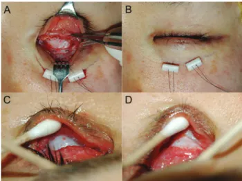
Fig. 1. The surgical procedure using the Permacol implant in anophthalmic socket reconstruction. (A) Intraoperative appear- ance of Permacol implant. The implant edge is sutured to the con- junctival edge. (B) External appearance of anophthalmic socket with fornix deepening sutures and retinal bolsters in place. (C) Appearance of graft at 1 week postoperatively. (D) Appearance of graft at 1 month postoperatively with superficial vasculariza- tion.
UK). A temporary traction suture was maintained for 3 days postoperatively in all patients. Two double 5-0 non-absorbable monofilaments polyamide 6 (Nylon;
WooRhi Medical, Namyangju, Korea) were used to anchor a silicone band bolster to deepen the inferior fornix. Each arm was passed through the silicone band and grafted mu- cosa, the periosteum of the inferior orbital rim, and out through the lower eyelid. Retinal sponge bolsters were used for the skin. A conformer was inserted in the socket for at least 4 weeks postoperatively. The surgical technique is presented in Fig. 1A-1D.
Statistical analysis
Patient characteristics, including age, duration of pros- thesis wear, calculated fornix length, preoperative fornix depth, vertical length of implanted graft, and postoperative outcomes, including time to vascularization and postoper- ative fornix depth were compared between the groups us- ing the Mann-Whitney test. Preoperative and postoperative fornix depth in each group was compared using Wilcoxon signed-rank test. All statistical tests were two-sided with an α-level of 0.05, and were performed using SPSS Statis- tics ver. 20.0. (IBM Co., Armonk, NY, USA).
Results
Permacol group
Ten patients underwent Permacol graft reconstruction.
Patient demographics and clinical characteristics are de- scribed in Table 1. The mean age was 38.2 years (range, 7 to 69 years), with an equal number of males and females (Table 2). They were followed for 12.5 ± 4.9 months with an average duration of prosthesis wear of 24.4 years (range, 1 to 55 years). The causes of anophthalmia were trauma (n
= 3, 30%), congenital microphthalmia (n = 3, 30%), previ- ous malignancy (n = 2, 20%), enucleation of eyeball after loss of vision by fever (n = 1, 10%), and unknown (n = 1, 10%).
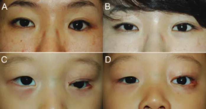
Seven patients (70%) were classified as having a good outcome due to improvement in the lower eyelid position with prosthesis retention (Fig. 2A-2D). Three patients (pa- tients 1, 8, and, 10) had a poor outcome. Two of these pa- tients (patients 1 and 10) had persistent socket inflamma-
tion with recurrent socket contracture, requiring a second fornix deepening procedure with oral mucosal grafting.
One patient (patient 8) was unable to retain an ocular pros- thesis at the end of the follow-up period, despite a deep- ened fornix.
Nine out of 10 eyelids showed complete vascularization of the graft within 2 to 2.5 months of surgery (Fig. 3A). In one case (patient 5), the Permacol graft in the remaining eyelid did not vascularize well even at 8 months postoper- atively (Fig. 3B). However, the unvascularized remnant is- lands were completely excised without complications.
The mean preoperative fornix depth was 4.4 ± 2.5 mm.
The postoperative fornix depth at 1 month postoperatively was 11.4 ± 2.6 mm (p < 0.01), though this became shallow- er at 9.1 ± 2.2 mm (p < 0.01) at postoperative 6 months.
Autologous buccal mucosal graft group
Patient ages ranged from 10 to 79 years with a mean age of 45.9 years, 17 patients (38.6%) were male (Table 2). The mean follow-up period was 8.4 months after the recon- Fig. 3. Conjunctivalization of the Permacol graft. (A) Good out- comes showing complete vascularization of the Permacol graft.
(B) Poor vascularization of the Permacol graft at 8 months post- operatively, showing unvascularized islands.
Fig. 2. Representative patients in preoperative (left) and post- operative (right) photographs, who underwent reconstructive surgery with the Permacol graft. Photographs of adult (A,B) and pediatric (C,D) anophthalmic socket patients.
Table 1. Demographic and clinical characteristics of patients in the Permacol group Patient no.AgeSex Cause of blindness Duration of prosthesis wear (yr)
OD / OS
Risk of socket contracture Prosthesis fitting (pre Combined surgery / further surgery / post)
Vascularization of graft (mon) Inferior fornix depth (mm)
(pre / post 1 mon / post 6 mon) Outcome Follow- up period (mon)
169F
Childhood infection, fever
55OS
Long duration of no prosthesis fitting and socket inflammation (35 yr)
N / Y
None / oral mucosa graft for lower and upper fornix reconstruction
2 1 / 8 / 7Poor12 250FTrauma30ODNoneN / YWedge implantation / none
2 3 / 8 / 7Good8 37MCM 6OS
Cowngenital small eyelid fissure, small silicone ball 13 mm
N / Y
Lateral canthoplasty / none
24 / 10 / 8Good9 455FCM40ODNoneY / YNone/ none2 7 / 14 / 13Good10 533MUnknown 8OSNoneN / YNone/ none8 4 / 13 / 10Good24 620M
Ocular tumor
19OSNoneY / YWedge implantation/ removal of remnant graft
2 4 / 9 / 7Good13 735FTrauma15ODNoneY / YNone/ none2 8 /15 /12Good16 833MTrauma25OD
Multiple orbital blows in fracture, facial nerve palsy
, severe
contracture with no implant
N / N
Dermis fat graft / Alloderm graft (his oral hygiene was very poor)
2.5 0 / 13 / 8Poor9 930F
Ocular tumor 1OSNoneY / YNone/ none2 5 / 13 / 11Good15 1050MCM45ODSocket inflammationY / Y
None / oral mucosa graft 2.57 / 11 / 8Poor9 OD = right eye; OS = left eye; F = female; N = prosthesis wear impossible; Y = prosthesis wear possible; M = male; CM = congenital microphthalmia.
structive surgery. Patients used an ocular prosthesis for an average of 15.6 years. The causes of anophthalmos includ- ed trauma (24 patients), tumor (five patients), congenital anomalies such as microphthalmos or anophthalmos (eight patients), and other disorders causing phthisis (e.g., retinal detachment, glaucoma, and uveitis) (seven patients).
In this group, 14 had severe socket contraction prior to the surgery; two had history of radiotherapy before enu- cleation for tumors; nine had previous complicated implant surgeries (e.g., exposure, infection, or small implant); three had a previous peg insertion; and two had prolonged sock- et inflammation for more than 1 month after socket recon- struction surgery.
The grafted buccal mucosa was fully vascularized ap- proximately 1 month after surgery (mean ± standard devi- ation, 1.1 ± 0.3 months). At preoperative and postoperative 6 months, fornix depths were 5.8 ± 2.4 and 14.9 ± 4.5 mm, respectively (p < 0.01).
Comparison between Permacol and autologous buccal mucosal graft groups
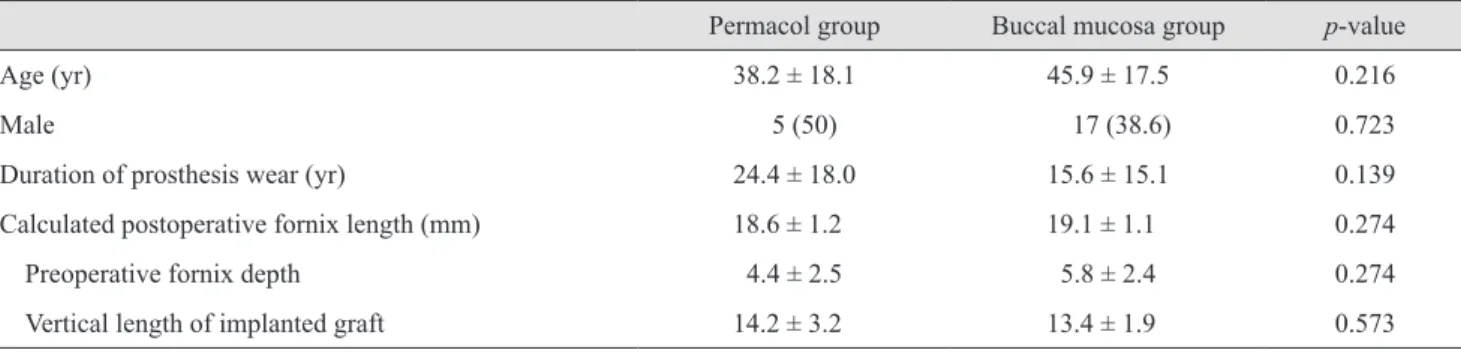
Age, sex, and duration of prosthesis wear were not sig- nificantly different between the two groups (Table 2). The Permacol graft took significantly longer (2.6 ± 1.9 months) to vascularize, compared to the buccal mucosal graft (1.1 ± 0.3 months) (p < 0.01). The preoperative fornix depths were not statistically different, as was the vertical length of the implanted graft (p = 0.274 and p = 0.573, respective- ly). The calculated postoperative fornix length did not dif- fer between the groups (p = 0.274). In contrast, the fornix depth in the Permacol group was significantly shorter 9.1 ± 2.2 mm at the end of the follow-up period, compared to the fornix depth in the buccal mucosal graft group 14.9 ± 4.5
mm (p < 0.01) (Fig. 4). Mean increases in fornix depth af- ter surgery were 4.4 mm and 9.1 mm, and they correspond- ed with 33.1% and 67.9% of the mean vertical lengths of the implanted graft, respectively.
Discussion
The Permacol surgical implant is a bioengineered, por- cine-derived dermal collagen implant, from which cells are removed in a gentle process to reduce immunogenicity.
The resulting acellular collagen matrix is then cross-linked for enhanced durability, which allows the formation of new collagen in the matrix. Permacol was first described in the literature for use in reconstructive hand surgery [4].
More recently, it has been used in the repair of abdominal wall defects [5], orbital fractures [6], and eyelid recon- structions [3,7].
Table 2. Comparison of clinical variables between Permacol and buccal mucosa groups
Permacol group Buccal mucosa group p-value
Age (yr) 38.2 ± 18.1 45.9 ± 17.5 0.216
Male 5 (50) 17 (38.6) 0.723
Duration of prosthesis wear (yr) 24.4 ± 18.0 15.6 ± 15.1 0.139
Calculated postoperative fornix length (mm) 18.6 ± 1.2 19.1 ± 1.1 0.274
Preoperative fornix depth 4.4 ± 2.5 5.8 ± 2.4 0.274
Vertical length of implanted graft 14.2 ± 3.2 13.4 ± 1.9 0.573
Values are presented as mean ± standard deviation or number (%).
* 18
16 14 12 10 8 6 4 2
0 Preop
Fornix depth (mm)
Postop 6 mon
Buccal mucosa Permacol
Fig. 4. Comparison of preoperative (preop) and postoperative (postop) fornix depth in anophthalmic socket patients receiving Permacol and undergoing buccal mucosal graft reconstructive surgery. *p < 0.05, both groups.
McCord et al. [3] have described a porcine acellular der- mal collagen matrix manufactured by Tissue Science Lab- oratories as spacer material for the reconstruction of the upper and lower eyelid, as well as for lateral canthal sus- pension surgery.
To our knowledge, this is the first study to examine the clinical characteristics, as well as the vascularization and contracture of the Permacol graft, in detail in context of implantation in the lower eyelid as spacer material in anophthalmic socket reconstruction. We chose to compare it with buccal mucosal tissue, which is the preferred lower eyelid spacer material in our practice for anophthalmic socket reconstruction.
One major advantage of Permacol is that it is readily available to the surgeon and does not inflict any donor site morbidity. Since there is no need to harvest any donor tis- sue and the tissue does not require hydration prior to use, surgical time is reduced. The buccal mucosal graft, on the other hand, results in donor site morbidity, patient discom- fort and prolongs surgical time as a result of donor har- vesting [8,9]. One possible disadvantage of Permacol is that, like other xenografts (e.g., Tarsys), it may carry with it a potential risk of allergic reaction and inflammation [10,11], though this was not encountered in our study popu- lation. It has also been reported to elicit a chronic granulo- matous reaction, similar to a foreign body reaction, when implanted in the orbit for orbital floor fracture repair [6].
The buccal mucosal graft, on the other hand, is an autoge- nous source, and hence avoids the risk of tissue rejection and disease transmission.
In our clinical trial, the Permacol graft provided a good clinical outcome in seven out of 10 patients. The poor out- come in two patients was attributed to persistent socket in- flammation with recurrent socket contracture. The third patient had a poor outcome despite a deepened fornix due to multiple factors, such as like facial nerve palsy, trau- matic blow in fracture of the orbit, and absent orbital im- plant, which made prosthesis fitting impossible. The Per- macol graft was completely vascularized in nine out of 10 patients within 2.5 months. This is not surprising, as the vascularization process of the implanted porcine acellular dermis graft has been demonstrated to take place as early as the third postoperative day in rat models [12]. However, vascularization of the graft was poor in one patient (patient 5). It is possible that vascularization is dependent on the surface area in contact with the host conjunctival tissue, as
demonstrated in human acellular dermis grafts [13]. Any local host tissue factors that interfere with this interaction could affect the vascularization of the graft (e.g., an unde- tected hematoma, graft slippage, infection, or an irradiated socket with poor vascular supply). Systemic conditions like poorly controlled diabetes with poor wound healing re- sponse may also affect the vascularization of the graft.
The duration to vascularization in the Permacol group was significantly longer than in the buccal mucosal group. Ani- mal histologic studies have shown that small vessels and inflammatory cells are present up to 5 weeks after implan- tation [14], suggesting that the host remodeling response is ongoing at this stage. This delay in vascularization could be attributed to delayed host cell recognition of the acellu- lar xenograft, in contrast with an autogenous buccal muco- sal graft.
In this study, direct measurement of the immediate post- operative fornix depth could not be completed due to post- operative discomfort. Instead, we used the calculated post- operative fornix length to estimate it. The calculated postoperative fornix length is computed as the sum of the preoperative fornix depth and the vertical length of the grafted material, but is not the actual postoperative fornix depth because the angulation of the fornix was formed on graft material, duplicating the graft length in the fornix; in other words, the actual fornix depth may be shorter than the calculated fornix length. However, the calculated for- nix length was not statistically different between the two groups, and a fornix depth difference at 6 months after surgery implies clinical significance.
The authors feel that Permacol is not as effective as buc- cal mucosal graft as a fornix-deepening spacer material.
There was an improvement in fornix depth in the Perma- col group, but the improvement was significantly less than that afforded by the buccal mucosa group. This is most likely due to fibrosis and contraction of the Permacol graft over time. Significant graft shrinkage of the porcine acel- lular dermal matrix was noted in the in vivo rat model [14].
Graft shrinkage of the acellular human dermis graft has previously been described [15-17], with resorption appear- ing to be a primary disadvantage. The rate and occurrence of resorption is unpredictable, and has been suggested to be due to inadequate exposure to vascular tissue and dehy- dration [13]. It seems likely that the porcine acellular der- mis graft undergoes a similar process of fibrosis, contrac- tion, and resorption. As such, the authors recommend
oversizing the Permacol graft intraoperatively, in order to account for graft shrinkage over time.
The limitations of this study are its retrospective design and limited duration of follow-up. It also has a small sam- ple size, and reflects only a single surgeon’s experience.
In summary, the Permacol graft can be a useful spacer graft material in patients with anophthalmic sockets. Care- ful selection of patients with no risk factors for recurrent socket contracture and poor vascularization is important.
Meticulous surgical technique will optimize the surgical outcome, and oversizing the graft intraoperatively will help to compensate for postoperative graft shrinkage.
Conflict of Interest
No potential conflict of interest relevant to this article was reported.
References
1. Hashikawa K, Terashi H, Tahara S. Therapeutic strategy for the triad of acquired anophthalmic orbit. Plast Reconstr Surg 2007;119:2182-8.
2. Yoshizawa M, Feinberg SE, Marcelo CL, Elner VM. Ex vivo produced human conjunctiva and oral mucosa equiva- lents grown in a serum-free culture system. J Oral Maxil- lofac Surg 2004;62:980-8.
3. McCord C, Nahai FR, Codner MA, et al. Use of porcine acellular dermal matrix (Enduragen) grafts in eyelids: a re- view of 69 patients and 129 eyelids. Plast Reconstr Surg 2008;122:1206-13.
4. Belcher HJ, Zic R. Adverse effect of porcine collagen inter- position after trapeziectomy: a comparative study. J Hand Surg Br 2001;26:159-64.
5. Liyanage SH, Purohit GS, Frye JN, Giordano P. Anterior abdominal wall reconstruction with a Permacol implant.
J Plast Reconstr Aesthet Surg 2006;59:553-5.
6. Cheung D, Brown L, Sampath R. Localized inferior orbital fibrosis associated with porcine dermal collagen xenograft orbital f loor implant. Ophthal Plast Reconstr Surg 2004;20:257-9.
7. Peter NM, Kumar B. Permacol in eyelid reconstruction: a novel use. Orbit 2013;32:57-9.
8. Klein M, Menneking H, Bier J. Reconstruction of the con- tracted ocular socket with free full-thickness mucosa graft.
Int J Oral Maxillofac Surg 2000;29:96-8.
9. Molgat YM, Hurwitz JJ, Webb MC. Buccal mucous mem- brane-fat graft in the management of the contracted socket.
Ophthal Plast Reconstr Surg 1993;9:267-72.
10. Liao SL, Wei YH. Correction of lower lid retraction using tarSys bioengineered grafts for graves ophthalmopathy. Am J Ophthalmol 2013;156:387-92.e1.
11. Kim HJ, Grossniklaus HE, Wojno TH. A cyst-like foreign body reaction to porcine decellularized membrane (TarSys).
Ophthal Plast Reconstr Surg 2014;30:e100-2.
12. Xie WG, Tan H, Zhao CL, Wang H. The histological changes and the revascularization process in the grafted dermal substitutes. Zhonghua Shao Shang Za Zhi 2005;21:37-9.
13. Rubin PA, Fay AM, Remulla HD, Maus M. Ophthalmic plastic applications of acellular dermal allografts. Ophthal- mology 1999;106:2091-7.
14. Liu Z, Tang R, Zhou Z, et al. Comparison of two por- cine-derived materials for repairing abdominal wall defects in rats. PLoS One 2011;6:e20520.
15. Sullivan SA, Dailey RA. Graft contraction: a comparison of acellular dermis versus hard palate mucosa in lower eye- lid surgery. Ophthal Plast Reconstr Surg 2003;19:14-24.
16. Owens KW, Yukna RA. Collagen membrane resorption in dogs: a comparative study. Implant Dent 2001;10:49-58.
17. Gryskiewicz JM, Rohrich RJ, Reagan BJ, Schwartz BM.
The use of Alloderm for the correction of nasal contour de- formities. Plast Reconstr Surg 2001;107:571.