403
폐렴을 동반한 기관 및 주기관지의 확장 소견
가톨릭대학교 의과대학 내과학교실, 방사선과학교실1
주겨레, 옥주현, 이성은, 장석태, 김성경, 이상학, 송정섭, 박성학, 문화식, 이배영1, 김현숙1
A Case of Tracheobronchomegaly with Pneumonia
Kyu Re Joo, M.D., Ju Hyun Oak, M.D., Sung Eun Lee, M.D., Suk Tae Jang, M.D., Sung Kyoung Kim, M.D., Sang Haak Lee, M.D., Jeong Sup Song, M.D., Sung Hak Park, M.D., Hwa Sik Moon, M.D., Bae Young Lee, M.D.1, Hyeon Sook Kim, M.D.1
Department of Internal Medicine & Radiology1, The Catholic University of Korea, College of Medicine, Seoul, Korea
□ 이달의 X선 □
A 66-years-old man was refered to our hospital because of cough, sputum, chill and fever. Enlargement of the trachea and main bronchi on radiography and bronchoscopy is compatible with Mounier-Kuhn syndrome. Mounier-Kuhn syndrome or tracheobronchomegaly is a rare disorder of uncertain etiology, characterized by marked dilatation of the trachea and major bronchi. This syndrome is associated with tracheal diverticulosis, bronchiectasis and recurrent respiratory tract infection.
We report a rare case of Mounier-Kuhn syndrome with pneumonia and literature reviews.
(Tuberc Respir Dis 2006; 61: 403-406)
Key words: Mounier-Kuhn syndrome, Tracheobronchomegaly.
Address for correspondence : Hwa Sik Moon, M.D.
Department of Internal Medicine, St. Paul's Hospital The Catholic University of Korea, 620-56, Jeonnong-dong, Dongdeamoon-gu Seoul, 130-709, Korea
Phone : 82-2-958-2463 Fax : 82-2-968-7250 E-mail : [email protected]
Received : Jun. 16. 2006 Accepted : Jul. 18. 2006
증 례
환 자: 김 O 태, 남자 66세 주 소: 2주 동안의 발열과 기침
현병력: 환자는 내원 2 주 전부터 간헐적으로 발열, 오한, 기침, 객담 있어 개인 의원 방문하여 경구 항생제 복용하였으나 증상 호전 없어 본원으로 내원하였다.
과거력, 사회력 및 가족력: 한 주에 소주 한 병 정 도의 음주력과 40 갑년의 흡연력 이외의 특이 사항은 없었다.
이학적 소견: 입원 당시 혈압은 110/60 mmHg, 맥 박수 87회/분, 호흡수 20회/분, 체온 38.4℃ 이었다. 의 식은 명료하였으나 급성병색을 띄고 있었다. 환자의 피부 및 두부 이학적 검사상에서 이상이 없었고 경부 임파절은 촉지 되지 않았다. 흉부 청진시 우측 하엽에 서 기관지 호흡음과 흡기시 수포음이 들렸다. 심음은
정상이었고 기타 부위의 진찰상 특이 소견은 없었다.
검사실 소견: 말초혈액검사에서 백혈구 20,300/㎣
(호중구 84.6%, 림프구 9.4%, 단핵구 4.1%, 호산구 0.8%, 호염기구 1.1%), 혈색소 13.3 g/dL, 그리고 혈소 판 256,000/μL 이었다. 혈액화학 검사 소견에서는 혈 중요소질소 9.7 mg/dL, 혈청 크레아티닌 0.9 mg/dL, AST 58 IU/L, ALT 66 IU/L, 총 빌리루빈 0.9 mg/dL 이었고 적혈구 침강속도 40 mm/hr (정상치 ; 0-10 mm/hr), C-reactive protein 28 mm/hr (정상치 ; 0-20 mm/hr)로 상승되었다. 동맥혈가스검사는 pH 7.418, PaCO₂26.3 mmHg, PaO₂73.9 mmHg, HCO3
17.0 mEq/L, 산소 포화도 96 %이었다. 객담의 결핵균 도말검사 및 음성 이었다.
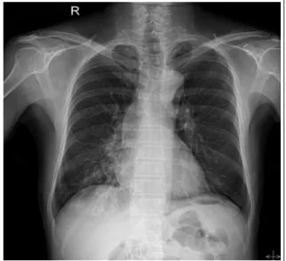
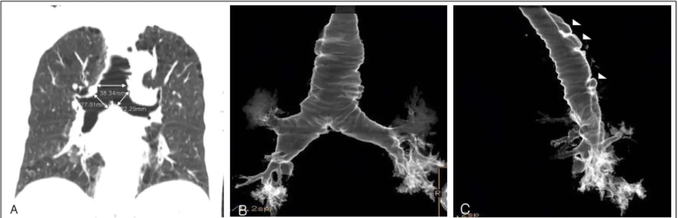
방사선 소견: 내원 당시 촬영한 단순흉부방사선 검 사에서 우측하폐하에 경계가 불분명한 미만성의 폐침 윤 소견, 기관과 양측 주기관지의 확장 소견이 관찰되 었다(Figure 1). 흉부 전산화 단층촬영에서 기관과 양 측 주기관지의 확장, 기관의 중격과 (Figure 2A) 우측 하엽의 불분명한 음영의 경화와 소결절이 관찰되었다 (Figure 2B). 흉부 전산화 단층촬영의 영상을 재구 성하여 만든 관상영상과 (Figure 3A), 정면(Figure 3B), 측면 (Figure 3C) 3차원 영상에서 기관과 양측 주기관의 확장 소견과 기관 게실 소견이 관찰되었다.
KR Joo et al: A case of tracheobronchomegaly with pneumonia
404
Figure 1. Chest X-ray shows ill-defined infiltrations in the right lower lobe. Dilatation of trachea and both bronchi are noted.
A
A B B
Figure 2. Chest CT scan in axial plane at the level of aortic arch shows dilatation of trachea, septation(arrowhead) in trachea and adjacent diverticula(arrow)(A). Ill-defined consolidation and small nodules are noted in right lower lobe(B).
기관지 내시경 소견: 기관지 내시경 검사에서는 성 대로부터 약 5 Cm 하부에 기관의 후면으로 기관게실 이 관찰되고 있었으며 게실 내부에는 많은 양의 분비 물이 관찰되었다(Figure 4). 기관지 솔질 검체, 기관지 세척액 검체로 시행한 그람염색, 항산균 염색, 세포진 검사 및 배양 검사 결과는 음성이었다.
임상경과: 입원 당시 임상소견과 방사선적 소견으 로 지역획득폐렴으로 진단하고 moxifloxacin 400mg 하루 한번 정주 치료 시작하였으며 치료시작 3일부터 발열, 기침, 객담과 같은 임상증상 호전과 함께 단순 흉부방사선 소견도 호전되어 정주 항생제 치료 13일 째 경구 항생제로 교체한 후 퇴원하였으며 외래에서 추적 관찰 중이다.
고 찰
Mounier-Kuhn syndrome은 1932년 Mounier-Kuhn 에 의하여 처음으로 기술된, 기관과 주기관지의 현저 한 확장을 특징으로 하는 드문 질환으로서 기관 게실, 기관지 확장증과 재발성 하부기도의 감염증을 동반할 수 있다1. 주로 30-40대의 남자에게 주로 발병되는 것 으로 보고되어 있으며 기도의 확장은 주로 기관지 4 번째 분지까지 확장 되어 있고 이후 말초기도는 정상 직경을 유지하고 있다2.
Mounier-Kuhn syndrome의 병인론은 명백하지 않 으나 기관과 주기관지의 민무늬근과 탄성조직의 선천 성 결함 또는 위축에 의하여 발병되는 것으로 알려져
Tuberculosis and Respiratory Diseases Vol. 61. No.4, Oct. 2006
405 Figure 4. Bronchoscopy shows the a divercitulum in posterior wall of trachea at the level of 5cm below vocal cord.
There was some secretions in this diverticulum(A). This secretions were disappeared after bronchoscopic suction(B). This images were reconstructed by CT virtual bronchoscopy(C). Arrow points this diverticulum(C).
38.34mm 27.01mm
22.29mm
A
38.34mm 27.01mm
22.29mm
A BB CC
Figure 3. Chest CT scan in coronal plane shows tracheomegaly and bronchomegaly(A). In three dimensional reconstruction CT bronchogram, anterior view(B) and lateral view(C) show diverticula(arrowhead).
있으며 이로 인하여 발생한 기관연골고리 사이의 점 막의 탈출증으로 기관 게실이 발생하게 된다3. 대부분 의 경우 돌발성으로 발생되나 열성 유전에 의하여 발 병할 수 있으며1 후천적으로는 성인에 있어서 폐섬유 증의 합병증과4 조산아의 기계호흡의 후유증으로 발 병할 수 있다5. 이차성으로 발병할 경우 Ehlers- Danlos syndrome, Marfan syndrome, Kenny-Caffey syndrome, Brachmann de Lange syndrome, 결합조 직질환, 모세혈관 확장성조화운동불능, Bruton형 무 감마글로불린혈증, 강직척추염, 이완피부증 그리고 경쇄침착질환과 연관이 있는 것으로 알려져 있다6,7.
임상증상은 비특이적이며 주로 하부기도 감염에 의 한 발열, 오한, 기침과 객담이며 과도한 양의 객담발 생으로 호흡곤란이 나타날 수도 있다. 재발성의 하부
기도 감염은 확장되어 있으나 취약한 기도의 정상적 이지 못한 기침 반응 기전에 의하여 점액섬모 청소능 이 감소되어 기도에서 발생한 점액이 정체되어 재발 성의 폐렴, 기관지확장증과 섬유화를 유발하게 한다.
점차적인 폐손상은 자발성 기흉, 객혈, 재발성 폐렴, 곤봉지와 호흡부전까지도 유발시킬 수 있다8.
단순흉부방사선 소견상 중심기도 직경의 증가를 확 인할 수 있으며 이는 후전 촬영보다 측면 촬영에서 더 쉽게 관찰 할 수 있다. 성인의 단순흉부방사선이나 기 관지조영상에서 기관, 우측 주 기관지, 좌측 주 기관 지의 직경이 각각 3.0 ㎝, 2.4 ㎝, 2.3 ㎝ 이상일 경우 흉부 전산화 단층촬영에서 각각 3.0 ㎝, 2.0 ㎝, 1.8 ㎝ 이상일 경우 진단할 수 있다. 기관게실은 대략 환자의 1/3에서 관찰되는데 주로 기관의 우측후측방벽에서
KR Joo et al: A case of tracheobronchomegaly with pneumonia
406
발생한다2. 폐기능검사상 폐쇄성 환기장애와 잔기량 의 증가 소견을 보이게 된다.
치료는 분비물의 제거를 위한 물리요법과 감염 악 화 시 적절한 항생제 요법이다9. 최근 들어서 지속적 인 폐 손상으로 폐기능이 상실된 환자에게 폐이식을 시행하여 성공적으로 치료한 예들도 보고되고 있다
10,11
.
Mounier-Kuhn syndrome 환자에게 기관 내 삽관 후 후유증으로 기관 협착이 유발될 수 있으므로 기계 호흡이 필요할 경우 커프가 없는 기관내관을 사용하 는 것이 추천된다. 기관 내 삽관 후 후유증에 의한 기 관 협착이 발생된 경우 기관지내시경을 통한 기관 내 스텐트 삽입술에 의하여 성공적으로 치료한 예들도 보고 되어 있다12.
요 약
저자들은 폐렴을 동반한 기도의 드문 질환인 Mounier-Kuhn syndrome을 가진 환자의 단순흉부방 사선, 기도내시경 소견과 함께 고식적인 흉부 전산화 단층 촬영을 시행 후 3차원적인 입체 영상으로 재구 성한 소견을 고찰과 함께 1예 보고하는 바이다.
참 고 문 헌
1. Schwartz M, Rossoff L. Tracheobronchomegaly. Chest 1994;106:1589-90.
2. Lazzarini-de-Oliveira LC, Costa de Barros Franco CA, Gomes de Salles CL, de Oliveira AC Jr. A 38 year old man with tracheomegaly, tracheal diverticulosis, and
bronchiectasis. Chest 2001;120:1018-20.
3. Spencer H. Congenital abnormalities of the lung: con- genital tracheobronchomegaly. In: Spencer H, editor.
Pathology of the lung. 4th ed. Oxford, UK: Pergamon Press; 1985. p. 129-30.
4. Woodring JH, Barrett PA, Rehm SR, Nurenberg P.
Acquired tracheomegaly in adults as a complication of diffuse pulmonary fibrosis. AJR Am J Roentgenol 1989;152:743-7.
5. Bhutani VK, Ritchie WG, Schaffer TH. Acquired tracheomegaly in very preterm neonates. Am J Dis Child 1986;140:449-52.
6. Blake MA, Clarke PD, Fenlon HM. Thoracic case of the day: Mounier-Kuhn syndrome (tracheobronchome- galy). AJR Am J Roentgenol 1999;173:822, 824–5.
7. Sane AC, Effmann EL, Brown SD. Tracheobronchio- megaly: the Mounier-Kuhn syndrome in a patient with the Kenny-Caffey syndrome. Chest 1992;102:
618-9.
8. van Schoor J, Joos G, Pauwels R. Tracheobron- chomegaly: the Mounier-Kuhn syndrome: report of two cases and review of the literature. Eur Respir J 1991;4:1303-6.
9. Guest JL Jr, Anderson JN. Tracheobronchomegaly (Mounier-Kuhn Syndrome). JAMA 1977;238:1754–5.
10. Shah SS, Karnak D, Mason D, Murthy S, Pattersson G, Budev M, et al. Pulmonary transplantation in Mounier-Kuhn syndrome: a case report. J Thorac Cardiovasc Surg 2006;131:757-8.
11. Drain AJ, Perrin F, Tasker A, Stewart S, Wells F, Tsui S, et al. Double lung transplantation in a patient with tracheobronchomegaly (Mounier-Kuhn syndrome).
J Heart Lung Transplant 2006;25:134-6.
12. Giannoni S, Benassai C, Allori O, Valeri E, Ferri L, Dragotto A. Tracheomalacia associated with Mo- unier-Kuhn syndrome in the Intensive Care Unit:
treatment with Freitag stent: a case report. Minerva Anestesiol 2004;70:651-9.