ISSN 1225-6552, eISSN 2287-7630 https://doi.org/10.7853/kjvs.2018.41.2.105
< Original Article >
Veterinary Service
Available online at http://kjves.org
*Corresponding author: Jae-Hoon Kim, Tel. +82-64-754-3387, Fax. +82-64-702-9920, E-mail. [email protected]
Identification of Salmonella spp. from porcine salmonellosis by matrix-assisted laser desorption ionization-time of
flight mass spectrometry
Hyoung-Seok Yang1, Jae-Hoon Kim2*
1Jeju Self-Governing Provincial Veterinary Research Institute, Jeju 63344, Korea
2College of Veterinary Medicine and Veterinary Medical Research Institute, Jeju National University, Jeju 63243, Korea (Received 16 March 2018; revised 26 April 2018; accepted 30 April 2018)
Abstract
A total of 41 Salmonella (S.) strains were isolated from pigs suffered with severe watery diarrhea and were tried to identify by both matrix-assisted laser desorption ionization-time of flight mass spectrome- try (MALDI-TOF MS) and polymerase chain reaction (PCR) analysis. Fibrinous exudate and ulceration in the large intestine were prevalent in gross observation, and variable degrees of enteritis were observed in the histology of large intestines. Subsequent polymerase chain reaction (PCR) analyses demonstrated that 41 strains were identified as S. Typhimurium (39 strains), though 2 stains were failed to identify.
Further identification was performed using both direct smear and protein extraction method by MALDI-TOF MS analyses. In terms of extraction methods, 100% (41/41) of isolates were identified to species level of S. spp. Whereas only 43.9% (18/41) were identified to species level using the direct method. These results thus suggest that rapid and accurate diagnosis of porcine salmonellosis can be guaranteed by MALDI-TOF MS combined with protein extraction method.
Key words : Porcine, Salmonellosis, Identification, PCR, MALDI-TOF MS
INTRODUCTION
Salmonella (S.) spp. are Gram-negative, motile, facul- tative anaerobic and flagellated rod-shape bacilli that cause gastroenteritis, septicemia and foodborne poison- ing in human and animal (van Duijkeren et al, 2002).
Although more than 2,400 serotypes are widely dis- tributed with broad host ranges, notable serotypes of salmonella in pigs are S. Typhimurium, S. Choleraesuis, and S. Typhisuis (Griffith et al, 2006; Brown et al, 2007). The pathology of porcine salmonellosis can be varied according to the causative agents (Wilcock et al, 1976; Griffith et al, 2006; Brown et al, 2007). For in- stance, pigs infected with S. Choleraesuis are reluctant to move, and mortal with cyanosis of extremities and
abdomens. However, enterocolitis is the major patho- logical observation in pigs infected with S. Typhimurium.
Studies have indicated that immunologically naïve pigs in poor hygiene are largely responsible for the outbreak of porcine salmonellosis (Griffith et al, 2006).
Since S. spp. has great economic importance in global swine industry, rapid diagnosis is essential for public health, cost-effectiveness and corresponding therapy (Griffith et al, 2006; Abubakar et al, 2007). Many stud- ies have shown that methods including biochemical tests, VITEK 2 system, enzyme-linked immunosorbent assay (ELISA), and polymerase chain reaction (PCR) are effective to diagnosis for porcine salmonellosis (Baggesen et al, 1996; Griffith et al, 2006). Recently, matrix-assisted laser desorption ionization-time of flight mass spectrometry (MALDI-TOF MS), has been em- ployed for the identification of the pathogenic bacteria
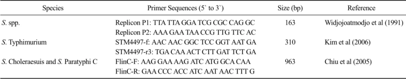
Table 1. Oligonucleotide primer sets for the detection of Salmonella spp.
Species Primer Sequences (5` to 3`) Size (bp) Reference
S. spp. Replicon P1: TTA TTA GGA TCG CGC CAG GC 163 Widjojoatmodjo et al (1991)
Replicon P2: AAA GAA TAA CCG TTG TTC AC
S. Typhimurium STM4497-f: AAC AAC GGC TCC GGT AAT GA 310 Kim et al (2006)
STM4497-r3: TGA CAA ACT CTT GAT TCT GA
S. Choleraesuis and S. Paratyphi C FlinC-F: AAG GAA AAG ATC ATG GCA CAA 963 Chiu et al (2005) FlinC-R: GAA CCC ACC ATC AAT AAC TTT G
in both human and veterinary medicine (Mellmann et al, 2008; Seng et al, 2009; Emonet et al, 2010; van Veen et al, 2010; Saffert et al, 2011; Welker and Moore, 2011). This technique is based on the generation of the spectral profile of proteins and peptides mostly derived from bacterial ribosome. Since specific peak patterns can be acquired from various sample preparation meth- ods such as whole cells, cell lysates, and bacterial ex- tracts from different bacterial species, MALDI- TOF MS has been shown to be a useful and simple method for not only rapid identification of bacteria but also discrim- ination among different clusters of microorganism based on proteome type (Hsieh et al, 2008; Sparbier et al, 2012). In this study, Salmonella spp. strains were iso- lated from large intestinal contents diagnosed as porcine salmonellosis by pathologic examination. Further identi- fication of Salmonella spp. by MALDI-TOF MS was performed to compare with the results of PCR. The re- sults indicated that MALDI-TOF MS is effective tools for identification of Salmonella spp. that responsible for porcine salmonellosis.
MATERIALS AND METHODS
Pathology and Microbiology
From 2007 to 2011, a total of 41 strains of S. spp.
were isolated from the cases of porcine salmonellosis submitted to Pathology Department of Veterinary Medicine, Jeju National University. All 41 pigs showed wasting and watery or soft stool diarrhea. For bacterial culture, aseptically collected feces from large intestine of 41 pigs were inoculated on Rambach agar plates (Merck, Germany) and aerobically incubated for 48 h at
37°C. Subsequent biochemical test and Gram staining was performed to clarify the isolated bacteria. After nec- ropsy of pigs, all major parenchymal organs were fixed in 10% phosphate-buffered formalin, routinely proc- essed, embedded in paraffin, and stained with hema- toxlyin and eosin (H&E) for light microscopic exam- ination. Notably, intensive observation was carried out for the slides from large intestines, the target organ of porcine salmonellosis.
PCR analysis
Bacterial colonies from Rambach agar were dissolved in DNase-free distilled water (Invitrogen, USA), and centrifuged at 14,000×g for 10 min to discard the supernatant. The pellets were then re-suspended in 300 L of water to incubate for 10 min at 100°C and kept at
−20°C. After defrosting at room temperature, samples were centrifuged at 14,000×g for 10 min and super- natants were used for the template DNA. The three PCR analysis was carried out to differentiate S. spp. using oli- gonucleotide primer sets in Table 1 (Widjojoatmodjo et al, 1991; Chiu et al, 2005; Kim et al, 2006). All analy- ses were performed using a Dice TP600 PCR Thermal Cycler (TaKaRa, Japan). PCR amplification for S. spp.
was an initial denaturation at 94°C for 5 min, followed by 30 cycles of 95°C for 30 sec, annealing at 52°C for 30 sec, 72°C for 1 min, and finished with a final ex- tension at 72°C for 5 min and stored at 4°C (Widjojoatmodjo et al, 1991). PCR amplification for S.
Typhimurium was performed with an initial denaturation at 94°C for 5 min, followed by 30 cycles of 94°C for 45 sec, annealing at 63°C for 30 sec, 72°C for 30 sec, and finished with a final extension at 72°C for 3 min and stored at 4°C (Kim et al, 2006). PCR amplification
Fig. 1.Gross and histopathologic findings of large intestine in pig.
(A) Note the fibrinous exudates and multifocal to coalescing ulcers (arrows) on colonic mucosa. (B) Note mucosal ulceration (arrows) and submucosal infiltration of in- flammatory cells in colon. H&E.
Bar = 200 µm.
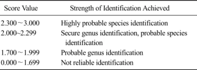
Table 2. Meaning of score values by MALDI-TOF MS Score Value Strength of Identification Achieved 2.300∼3.000 Highly probable species identification 2.000~2.299 Secure genus identification, probable species
identification
1.700∼1.999 Probable genus identification 0.000∼1.699 Not reliable identification
for S. Choleraesuis and S. Paratyphi C were performed with an initial denaturation at 94°C for 10 min, fol- lowed by 35 cycles of 94°C for 45 sec, annealing at 55°C for 30 sec, 72°C for 1 min, and finished with a final extension at 72°C for 7 min and stored at 4°C (Chiu et al, 2005). The amplified products were vi- sualized by electrophoresis on a 1.2% agarose gel con- taining ethidium bromide.
MALDI-TOF MS analysis
MALDI-TOF MS analysis was performed using the colonies from Rambach agar plates by both direct smear and protein extraction methods. For direct smear meth- od, one to two colonies of each isolate were picked us- ing a wooden applicator. The wooden applicator was then smeared over an individual spot on the MSP 96 target plate, creating a confluent layer of bacterial colonies.
The bacterial colonies were dried for 5 min, after that 1 L of matrix solution (-cyano-4-hydroxycinnamic acid sol- ution; HCCA) was applied to the spot and allowed to dry. This method was performed in twice for each isolate. For protein extraction method, one to two colo- nies were picked from solid media and inoculated into a 1.5 tube containing 300 L of deionized water, and then added 600 L of 100% ethanol. Each tube was centri-
fuged at 16,000×g for 2 min. After discard the super- natant, the pellets were allowed to dry and then were resuspended in 50 L of 70% formic acid and 50 L of 100% acetonitrile. The suspension was centrifuged again at 16,000×g for 2 min. Following centrifugation, 2 L of the resultant supernatant containing the extracted pro- teins was applied to each well and dried for 5 min. This method was performed in twice for each isolate.
MALDI-TOF analysis was performed using the MALDI Microflex LT instrument and Biotyper 3.0 software (Bruker Daltonik, Bremen, Germany). Each 96 target plate was tested using the automated analysis feature of the Biotyper software. Results are given as a score val- ue between 0.000 and 3.000 and the criteria applied for accepting results were shown in Table 2. Confidence scores of less than 1.699 corresponded to “Not reliable identification”, scores of 1.700 to 1.999 corresponded to
“probable genus identification”, scores of 2.000 to 2.299 corresponded to “secure genus identification, probable species identification”, and scores of 2.300 to 3.000 cor- responded to “highly probable species identification”.
RESULTS
Pathology and Microbiology
The pigs with salmonellosis were weaned pigs (39 pigs; 95.1%) ranged from 4 to 12 weeks. These pigs were composed of 25 pigs (61.0%) ranged from 4 to 8 weeks and 14 pigs (34.1%) ranged from 8 to 12 weeks.
The other 2 pigs (4.9%) were 25-day-old suckling pig and 130 days old growing pig. Clinically, most pigs showed watery diarrhea (35 pigs; 85.4%) and wasting
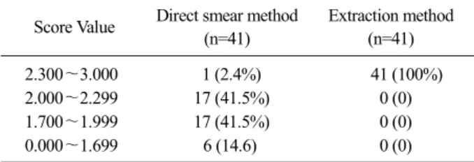
Table 3. The number of isolates identified as Salmonella spp. by MALDI-TOF MS
Score Value Direct smear method (n=41)
Extraction method (n=41)
2.300∼3.000 1 (2.4%) 41 (100%)
2.000∼2.299 17 (41.5%) 0 (0)
1.700∼1.999 17 (41.5%) 0 (0)
0.000∼1.699 6 (14.6) 0 (0)
(31 pigs; 75.6%). Grossly, fibrinous exudate (13 pigs;
31.7%) and ulceration (16 pigs, 39.0%) in the large in- testine were prevalent lesions in porcine salmonellosis (Fig. 1A). Histopathologically, variable degrees of enter- itis were observed in the large intestine from all pigs.
Focal to diffuse, mild to moderate necrosis of intestinal crypt, lamina propria, and surface enterocytes were ob- served in 9 pigs (22.0%). Multifocal to diffuse ulcer- ative colitis with/without colitis cystica profunda were frequently observed in 32 pigs (78.0%) (Fig. 1B).
Vascular thrombosis composed of fibrin and neutrophils were also frequently presented in the submucosa of large intestine. Among these 32 pigs, Balantidium coli were also observed at the ulcerated mucosa or crypts in 9 cases. In bacterial culture, red round colonies were isolated from 41 pigs using Rambach agar plates. And isolated bacteria were confirmed as gram-negative bacilli using Gram staining.
PCR
The results of PCR demonstrated that all 41 isolates were positive for S. spp. and 39 isolates (95.1%) were positive for S. Typhimurium. However, 2 isolates (4.9%) were all negative for S. Typhimurium, S. Choleraesuis and S. Paratyphi C.
MALDI-TOF MS
MALDI-TOF MS analyses for the isolates were per- formed both direct smear method and protein extraction method. The level of identification was determined by 0.000 to 3.000 score provided by the Biotyper database.
Results for each isolate of S. spp. were shown in Table 3.
Using the direct method, 85.4% (35/41) of isolates
were identified to the genus level and 43.9% (18/41) to the species level. But only one (2.4%) out of 41 isolates was identified to “Highly probable species identi- fication”. Surprisingly, all tested 41 isolates (100% to the species level) got score values above 2.300 (high probable species identification) by protein extraction method (Table 3).
DISCUSSION
One of the well-known diagnostic methods for por- cine salmonellosis is bacterial identification combined with pathological observations (Griffith et al, 2006). In this study, 41 pigs were diagnosed as salmonella enter- ocolitis based on the gross findings such as watery diar- rhea, fibrinous exudates and ulceration in large intestine, and histopathologic evidences in variable degree of ne- crotic or ulcerative typhlitis and colitis. Furthermore, all red round colonies in selective Rambach agar were identified as S. spp. by PCR analysis.
Identification of the causative agents is important for herd management in food animal industries. Manually, identification of Salmonella spp. has performed by bio- chemical, serological and phenotypic analysis after bac- terial growth on selective or enrichment media (Saffert et al, 2011). Among the manual bacterial identification procedure, serotyping of S. spp. is essential for catego- rizing Salmonella based on the surface antigen differ- ences of somatic (O) and flagellar (H) antigens (Kauffmann-White scheme) (Baggesen et al, 1996; Kim et al, 2006). Although studies have indicated that man- ual methods are still critical for identification for S.
spp., it is considered labor-intensive, expensive, compli- cated, and time-consuming (Abubakar et al, 2007; van Veen et al, 2010; Sparbier et al, 2012). In fact, recent molecular techniques such as real-time (RT) PCR, se- quencing analysis, and microarray have introduced for rapid diagnosis of porcine salmonellosis. In particular, PCR methods are highly recommended for bacterial identification since the methods enable to guarantee ease of use, relatively low cost, rapid detection and increased accuracy by amplification of targeted regions from bac- terial genome (Kim et al, 2006; Lay, 2001). In this
study, all strains were identified as Salmonella spp., though two strains were failed to identify in the genus level.
Recently, MALDI-TOF MS are widely used for accu- rate and rapid identification of various microorganisms including Enterobacteriaceae, non-fermenting bacteria, mycobacteria, anaerobes, and even yeasts (van Veen et al, 2010). This method provides probability of identi- fication at the genus and species levels of micro- organisms by measuring of peptide mass fingerprinting from each bacterial species (Table 2) and enable to de- termine the species of bacteria within few minutes de- pending on preparation of bacterial protein such as whole cell lysates and bacterial extraction (Mellmann et al, 2008; Sauer and Kliem, 2010; Welker and Moore, 2011). Indeed, a commercial system (MALDI Microflex LT instrument equipped with Biotyper 3.0 software) provides comprehensive, secure databases, and user- friendly software and considered as high-throughput technology for efficient bacterial identification (Emonet et al, 2010).
There are two well-known sample preparation meth- ods for MALDI-TOF MS, which were direct method and extraction method (Anderson et al, 2012). For sim- ple direct method, a single colony can be picked from solid culture media using a swab or toothpick and smeared directly onto a polished steel target plate for identification (Emonet et al, 2010; Anderson et al, 2012). Alternatively in extraction method, isolates re- quire processing through a short formic acid-acetonitrile extraction for 5 min and centrifugation step prior to ap- plication on the target (Emonet et al, 2010; Anderson et al, 2012). A study suggested that MALDI-TOF MS guarantee high-throughput and rapid diagnostics at low costs and can be considered as an alternative tool in- stead of conventional biochemical and molecular identi- fication systems (van Veen et al, 2010).
Previously, identification of S. spp. by means of a combination of selective enrichment broth and MALDI- TOF MS was performed using human clinical stool samples (Sparbier et al, 2012). The application of MALDI-TOF MS analysis using the colonies from se- lective selenite enrichment media from stool samples re- vealed that a significant number of samples can be iden-
tified with S. spp. one day earlier than the standard mi- crobiological procedure. In addition, 60 S. spp. isolates from pigs with diarrhea were applied in MALDI-TOF MS based on the direct method and RT PCR, in which the identification rate were 98.3% (59/60) and 100%, re- spectively (Sohn et al, 2016). However, our study showed that 95.1% (39/41) isolates were confirmed by PCR analysis and 100% (41/41) of isolates were identi- fied to species level of S. spp using MALDI-TOF MS based on the protein extraction method. Only 43.9%
(18/41) were identified to species level using the direct method. In fact, results suggest that protein extraction method for bacterial isolates enhance the identification rate of bacteria than the direct smear method in MALDI-TOF MS analysis for porcine salmonellosis.
Although applications of MALDI-TOF MS are widely expended, limitations are quite evident. These include several issues such as analyses of uncultivable micro- organisms, analyses of samples of mixed strains and dif- ferentiation of very closely related taxa (Welker and Moore, 2011). Due to the difficulties with the reproduci- bility of results by different cultivation conditions be- tween laboratories and the limited of reference spectral sets, MALDI-TOF MS still needs to be improved for species identification of microorganisms (Mellmann et al, 2008). However, with in-depth advances in technol- ogy, as well as more accurate methods of sample prepa- ration, some of the current limitations will be overcome in the near future (van Veen et al, 2010; Saffert et al, 2011; Welker and Moore, 2011). Therefore, future appli- cations of MALDI-TOF MS for diagnosis in veterinary medicine will be relied on the expansion of databases such as relevant reference strains of microorganisms for various microbiological fields. In conclusion, the causa- tive agent for porcine salmonellosis can be diagnosed based on the pathological observation, isolation of bac- teria by selective media and identification using PCR and MALDI-TOF MS. This study suggests that MALDI-TOF MS is a useful method for salmonellosis in swine industry.
ACKNOWLEDGEMENTS
This research was supported by the 2017 scientific promotion program funded by Jeju National University.
REFERENCES
Abubakar I, Irvine L, Aldus CF, Wyatt GM, Fordham R, Schelenz S, Shepstone L, Howe A, Peck M, Hunter PR. 2007. A systematic review of the clinical, public health and cost-effectiveness of rapid diagnostic tests for the de- tection and identification of bacterial intestinal pathogens in faeces and food. Health Technol Assess 11: 1-216.
Anderson NW, Buchan BW, Riebe KM, Parsons LN, Gnacinski S, Ledeboer NA. 2012. Effects of solid-medium type on routine identification of bacterial isolates by use of ma- trix-assisted laser desorption ionization-time of flight mass spectrometry. J Clin Microbiol 50: 1008-1013.
Baggesen DL, Wegener HC, Bager F, Stege H, Christensen J.
1996. Herd prevalence of Salmonella enterica infections in Danish slaughter pigs determined by microbiological testing. Prev Vet Med 26: 201-213.
Brown CC, Baker DC and Barker IK. 2007. Alimentary system.
pp. 193-199. In: Maxie MG(ed.). Jubb, Kennedy, and Palmer’s Pathology of Domestic Animals Vol 2. 5th ed.
Saunders Elsevier, Toronto.
Chiu TH, Pang JC, Hwang WZ, Tsen HY. 2005. Development of PCR primers for the detection of Salmonella enterica se- rovar Choleraesuis based on the fliC gene. J Food Prot 68: 1575-1580.
Emonet S, Shah HN, Cherkaoui A, Schrenzel J. 2010. Application and use of various mass spectrometry methods in clin- ical microbiology. Clin Microbiol Infect 16: 1604–1613.
Griffith RW, Schwartz KJ, Meyerholz DK. 2006. Salmonella. pp.
739-754. In: Straw BE, Zimmermann JJ, D’Allaire S, Taylor DJ (eds), Diseases of Swine, 9th ed. Blackwell Publishing, Ames, USA.
Hsieh SY, Tseng CL, Lee YS, Kuo AJ, Sun CF, Lin YH, Chen JK. 2008. Highly efficient classification and identification of human pathogenic bacteria by MALDI-TOF MS. Mol Cell Proteomics 7: 448-456.
Kim HJ, Park SH, Kim HY. 2006. Comparison of Salmonella en- terica serovar Typhimurium LT2 and non-LT2 Salmonella genomic sequences, and genotyping of salmonellae by using PCR. Appl Environ Microbiol 72: 6142-6151.
Lay JO Jr. 2001. MALDI-TOF mass spectrometry of bacteria.
Mass Spectrom Rev 20: 172-194.
Mellmann A, Cloud J, Maier T, Keckevoet U, Ramminger I, Iwen P, Dunn J, Hall G, Wilson D, LaSala P, Kostrzewa M, Harmsen D. 2008. Evaluation of matrix-assisted laser desorption ionization-time-of-flight mass spectrometry in comparison to 16S rRNA gene sequencing for species identification of nonfermenting bacteria. J Clin Microbiol 46: 1946-1954.
Saffert RT, Cunningham SA, Ihde SM, Jobe KE, Mandrekar J, Patel R. 2011. Comparison of Bruker Biotyper matrix-as- sisted laser desorption ionization–time of flight mass spectrometer to BD Phoenix automated microbiology system for identification of gram-negative bacilli. J Clin Microbiol 49: 887-892.
Sauer S, Kliem M. 2010. Mass spectrometry tools for the classi- fication and identification of bacteria. Nat Rev Microbiol 8: 74-82.
Seng P, Drancourt M, Gouriet F, La Scola B, Fournier PE, Rolain JM, Raoult D. 2009. Ongoing revolution in bacteriology:
routine identification of bacteria by matrix-assisted laser desorption ionization time-of-flight mass spectrometry.
Clin Infect Dis 49: 543-551.
Sohn JH, Jeon WJ, Lee YM, Kim SS. 2016. MALDI TOF MS for the identification of Salmonella spp. from swine.
Korean J Vet Serv. 39: 247-251.
Sparbier K, Weller U, Boogen C, Kostrzewa M. 2012. Rapid de- tection of Salmonella sp. by means of a combination of selective enrichment broth and MALDI-TOF MS. Eur J Clin Microbiol Infect Dis 31: 767-773.
van Duijkeren E, Wannet WJ, Houwers DJ, van Pelt W. 2002.
Serotype and phage type distribution of Salmonella strains isolated from humans, cattle, pigs, and chickens in the Netherlands from 1984 to 2001. J Clin Microbiol 40: 3980-3985.
van Veen SQ, Claas EC, Kuijper EJ. 2010. High-throughput iden- tification of bacteria and yeast by matrix-assisted laser desorption ionization–time of flight mass spectrometry in conventional medical microbiology laboratories. J Clin Microbiol 48: 900-907.
Welker M, Moore ER. 2011. Applications of whole-cell ma- trix-assisted laser-desorption/ionization time-of-flight mass spectrometry in systematic microbiology. Syst Appl Microbiol 34: 2-11.
Widjojoatmodjo MN, Fluit AC, Torensma R, Keller BH, Verhoef J. 1991. Evaluation of the magnetic immuno PCR assay for rapid detection of Salmonella. Eur J Clin Microbiol Infect Dis 10: 935-938.
Wilcock BP, Armstrong CH, Olander HJ. 1976. The significance of the serotype in the clinical and pathological features of naturally occurring porcine salmonellosis. Can J Comp Med 40: 80-88.