J Kor Soc Fish Technol, 50 (4), 447―454, 2014 http://dx.doi.org/10.3796/KSFT.2014.50.4.447
<Original Article>
LED (Light-Emitting Diode)를 이용한 미세조류 (Chaetoceros calcitrans)의 성장 및 생화학적 조성 변화
안희춘⋅배재현⋅권오남1⋅박흠기2⋅박진철*2
국립수산과학원 동해수산연구소 해역산업과, 1강릉원주대학교 동해안생명과학연구소,
2강릉원주대학교 해양자원육성학과
Changes in the growth and biochemical composition of Chaetoceros calcitrans cultures using light-emitting diodes
Heui-Chun AN, Jae-Hyun BAE, O-Nam KWON1, Heum-Gi PARK2 and Jin-Chul PARK*2 Aquaculture Industry Division, East Sea Fisheries Research Institute, NFRDI, Gangneung 210-861, Korea
1East Costal Life Science Institute Gangneung-Wonju National University, Gangneung 210-702, Korea
2Department of Marine Bioscience Gangneung-Wonju National University, Gangneung 210-702, Korea
The marine microalgae Chaetoceros calcitrans was cultured under a fluorescent lamp (CON) and light-emitting diodes (LEDs) of various wavelengths (blue, LB; red, LR; green, LG; white, LW); changes in growth, fucoxanthin, chlorophyll-a, amino acid and fatty acid profiles were investigated. LR-exposed cultures exhibited the highest specific growth rate (SGR) (0.34), whereas LG-exposed cultures showed the lowest SGR (0.26). After cultivation for 10 days, the maximum dry cell weight (g/L) of LR-exposed cultures was significantly higher than that of those exposed to other light conditions (LR≧
CON>LB≧LW≧LG). Eicosapentaenoic acid (EPA) levels were significantly higher in CON-exposed cultures compared to those exposed to LW (P<0.05), with no marked difference compared to those exposed to LB, LR and LG (P>0.05). The fucoxanthin content was highest in LB-exposed cultures (6.3㎍/mL), whereas LW showed the lowest (3.6㎍/mL; P<0.05).
Chlorophyll-a content was highest in cultures exposed to LB compared to other light sources. These results suggest consistent differences in growth and biochemical composition after exposure to light of different wavelengths.
Keywords: Chaetoceros calcitrans, LED, Specific growth rate, Fatty acid, Fucoxanthin
*Corresponding author: [email protected], Tel: 82-33-640-2345, Fax: 82-33-640-2340
서 론
최근 미세조류는 다양한 영양소 및 생리활성물질의 생산뿐만 아니라 광합성 능력에 기인한 고효율의 이산 화탄소 제거와 화석연료를 대체하기 위한 바이오에너
지원으로써 최근 각광을 받고 있다 (Oh et al., 2003;
Park et al., 2010). 하지만 미세조류가 산업적으로 활용 되기 위해서는 반드시 대량생산에 따른 바이오매스 (biomass)가 확보되어야 하는데 불행히도 미세조류를
대량 배양하는데 있어 소요되는 비용은 상당히 높은 편 이다 (Coutteau and Sorgeloos, 1993). 이러한 경제적인 문제점을 해결하기 위해서는 기존 배양방법보다 배양 기간을 단축시키면서 동시에 단위시간당 생산량을 높 을 수 있는 배양기술을 개발해야만 한다. 이를 위해서 는 다양한 환경 조건하 (수온, 염분, pH, 이산화탄소 및 광 조건 등)에서 최적 성장조건을 규명하는 것이 가장 우선시 되어야만 하는데, 그 중에서도 광 관련인자는 무엇보다도 고려되어야 할 중요한 사항이다 (Park et al., 2013). 특히, 광의 파장은 미세조류의 양적 성장과 질적 함유량 변화를 유도할 수 있기 때문에 광원의 선 택은 인위적인 미세조류 배양에 있어 매우 중요한 요인 이다 (Mouget et al., 2004; Sánchez-Saavedra and Voltolina, 2006).
한편, 차세대 에너지절약형의 대체 광원인 발광다이 오드 (Light-Emitting Diode, LED)는 다른 발광체보다 친환경적으로 수명이 길고 소비전력이 낮으며 열 발생 이 없는 장점을 지니고 있다 (Oh et al., 2007). 또한 가 시광선대의 모든 파장을 구현할 수 있어 해당 미세조류 의 광합성에 필요한 특정 파장만을 공급해 줄 수 있기 때문에 최근 유용 미세조류의 대량배양에 효율적인 광 원으로 주목받고 있다 (Das et al., 2011). 하지만 현재 대부분의 미세조류 배양장에서는 이러한 빛 파장에 따 른 광원은 고려하지 않은 채 단일 형광등 (실내)과 자 연광 (실외)만을 이용하여 배양을 행하고 있으므로 이 를 개선할 필요성이 있다.
따라서 본 연구에서는 다양한 파장의 LED 광원을 준 비하여 대량배양이 용이하고 영양소가 풍부하여 패류 양식산업에 주로 이용되어지고 있는 유용 미세조류 Chaetoceros calcitrans (Raghavan et al., 2008)의 성장, 클로로필-a (chlorophyll-a), 푸코잔틴 (fucoxanthin), 지 방산 (fatty acid) 및 아미노산 (amino acid) 함량의 변화 를 조사하여 최적 배양을 위한 광원을 선택해 보고자 한다.
재료 및 방법 실험 종
본 연구에 사용된 미세조류 (C. calcitrans)는 2013년 3월에 국내 생산업체인 (주)NLP에서 종을 분양받아 지 속적으로 계대배양 (sub-culture)된 것을 이용하였다.
실험 장치 및 실험구
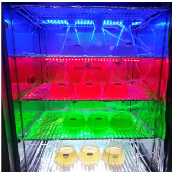
인큐베이터 (MIR-553, SANYO, Japan) 내부에 가로 60 cm, 세로 65 cm의 공간을 확보하여 bar 형태의 LED 램프 (SS-light 0.75 W, LED-SYSTEM Inc., Korea)를 고정시켜 설치하였다 (Fig. 1). Bar 형태인 LED 램프를 각 공간에 설치한 것이며, 이 때 투입된 전력은 각각 3W로 동일하게 공급되었다. 설치된 각각의 LED 실험 구는 파장대가 다른 청색 (460 nm, LB), 녹색 (520 nm, LG), 적색 (630 nm, LR) 및 흰색 (복합파장, LW)이였 으며, 여기에 실내 배양에서 주로 이용되고 있는 형광 등 (FL10CW-10W, Wooree Inc., Korea)를 대조구 (CON)로 하여 3회 반복 실험하였다. LED 광원은 상부 에 설치된 것으로 위에서 아래로 광이 공급된 것이며, 각각의 실험 공간은 칸막이로 설치되었기 때문에 해당 LED 광원만이 조명되었다.
Fig. 1. Experiment view of LED light treatment.
실험 조건
실험을 위해 멸균 해수가 담겨진 1L 유리플라스크 (배양수 700 mL)에 C. calcitrans를 접종하고 conway 배 지와 함께 추가적으로 규산염을 넣어주었다. 또한 LED 램프가 설치된 곳에 미세조류가 접종된 유리플라스크 를 넣고 에어라인을 통해 폭기를 시켜주었으며, 이후 인큐베이터의 온도를 22±1℃로 맞춰주어 10일간 회분 (batch) 배양방법으로 실험을 행하였다.
측정 항목의 분석 방법
배양일수에 따른 양적 성장을 알아보고자 건조중량 값을 산출하였다. 건조중량은 GF/C 여과지 (47 mm, WhatmanTM, UK)를 이용하여 20 mL의 배양수를 여과 하고 증류수로 남아있는 염을 녹인 후 100℃에서 1시간 동안 건조하여 무게를 매일 측정하여 L당 g으로 환산하 여 값을 나타내었다. 또한 배양일수 경과에 따른 상대성 장률 (Specific growth rate, SGR) 값은 Rico-Martinez and Dodson (1992)의 방법에 따라 구하였다.
한편, 클로로필-a 및 푸코잔틴과 같은 색소 함유량의 변화는 Wellburn (1994)의 방법에 따라 분광광도계 (V-550, JASCO, Japan)로 값을 측정한 후 공식에 대입 하여 구하였으며, 그 외 지방산과 아미노산은 Park et al. (2013)의 방법에 따라 전처리를 행한 후 gas chromatography (HP6890 plus, Agilent, USA)와 아미노 산 분석기 (L-8800, Hitachi, Japan)를 이용하여 구하였다.
통계처리
본 실험의 모든 결과는 일원분산분석 (one-way ANOVA-test)의 방법을 이용하였는데, 이는 독립변수 (빛 광원)에 의해 종속변수 (세포 건조중량, 색소, 지방 산 및 아미노산 등)에 대한 평균치의 차이를 살펴본 것 이며, 이를 통해 얻어진 결과 값은 Duncan's multiple range test (Duncan, 1955)를 실시하여 유의성을 SPSS program (Ver. 14.0)으로 검정하여 나타내었다. 이 때 그림 및 표에 나타낸 상이한 문자나 윗첨자는 유의성 (P<0.05, 신뢰수준 95%)에서 서로 간에 유의적인 차이 가 있다는 것을 의미한다.
결 과 광원에 따른 C. calcitrans의 성장
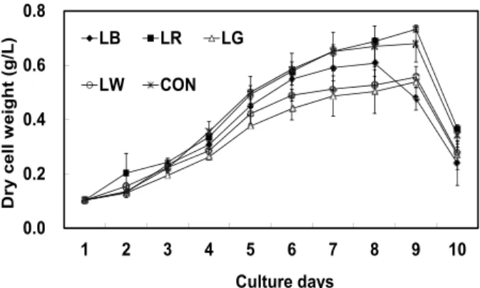
세포성장에 따른 건조중량과 상대성장률 (SGR)을 Fig. 2과 Fig. 3에 나타내었다. 세포성장에 따른 건조중 량은 4일째부터 실험구간 내 유의적인 차이가 나타나 기 시작하여 8일째 LR 실험구에서 가장 높은 건조중량 값 (0.73 g/L)을 보여 다른 LED 실험구와의 유의적인 차이를 나타냈다 (P<0.05). 하지만 대조구인 CON (0.68 g/L)과 유의적인 차이는 보이지 않았다 (P>0.05). 반면 LG는 0.54 g/L로 유의적으로 가장 낮은 값을 보였다 (P<0.05). 또한 상대성장률인 SGR 값도 동일한 경향이
나타나 LR에서 0.34로 가장 높게 나타났으며, LG에서 는 0.26으로 가장 낮게 나타났다 (P<0.05).
0.0 0.2 0.4 0.6 0.8
1 2 3 4 5 6 7 8 9 10
Dry cell weight (g/L)
Culture days
LB LR LG
LW CON
Fig. 2. Dry cell weight (g/L) of Chaetoceros calcitrans cultured 10 days under various light sources. LB, LED blue; LR, LED red; LG, LED green; LW, LED white; CON, fluorescent lamp.
bc
d
a
ab cd
0.0 0.1 0.2 0.3 0.4
LB LR LG LW CON
Specific growth rate (SGR)
Light conditions
Fig. 3. Specific growth rate (SGR) of Chaetoceros calcitrans cultured at the different light sources. Letters of "a" to "d" on bars indicate significant differences as non-order symbols by Duncan's multiple range test. Totally different letters (ex. a:b or a:c etc) between light conditions indicate significant differences (P<0.05) while a same superscript (ex. a:ab or b:ab) indicate non-significant differences (P>0.05).
색소 함량의 변화
광원에 따른 C. calcitrans의 클로로필-a와 푸코잔틴 함량의 변화를 Fig. 4와 Fig. 5에 나타내었다. 클로로필 -a의 최대값은 LB에서 3.9 mg/L로 대조구인 CON의 2.8 mg/L 보다도 유의적으로 높게 나타났다. 그 뒤로 LR, LG 순으로 높게 나타났으나 LW는 1.3 mg/L로 가 장 낮은 값을 보였다 (P<0.05). 푸코잔틴의 함량도 LB 에서 가장 높은 6.3 ㎍/ mL로 나타났으며, CON과 LG 가 다음 그룹으로 높게 나타났다 (P<0.05). 반면 LW와
LR는 각각 3.6, 3.7 ㎍/mL으로 가장 낮은 값을 나타냈 다 (P<0.05).
c
b
a a
b
0 1 2 3 4 5
LB LR LG LW CON
Chlorophyll-a content (mg/L)
Light conditions
Fig. 4. Comparison of chlorophyll-a content (mg/L) of Chaetoceros calcitrans at various light sources. Symbols are same with Fig. 2. The different letters on bars denote significant differences as Duncan's multiple range test (P<0.05) refer to Fig. 3.
c
a ab a
b
0 1 2 3 4 5 6 7 8
LB LR LG LW CON
Fucoxanthin content (μg/mL)
Light conditions
Fig. 5. Comparison of fucoxanthin content (μg/L) of Chaetoceros calcitrans at various light sources. Symbols are same with Fig. 2. The different letters on bars denote significant differences as Duncan's multiple range test (P<0.05) refer to Fig. 3.
지방산 및 아미노산 함량의 변화
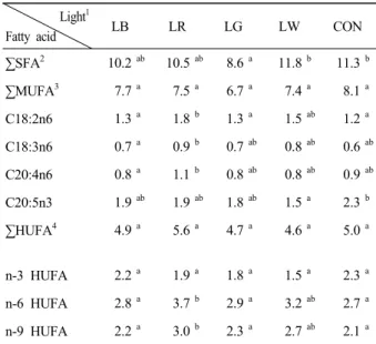
각기 다른 광원에 따른 C. calcitrans의 지방산 조성 변화를 Table 1에 나타내었다. SFA (saturated fatty acids)은 LW 및 CON에서 유의적으로 높았으나 LG를 제외하곤 차이는 보이지 않았다 (P>0.05). MUFA (mono unsaturated fatty acids)는 모든 실험구간에서 유 의적인 차이가 없었다. C18:2n6 (linoleic acid) 및 C18:3n6 (gamma-linolenic acid)은 LR가 다른 실험구에 비해 유의적으로 가장 높게 나타났다 (P<0.05). 또한 C20:4n6 (arachidonic acid, ARA)은 LR에서 유의적으
로 가장 높게 나타났으나 LB를 제외한 다른 실험구들 과의 유의적인 차이는 보이지 않았다 (P>0.05). 본 종에 있어 가장 중요한 지방산 중에 하나인 고도불포화지방 산 20:5n3 (eicosapetaenoic acid, EPA)은 CON에서 2.3 (㎍/mg dry matter)으로 높게 나타났으나 LW를 제외한 다른 LED 실험구와의 유의적인 차이는 보이지 않았다 (P>0.05). n-3 HUFA (highly unsaturated fatty acids)의 경우, 대조구를 포함한 모든 실험구간에서 유의적인 차 이가 없는 것으로 나타났다 (P>0.05).
Table 1. Fatty acid compositions (㎍/mg dry matter) of Chaetoceros calcitrans at various light sources*
Light1
Fatty acid LB LR LG LW CON
∑SFA2 10.2ab 10.5ab 8.6a 11.8b 11.3b
∑MUFA3 7.7a 7.5a 6.7a 7.4a 8.1a
C18:2n6 1.3a 1.8b 1.3a 1.5ab 1.2a
C18:3n6 0.7a 0.9b 0.7ab 0.8ab 0.6ab C20:4n6 0.8a 1.1b 0.8ab 0.8ab 0.9ab C20:5n3 1.9ab 1.9ab 1.8ab 1.5a 2.3b
∑HUFA4 4.9a 5.6a 4.7a 4.6a 5.0a
n-3 HUFA 2.2a 1.9a 1.8a 1.5a 2.3a
n-6 HUFA 2.8a 3.7b 2.9a 3.2ab 2.7a n-9 HUFA 2.2a 3.0b 2.3a 2.7ab 2.1a
*Values are means of triplicate groups in the same row and different letter superscripts are significantly different as determined by Duncan's multiple range test (P<0.05)
1Refer to Fig. 2
2Saturated fatty acid; 6:0, 8:0, 10:0, 11:0, 12:0, 13:0, 14:0, 15:0, 16:0, 17:0, 18:0, 20:0, 21:0
3Mono unsaturated fatty acid; 15:1, 16:1, 17:1, 18:1n9, 20:1, 22:1n-9, 24:1
4Highly unsaturated fatty acid; 18:2n-6, 18:3n-3, 20:2, 18:3n-6, 20:3n-6, 20:4n-6, 20:5n-3, 22:2, 22:6n-6
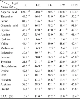
한편, 실험구에 따른 아미노산 함량 변화를 Table 2 에 나타내었다. 모든 실험구간을 비교했을 때, 필수아 미노산 및 비필수아미노산의 함량은 특정 파장대에서 뚜렷한 경향은 보이지 않았다. 하지만 세부 항목간을 비교해 볼 때, 파장에 따라 분명 필수아미노산 및 비필 수아미노산의 함량 변화에는 유의적인 차이가 나타나 는 것으로 조사되었다 (P<0.05).
Table 2. Amino acid compositions (ng/mg dry matter) of Chaetoceros calcitrans at various light sources*
Light1
Amino acid LB LR LG LW CON
Aspartic acid 124.3ab 120.8ab 130.5b 130.8b 110.4a Threonine 49.7 ab 46.4 a 51.9 b 50.0 ab 50.2 ab Serine 84.7 a 83.6 a 86.4 a 92.4 a 82.7 a Glutamic acid 86.9 a 86.8 a 98.2 b 88.4 a 91.0 a Glycine 43.2 ab 42.0 a 47.0 bc 43.1 ab 47.3 c Alanine 57.0 a 55.6 a 63.9 b 59.1 ab 61.4 ab Cysteine 7.0 a 6.6 a 7.1 a 7.4 a 7.7 a Valine 46.4 a 44.9 a 49.0 a 48.6 a 47.4 a Methionine 7.3 a 6.3 a 7.3 a 6.4 a 7.2 a Isoleucine 30.6 a 30.7 a 34.1 b 32.3 ab 34.8 b Leucine 59.9 a 60.0 a 66.3 b 62.6 ab 66.0 b Tyrosine 21.5 ab 21.1 a 23.0 ab 20.0 a 26.9 b Phenylalanine 47.7 ab 46.9 a 52.1 b 48.1 ab 50.8 ab Lysine 42.5 a 42.3 a 46.6 a 45.0 a 53.4 b HN3 19.4 a 18.1 a 20.3 a 19.9 a 18.6 a Histidine 12.7 a 13.3 a 15.6 b 13.4 a 16.4 b Arginine 38.5 a 38.3 a 42.1 b 38.8 a 45.0 b Proline 49.6 a 47.4 a 50.4 a 51.6 a 44.8 a EAA2 (%) 10.4a 11.0 a 12.2b 11.0ab 12.4b
*Values are means of triplicate groups in the same row and different letter superscripts are significantly different as Duncan's multiple range test (P<0.05)
1Refer to Fig. 2
2Essential amino acids
고 찰 광원에 따른 C. calcitrans의 성장
일반적으로 미세조류는 광합성을 위해 광원 중 400- 700 nm의 파장대를 이용하나 모든 범위 내에서 광합성 을 할 수 있는 것은 아니며, 주로 적색광 (600-700 nm) 과 청색광 (400-500 nm)에서 미세조류의 성장을 촉진 시키는 것으로 보고되고 있다 (Das et al., 2011). 또한 미세조류 종마다 선호하는 파장대가 각기 다른 것으로 알려져 있기 때문에 모든 파장대를 구현할 수 있는 LED를 미세조류 배양에 적용한다면 보다 효율적인 배 양시스템이 구축될 수 있을 것이다.
이러한 맥락으로 본 연구에도 LED를 적용해 본 결 과, 각기 다른 파장에 따른 C. calcitrans은 분명 성장에 영향을 미치는 것으로 나타났다. 세포의 성장률 (SGR) 및 세포성장에 따른 건조중량은 LR인 적색광에서 가장 높은 것으로 나타났는데, 이는 다른 연구결과와 유사하게
나타난 것이다. Markou (2014)는 Arthrospira platensis 의 바이오매스 생산량은 blue LED에서 4.68 mg/L/day 인 반면 red LED에서는 30.69 mg/L/day으로 차이를 보 인다고 하였다. 또한 Wang et al. (2007)의 Spirulina platensis와 Choi et al. (2013)의 Chlorella sp. 및 Lee and Palsson (1994)의 C. pyrenoidosa도 red LED에서 가장 높은 성장을 보인다고 보고하였다.
이처럼 적색광이 다른 LED 파장대와 형광등에 비해 성장이 높은 이유로는 몇 가지 의견이 제시되고 있다.
첫째로 광자 (photon)에 의한 빛 흡수의 전달 차이라 할 수 있다. Figueroa et al. (1994)는 빛 흡수를 행하여 광 합성을 위한 다음 단계로 전달함에 있어 적색광이 청색 광에 비해 1.2~1.5배 높은 효율성을 가지고 있어 보다 높은 성장률을 유도할 수 있다고 하였다. 둘째로 탄소 원의 축적률 차이를 들 수 있다. 세포 내 탄소 축적률이 높을 때 광합성률은 증가하는 것으로 알려져 있는데 (Marchetti et al., 2013), 특히 다른 파장대에 비해 적색 광 파장대에서 탄소 축적률이 높은 것으로 보고되고 있 다 (Figueroa et al., 1995). Aguilera et al. (2000)는 Porphyra leucosticta에게 흰색, 청색 및 적색광에 따라 탄소 대사작용의 영향을 살펴본 결과, 적색광에 비해 청색광에서 탄소의 축적이 낮아 결국 성장률과 바이오 매스가 낮은 결과로 이어진다고 하였다. 셋째로 광합성 유효광량자 (umol/m2/s)의 차이를 들 수 있는데, Choi et al. (2013)의 실험에서 형광등은 64 W로 4,500 lux이 나 유사한 빛 세기를 가지고 있는 red LED에 비해 광 합성유효광량자속은 절반 수준이기 때문에 성장률은 2.5배 느리며, 생산량은 50% 감소한다고 하였다. 마지 막으로 세포 내 동화작용 및 대사작용의 차이를 들 수 있다. Figueroa et al. (1995)는 색소 당 산소 발생을 통 한 광합성률을 측정한 결과, red LED가 다른 광에 비 해 빛 흡수의 효율성이 높다고 하였으며, 이로 인해 광 합성 유도 유전자의 전사 및 효소활성과 같은 세포 내 동화작용 및 대사작용이 활발히 이루어진다고 보고하 였다 (Ruyters, 1984; Das et al., 2011). 이처럼 본 연구 에서도 red LED가 다른 LED와 형광등에 비해 높은 성 장률과 바이오매스를 보였는데, 이는 위에서 언급한 이 유들에 의한 복합적인 요인이 작용했기 때문인 것으로 판단되어진다.
한편, 본 실험에서 흰색계열인 LW (복합파장) 및 RGB 조합인 형광등 (대조구)과 green LED인 LG는
LR에 비해 상대적으로 낮은 성장을 보였을 뿐이지 성 장이 전혀 안 되었던 것은 아니다. Schofield et al.
(1990)는 C. gracilis를 RGB 광원인 white로 배양했을 때 성장 및 광합성이 향상된다고 하였는데 본 연구에서 도 부정적인 영향은 받지 않았으며, 성장률도 증가하는 경향을 보였기 때문에 본 종을 배양하는데 있어 문제가 되지는 않을 것으로 판단되어진다. 하지만 LG의 경우 성장률은 다른 조명등에 비해 유독 낮았는데, 이는 빛 을 효율적으로 받아들이지 못하고 부분적으로 빛 파장 을 수용하여 광합성을 행한 결과이므로 본 종을 배양하 기에는 다소 비효율적인 광원이라 판단되어진다.
색소 함량의 변화
광합성에 의해 축적되는 미세조류의 색소 함량 및 축 적할 수 있는 생합성 능력은 환경요인에 의해 달라진다 고 하였으며, 그 중에서도 빛의 파장은 매우 밀접한 관 계가 있다고 하였다 (Mouget et al., 2004). 실제 Figueroa et al. (1995)는 빛 파장에 의해 chlorophyll, phycocyanin, phycoerythrin와 같은 주색소 및 보조색소 의 함유량이 차이를 보인다고 하였으며, Katsuda et al.
(2006)는 Haematococcus pluvialis도 LED 광원에 따라 astaxanthin의 축적률이 달라진다고 보고하였다. 본 연 구에서 C. calcitrans의 chlorophyll-a 함유량은 blue 파 장에서 가장 높게 나타났다. 세포 성장이 높았던 red 파 장보다도 높게 나타난 것인데, 이는 파장대에 따른 chlorophyll-a 축적률의 차이 때문이라 판단되어진다.
광합성을 통한 세포 성장이 높다고 해서 반드시 chlorophyll-a이 높아야 하는 것은 아니다. 즉, 선호하는 파장대가 아니기에 광합성을 위해 빛을 더욱 활용하고 자 성장이 유리했던 red 파장대에 비하여 그렇지 않았 던 blue 파장대에서 chlorophyll-a 생합성이 더욱 유리 하게 작용했다는 것이다 (Kim et al., 2011). 또한 Seyfabadi et al. (2011)는 성장이 유리한 파장대에서 세 포분열이 빨리 진행됨에 따라 미처 chlrophyll을 축적할 만큼의 시간적 여유가 없었기 때문이라는 연구보고도 있어 이러한 영향도 작용했으리라 판단되어진다.
한편, 최근 fucoxanthin은 다양한 의약산업 및 식품산 업 분야에서 그 이용성이 넓어지고 있는 유용 색소로 현장 적용 연구가 이루어지고 있다 (Lamers et al., 2010; Peng et al., 2011). 본 연구결과, fucoxanthin의 함량은 chlorophyll-a 함량과 같은 경향을 보여 성장이
다소 낮았던 blue LED에서 가장 높은 것으로 나타났 다. 이처럼 성장과 색소함량에 대한 LED 광원의 불일 치는 빛에 의해 높은 스트레스를 받을 경우에는 세포수 의 감소와 함께 성장률 저하로 이어지고 (Lamers et al., 2010; Shariati and Hadi, 2011), 스트레스 환경 조건에 서 세포분열을 행하는 동안에는 최대한 빛 흡수의 양을 많이 하려는 방어기작이 작용하여 세포 당 fucoxanthin 축적률은 오히려 향상된다는 것으로 설명할 수 있다 (Shariati and Hadi, 2011). 실제 blue LED 파장은 자외 선에 가까운 단파장이므로 성장보다는 스트레스에 대 한 대사작용이 촉진되어 세포성장은 저해되나 (Ben- Amotz et al., 1989), fucoxanthin과 같은 색소의 함량은 성장에 유리했던 다른 파장에 비해 높게 축적되었던 것 으로 판단되어진다. 실제로 Mogedas et al. (2009)의 광 합성활성광 (400-700 nm)에 UV-A (320-400 nm)를 보 충해 주는 실험에서 carotenoid 축적량은 UV-A를 비춰 주지 않은 대조구에 비해 약 2배 정도 높아졌는데, 이는 UV 광원에 따른 산화 스트레스에 의한 미세조류의 반 응기작 때문이라고 보고하였다 (Mogedas et al., 2009).
지방산 및 아미노산 함량의 변화
미세조류의 지방산 및 아미노산과 같은 생화학적 조 성은 배양환경에 의해 결정되는데, 그 중에서도 빛 광 원은 매우 중요하게 작용하고 있다 (Pratoomyot et al., 2005; Park et al., 2013; Saha et al., 2013). 최근 미세조 류를 활용한 바이오디젤 연구가 각광을 받고 있는데 이 는 지방산 중 C16~C18 계열의 지방산이 많이 함유되 어 있기 때문이다 (Saha et al., 2013). 또한 높은 함량의 SFA 및 MUFA는 원활한 바이오디젤 생산을 이상적인 지방산으로 알려져 있기 때문에 이러한 지방산의 함량 변화 유도는 매우 중요한 연구가 될 수 있다 (Saha et al., 2013). 아울러, EPA 및 DHA와 같은 고도불포화지 방산도 다양한 산업분야에 이용되어지고 있는 고부가 치의 지방산이기 때문에 함량을 높이기 위한 연구는 현 재까지도 계속 진행 중에 있다 (Das et al., 2011;
Yoshioka et al., 2012; Saha et al., 2013).
이러한 맥락에서 본 연구를 수행한 결과, SFA 및 EPA와 같은 지방산의 함량 변화는 각기 다른 광원에 따라 변화가 있는 것으로 나타나 다른 연구자료에 의한 것처럼 빛 파장에 따른 영향을 확인할 수 있었다 (Das et al., 2011; Yoshioka et al., 2012; Saha et al., 2013).
Saha et al. (2013)는 H. pluvialis에게 빛 광원과 영양염의 조건을 달리하여 SFA와 MUFA와 같은 지방산 함량의 변화를 유도하였으며, Yoshioka et al. (2012)와 Das et al.
(2011)도 각각 Isochrysis galbana 및 Nannochloropsis sp.
에게 LED 파장대를 달리하여 EPA와 DHA의 함량변화 를 유도하여 그에 따른 영향력이 있는 것을 확인하였다.
한편, 단백질원으로서 사용하고자 하는 물질의 영양 성 평가는 구성아미노산의 조성 및 함량과 관련이 있으 며, 미세조류를 단백질원으로서 활용하기 위해서는 필 수아미노산의 함량은 높을수록 효율적이다 (Webb and Chu, 1983). 이러한 측면으로 본 연구를 수행한 결과, 각기 다른 광원에 따라 분명 아미노산 함량 변화가 유 도된 것을 확인할 수 있었다. 다만, 특정 파장대에서 필 수 및 비필수아미노산 함량 변화가 규칙적인 변화를 유 도하는 경향은 관찰되지 않았다. 하지만 필수아미노산 의 총량 (EAA)은 green LED에서 높게 나타났는데, 이 는 Park et al. (2013)에서 Nannochloropsis sp. 종과 동 일하게 나타난 결과로 green 파장대에서 보다 자극을 받아 이에 더욱 활발하게 합성했기 때문인 것으로 판단 되어진다.
결 론
최근 각광받고 있는 미세조류에 파장대가 다른 LED를 조명했을 때 세포성장, 색소 (chlorophyll-a, fucoxanthin), 아미노산 및 지방산 함량이 다르게 나타난다는 사실을 확인할 수 있었다. 단순 세포성장 및 성장률을 고려한 다면 red LED로 본 종을 배양하는 것이 유리하나 색소 는 blue LED가 더욱 효과적인 것으로 나타났다. 여기 에 특정 지방산 및 아미노산을 고려한다면 또 다른 적 합한 파장대를 선정해야 할 것으로 판단된다.
따라서 배양 목적이 다르다면 그에 맞는 특정 파장의 광원으로 본 종을 배양하여 원하고자 하는 물질을 얻는 것이 더욱 효율적일 것이라 판단되며, 추후 2개 파장대 의 혼합 광원을 이용한 실험을 행하여 그에 따른 결과 를 얻는다면 본 종을 배양함에 있어 보다 명확한 최적 의 광원을 제공할 수 있을 것이라 사료된다.
사 사
본 연구는 국립수산과학원 수산시험연구사업 (RP- 2014-FE-14)의 지원에 의해 수행되었습니다.
References
Aguilera J, Gordillo FJ, Karsten U, Figueroa FL and Niell FX.
2000. Light quality effect of photosynthesis and efficiency of carbon assimilation in the red alga Porphyra leucosticta.
J Plant Physiol 157, 86-92. (doi:10.1016/S0176-1617(00) 80140-6)
Ben-Amotz A, Shaish A and Avron M. 1989. Mode of action of the massively accumulated β-carotene of Dunaliella barda- wil in protecting the alga against damage by excess irradiation. Plant Physiol 91, 1040-1043.
Choi BR, Lim JH, Lee JK and Lee TY. 2013. Optimum conditions for cultivation of Chlorella sp. FC-21 using light emitting diodes. Korean J Chem Eng 30, 1614-1619. (doi:10.1007/
s11814-013-0081-0)
Coutteau P and Sorgeloos P. 1993. Substitute diets for live algae in the intensive rearing of bivalve mollusks a state of the art report. World Aquacult 24, 45-52.
Das P, Lei W, Aziz SS and Obbard JP. 2011. Enhanced algae growth in both phototrophic and mixotrophic culture under blue light. Bio Technol 102, 3883-3887. (doi:10.1016/
j.biortech.2010.11.102)
Duncan DB. 1955. Multiple-range and mutiple F tests. Biometrics 11, 1-42. (doi:10.2307/3001478)
Figueroa FL, Aguilera J and Niell FX. 1994. Red and blue light regulation of growth and photosynthetic metabolism in Porphyra umbilicalis. Eur J Phycol 30, 11-18. (doi:10.1080/
09670269500650761)
Figueroa FL, Aguilera J, Jimenez C, Vergara JJ, Robles MD and Niell FX. 1995. Growth, pigment synthesis and nitrogen as- similation in the red alga Porphyra sp. under blue and red light. Scientia Marina 59, 9-20.
Katsuda T, Shimahara K, Shiraishi H, Yamagami K, Ranjbar R and Katoh S. 2006. Effect of flashing light from blue light emitting diodes on cell growth and astaxanthin production of Haematococcus pluvialis. J Biosci Bioeng 102, 442-446.
(doi:10.1263/jbb.102.442)
Kim JY, Joo H and Lee JH. 2011. Carbon dioxide fixation and light source effects of Spirulina platensis NIES 39 for LED photobioreactor design. App Chem Eng 22, 301-307.
Lamers PP, van de Laak CC, Kaasenbrood PS, Lorier J, Janssen M, De Vos RC, Bino RJ and Wijffels RH. 2010. Carotenoid and fatty acid metabolism in light-stressed Dunaliella salina.
Bio Bioeng 106, 638-648. (doi:10.1002/ bit.22725) Lee CG and Palsson BØ. 1994. High-density algal photobioreactors
using light-emitting diodes. Biotechnol Bioeng 44, 1161-1167.
Marchetti J, Bougaran G, Jauffrais T, Lefebvre S, Rouxel C, Saint-Jean B, Lukomska E, Robert R and Cadoret JP. 2013.
Effects of blue light on the biochemical composition and photosynthetic activity of Isochrysis sp. (T-iso). J Appl Phycol 25, 109-119. (doi:10.1007/ s10811-012-9844-y) Markou G. 2014. Effect of various colors of light-emitting diodes
(LEDs) on the biomass composition of Arthrospira platensis cultivated in semi-continuous mode. Appl Biochem Biotechnol 172, 2758-2768. (doi:10.1007/ s12010-014-0727-3) Mogedas B, Casal C, Forjá E and Vilchez C. 2009. β-carotene production enhancement by UV-A radiation in Dunaliella bardawil cultivated in laboratory reactors. J Biosci Bioeng 108, 47-51. (doi:10.1016/j.jbiosc.2009.02.022)
Mouget JL, Rosa P and Tremblin G. 2004. Acclimation of Haslea strearia to light of different spectral qualities- confirmation of 'chromatic adaptation' in diatoms. J Photochem Photobiol B 75, 1-11. (doi:10.1016/j.jphotobiol. 2004.04.002) Oh HM, Choi AR and Mheen TI. 2003. High-value materials from
microalgae. Kor J Microbiol Biotechnol 31, 95-102.
Oh SJ, Park DS, Yang HS, Yoon YH and Honjo T. 2007.
Bioremediation on the benthic layer in polluted inner bay by promotion of Microphytobenthos growth using light emitting diode (LED). J Kor Soc Mar Env Eng 10, 93-101.
Park HJ, Jin EJ, Jung TM, Joo H and Lee JH. 2010. Optimal cul- ture conditions for photosynthetic microalgae Nannochloropsis oculata. App Chem Eng 21, 659-663.
Park JC, Kwon ON, Hong SE, An HC, Bae JH, Park MS and Park HG. 2013. Changes in the growth and biochemical composi- tion of Nannochloropsis sp. cultures using light-emitting diodes. Kor J Fish Aquat Sci 46, 259-265.
Peng J, Yuan JP, Wu CF and Wang JH. 2011. Fucoxanthin, a ma- rine carotenoid present in brown seaweeds and diatoms: me- tabolism and bioactivities relevant to human health. Marine Drugs 9, 1806-1828. (doi:10.3390/ md9101806)
Pratoomyot J, Srivilas P and Noiraksar T. 2005. Fatty acids compo- sition of 10 microalgal species. Songklan J Sci Technol 27, 1179-1187. (doi:10.1016/0022-0981(89)90029-4)
Raghavan G, Haridevi CK and Gopinathan CP. 2008. Growth and proximate composition of the Chaetoceros calcitrans f. pum- ilus under different temperature, salinity and carbon dioxide levels. Aquaculture Research 39, 1053-1058. (doi:10.1111/
j.1365-2109.2008.01964.x)
Rico-Martinez R and Dodson SI. 1992. Culture of the rotifer, Brachionus calyciflorus Pallas. Aquaculture 105, 191-199.
(doi:10.1016/0044-8486(92)90130-D)
Ruyters G. 1984. Effects of blue light on enzymes. Senger H, ed.
Springer Verlag, Berlin, Germany, 283-301.
Saha SK, McHugh E, Hayes J, Moane S, Walsh D and Murray P. 2013. Effect of various stress-regulatory factors on bio- mass and lipid production in microalga Haematococcus pluvialis. Bioresour Technol 128, 118-124. (doi:10.1016/
j.biortech.2012.10.049)
Sánchez-Saavedra MP and Voltolina D. 2006. The growth rate, bi- omass production and composition of Chaetoceros sp. grown with different light sources. Aquacult Eng 35, 161-165.
(doi:10.1016/j.aquaeng.2005.12.001)
Schofield O, Bidigare RR and Préelin BB. 1990. Spectral photo- synthesis quantum yield and blue-green light enhancement of productivity rates in the diatom Chaetoceros gracile and prymnesiophyte Emiliania huxleyi. Mar Ecol Prog Ser 64, 175-186.
Seyfabadi J, Ramezanpour Z and Zahra AK. 2011. Protein, fatty acid, and pigment content of Chlorella vulgaris under differ- ent light regimes. J Appl Phycol 23, 721-726. (doi:
10.1007/s10811-010-9569-8)
Shariati M and Hadi MR. 2011. Microalgae biotechnology and bio- energy in Dunaliella. Angelo C, ed. Progress in Molecular and Environmental Bioengineering, Intech, Rijeka, Chapter 22. (doi:10.5772/19046)
Wang CY, Fu CC and Liu YC. 2007. Effects of using light emit- ting diodes on the cultivation of Spirulina platensis. Biochem Eng 37, 21-25. (doi:10.1016/j.bej.2007.03.004)
Webb KL and Chu FE. 1983. Phytoplankton as a food source for bivalve larvae. Pruder GD, ed. World Mariculture Society Spcc. Publ., Louisiana State University, Lousiana, USA, 272-291.
Wellburn AR. 1994. The spectral determination of chlorophylls a and b, as well as total carotenoids, using various solvents with spectrophotometers of different resolution. J Plant Physiol 144, 307-313. (doi:10.1016/S0176-1617 (11)81192-2) Yoshioka M, Yago T, Yoshie-Stark Y, Arakawa H and Morinaga
T. 2012. Effect of high frequency of intermittent light.
Aquaculture 338-341, 111-117. (doi:10.1016/j.aquaculture.
2012.01.005)
2014. 8.29 Received 2014. 10.13 Revised 2014. 11.11 Accepted