보 문
Molecular diversity of endobacterial communities in edible part of King oyster mushroom (Pleurotus eryngii) based on 16S rRNA
Choung Kyu Lee
1, Md. Azizul Haque
2, Byoung Rock Choi
3, Hee Yul Lee
2, Chung Eun Hwang
2, Min Ju Ahn
2, and Kye Man Cho
2*
1
Department of Forest Resources, Gyeongnam National University of Science and Technology, Jinju 660-758, Republic of Korea
2
Department of Food Science, Gyeongnam National University of Science and Technology, Jinju 660-758, Republic of Korea
3
Department of Future Farming, Sacheon Agricultural Development & Technology Center, Sacheon 664-951, Republic Korea
16S rRNA 기초 새송이 버섯(Pleurotus eryngii)의 식용가능 부위 내생세균 군집 다양성
이총규
1・ 모하메드 아지줄 하크
2・ 최병록
3・ 이희율
2・ 황정은
2・ 안민주
2・ 조계만
2*
1
경남과학기술대학교 산림자원학과,
2경남과학기술대학교 식품과학부,
3사천시농업기술센터 미래농업과
(Received December 17, 2014; Accepted June 10, 2015)
ABSTRACT: The diversity of endobacteria in the edible part (cap and stipe) king oyster mushroom (Pleurotus eryngii) was investigated using 16S rRNA sequence analysis. The bacterial 16S rRNA libraries were constructed from the body cap (BC) and the body stipe (BS) of the king oyster mushroom. The twenty sequenced BC clones were divided into four groups and the largest group was affiliated with the Firmicutes (40% of clones). While, the twenty sequenced BS clones could be divided into six groups and the largest group was affiliated with the Actinobacteria (40% of clones). The predominant bacterial family from both the cap and stipe of the mushroom was corresponded with the Gram positive bacteria (62.5%).
Key words: Pleurotus eryngii, 16S rRNA gene, bacteria diversity, endobacteria, phylogenetic tree
*For correspondence. E-mail: [email protected];
Tel.: +82-55-751-3272; Fax: +82-55-751-3279
The edible king oyster mushroom (Pleurotus eryngii) is cultivated throughout the world and Pleurotus fungi as a group are linked to several agro-industrial activities of great economic importance, e.g., conversion of lignocellulosic residues to food and feed, biocontrol of plant diseases, degradation of noxious pollutants, and production of enzymes and medicinal compounds (Ruiz-Duenas and Martinez, 1996; Wasser and Weis, 1999;
Philippoussis et al., 2001; Wang and Ng, 2004; Kang and Cho, 2014).
Intracellular symbiosis is a widespread and biologically important phenomenon. Although host cells usually provide a suitable shelter and supply important material needs for
endosymbionts, the cytoplasm of a host cell may be considered to be a hostile environment for an invading organism (Morioka and Ishikawa, 1993; Mota et al., 2005). Thus endosymbionts must still overcome many difficulties to survive in the host.
Nevertheless, microorganisms can live in different tissues of healthy plants without causing symptoms of plant damage and give benefit to plants (Perotti, 1926; Gao et al., 2005; Hashiba and Narisawa, 2005). The root system of plants, in particular, is known to offer different microhabitats for microbial growth.
Bacterial endosymbiosis with fungi, both parasitic and mutualistic types, is characterized by the intracellular localization of the bacteria (Moran and Wernegreen, 2000).
Examples reported thus far are mainly for species belonging to
the phylum Glomeromycota (Schußler et al., 2001). Moreover,
except for the cyanobacteria colonizing Geosiphon pyriforme (Schußler and Kluge, 2000), known intra-fungal bacteria are nonculturable. Despite this difficulty, the bacterial nature of these endosymbionts has been confirmed using molecular methods. Previously, endobacteria (e.g. Burkholderia sp.) was identified from several species of endomycorrhizal fungi such as Gigaspora sp. and Scutellospora sp. (Bianciotto et al., 1996). Meanwhile, intracellular bacteria from wild types of Laccaria bicolor were detected using fluorescence in situ hybridization (FISH) in combination with confocal laser scanning microscopy (Bertaux et al., 2003, 2005). In fact, the bacterium was identified as Paenibacillus sp. by using a 16S rRNA-directed oligonucleotide probe (Bertaux et al., 2003).
While, Barbieri et al. (2000) identified bacteria belonging to Flexibacter-Cytophaga-Bacteroides (FCB) phylogroup of Bacteroidetes phylum from the cell wall of hyphae of Tuber borchii (ectomycorrhizal fungus) using FISH analyses. A molecular study aimed at surveying potential endobacteria within the mycelium of the ectomycorrhizal fungus Tuber borchii Vittad. revealed the presence of novel uncultured bacteria (Gazzanelli et al., 1999; Barbieri et al., 2000). In addition, the phylogenetic analysis placed the bacterial sequences in a single new rRNA branch in the Sphingobacterium subgroup of the Bacteroidetes phylum (Steyn et al., 1998;
Barbieri et al., 2002, 2005). Despite the above progress identifying fungal endosymbionts, little is known about the diversity and functions of bacterial populations living in the edible mushroom. Studies on the genetic and physiological diversity of the endomicrobial populations in edible mushrooms are important for understanding of their ecological role and for the development of biotechnological applications aimed at utilizing beneficial strains (Frey et al., 1997; Timonen et al., 1998).
The structure of mushroom have a stem (stipe), a cap (pileus), and gills (lamellae, sing. lamella) on the underside of the cap.
These gills produce microscopic spores that help the fungus spread across the ground or its occupant surface. The goal of this study is to examine the endobacterial community in edible part (cap and stipe) of king oyster mushroom using analysis of 16S rRNA sequences in a culture-independent manner. To date, this study first reveals the endobacterial community in edible part of king oyster mushroom.
Materials and Methods
Microbial strains and growth conditions
Escherichia coli DH5 and recombinant E. coli cells were cultured in LB containing ampicillin (50 µg/ml). The king oyster mushroom (P. eryngii) used in this study was kindly provided by the Laboratory of Mushroom, Gyeongnam Agricultural Research and Extension Services (GARES), Jinju, Republic of Korea.
Growth of mushroom
The king oyster mushroom is usually cultivated on a sawdust medium in a plastic bottle. Sawdust from poplar was used and rice bran (30%) was supplemented to promote mycelial growth.
The final moisture content of substrate materials was 65%.
Prepared and moisture-conditioned sawdust mixture was loaded into the bottle feeder. Bottles were sterilized at 121°C for 90 min. After the bottles were removed from the autoclave, they were cooled to 20°C in the cooling room. Inoculated bottles were hauled to an incubation room, where temperature and humidity were maintained at 22 to 24°C and 65 to 68%, respectively. When the substrate in the bottles was colonized, the bottles were transferred to a cultivation room for 20 days to obtain fruiting bodies. The formation of fruiting body was induced at low temperature (about 15°C) and high humidity (about 90 to 95%).
Total DNA extraction
The surface of mushroom was disinfected with 1% sodium hypochlorite for 10 min. The external fruiting body of mushroom was removed approximately 0.5 cm from the margin of fruiting body with sterile razor blades. The fruiting body tissue was triturated by pestle in sterile 10 mM phosphate buffer (pH 7.2). The triturate was subjected to DNA extraction using the DNAzol Extraction Kit (MRC Inc.). The mixture of the extracted DNA from the ten samples was used as template for PCR to amplify 16S rDNA.
Polymerase chain reaction
The PCR primers used to amplify 16S rDNA fragments were
the bacteria-specific primers, 5ʹ-CGGAGAGTTTGATCCTGG-
3 ʹ (forward) and 5ʹ-TACGGCTACCTTGTTACGAC-3ʹ (reverse) (Lane, 1991). Ribosomal DNAs were amplified by PCR using the extracted DNA, Super-Therm DNA polymerase (JMR), 1.5 mM MgCl
2, 2 mM dNTP, and primers in a final volume of 50 µl.
Fifteen cycles (denaturation at 94°C for 30 sec, annealing at 50°C for 30 sec, and extension at 72°C for 1 min 30 sec) were followed by a final incubation at 72°C for 10 min. The anticipated product of approximately 1,500 bp was isolated after agarose gel electrophoresis of the amplified mixture using a gel extraction kit (NucleoGen).
Cloning and sequencing
PCR products were directly cloned into the pGEM-T Easy vector (Promega) and recombinant colonies were randomly (20 colonies) picked from approximately 40 colonies. The recombinant plasmids were extracted using plasmid DNA isolation kit (iNtRON). Samples for nucleotide sequencing were prepared by the dideoxy-chain termination method using the PRISM Ready Reaction Dye terminator/primer cycle sequencing kit (Perkin-Elmer Corp.). The samples were analyzed in both directions and two repeats with an automated DNA sequencer (model 3100; Applied Biosystems). Assembly of the nucleotide sequences was performed with the DNAMAN analysis system (Lynnon Biosoft).
Sequence analysis
All reference sequences were obtained from the GenBank and Ribosomal Database Project (RDP) databases (Maidak et al., 2000). The sequences were analyzed using the CHECK_
CHIMERA program (Maidak et al., 2000) to exclude sequences from chimeric rDNA clones. The similarity search against database entries was done using online BLAST search (Madden et al., 1996). Sequences were aligned using the multiple sequence alignment program CLUSTAL W versions 1.6 (Tompson et al., 1994). Phylogenetic analysis was performed using the neighbor-joining method (Saitou and Nei, 1997). Gaps and positions with ambiguities were excluded from the phylogenetic analysis. Bootstrap analysis was performed on data resampled 1,000 times using the DNAMAN analysis system (Lynnon Biosoft).
Nucleotide sequence accession numbers and nomenclature
The 16S rDNA gene sequences of endobacteria isolated from the king oyster mushroom have been deposited in the GenBank database under the accession numbers AY838556, AY838458- AY838495. The names of the 16S rDNA gene library from body cap (BC) and body stipe (BS) of mushroom begins with the letters BC and BS (e.g., BC1 for clone 1 of the cap library).
Results
Cloning and detection of bacterial rDNA
The PCR amplification from the total DNAs by the cap and the stipe samples of king oyster mushroom with the bacteria- specific primers produced a single band of approximately 1.5 kb. The products were purified from an agarose gel and cloned in E. coli DH5 in the pGEM-T Easy vector. Twenty clones were obtained from the body cap library (BC clones) and twenty from the body stipe library (BS clones).
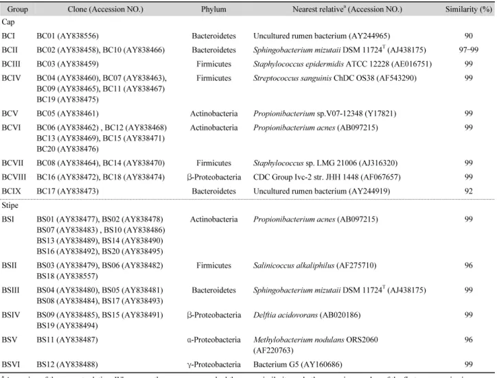
Similarity with database sequences
All clones from the two libraries were subjected to sequence analysis followed by online homology searches using two databases: GenBank which implements the BLAST algorithm, and the RDP database which implements the SIMILARITY_
RANK program (Table 1). Although there are no exact 16S
rDNA similarity limits for defining specific taxa such as genus
and species, in general, species definition requires sequence
similarities greater than 97%. Fourteen sequences (35%) from
the 40 clones in our libraries (6 from BC clones, 8 from BS
clones) can be identified as belonging to Actinobacteria. About
15% of the sequences have a similarity level with database
sequences in the range of 90–97% (Table 1). Twenty clones of
the BC library were analyzed for DNA similarity. Among them,
eighteen clones had the highest similarities with cultured
bacteria and two clones had the highest similarities to
uncultured rumen bacteria. Of the 20 clones of the BS library
that were analyzed, eight were highly similar to each other and
gave hits in the same similarity range (Table 1).
Table 1. Similarity value of 16S rDNA sequences retrieved from mushroom cap and stipe
Group Clone (Accession NO.) Phylum Nearest relative
a(Accession NO.) Similarity (%)
Cap
BCI BC01 (AY838556) Bacteroidetes Uncultured rumen bacterium (AY244965) 90
BCII BC02 (AY838458), BC10 (AY838466) Bacteroidetes Sphingobacterium mizutaii DSM 11724
T(AJ438175) 97 –99
BCIII BC03 (AY838459) Firmicutes Staphylococcus epidermidis ATCC 12228 (AE016751) 99
BCIV BC04 (AY838460), BC07 (AY838463), BC09 (AY838465), BC11 (AY838467) BC19 (AY838475)
Firmicutes Streptococcus sanguinis ChDC OS38 (AF543290) 99
BCV BC05 (AY838461) Actinobacteria Propionibacterium sp.V07-12348 (Y17821) 99
BCVI BC06 (AY838462) , BC12 (AY838468) BC13 (AY838469), BC15 (AY838471) BC20 (AY838476)
Actinobacteria Propionibacterium acnes (AB097215) 99
BCVII BC08 (AY838464), BC14 (AY838470) Firmicutes Staphylococcus sp. LMG 21006 (AJ316320) 99 BCVIII BC16 (AY838472), BC18 (AY838474) β-Proteobacteria CDC Group Ivc-2 str. JHH 1448 (AF067657) 99
BCIX BC17 (AY838473) Bacteroidetes Uncultured rumen bacterium (AY244919) 92
Stipe
BSI BS01 (AY838477), BS02 (AY838478) BS07 (AY838483) , BS10 (AY838486) BS13 (AY838489), BS14 (AY838490) BS16 (AY838492), BS20 (AY838495)
Actinobacteria Propionibacterium acnes (AB097215) 99
BSII BS03 (AY838479), BS06 (AY838482) BS18 (AY838557)
Firmicutes Salinicoccus alkaliphilus (AF275710) 96
BSIII BS04 (AY838480), BS05 (AY838481) BS08 (AY838484), BS17 (AY838493)
Bacteroidetes Sphingobacterium mizutaii DSM 11724
T(AJ438175) 99
BSIV BS09 (AY838485), BS15 (AY838491) BS19 (AY838494)
β-Proteobacteria Delftia acidovorans (AB020186) 99
BSV BS11 (AY838487) α-Proteobacteria Methylobacterium nodulans ORS2060 (AF220763)
96
BSVI BS12 (AY838488) γ-Proteobacteria Bacterium G5 (AY160686) 99
a