pISSN 1598-642X eISSN 2234-7305
16S rRNA Gene Sequence-based Microbial Diversity Analyses of the Geothermal Areas of Cisolok, Kamojang, and Likupang in Indonesia
Seo, Myung-Ji1,2*, Jeong-Nyeo Kim3, and Yu-Ryang Pyun3
1Fermentation and Functionality Research Group, Korea Food Research Institute (KFRI), Sungnam 463-746, Korea
2Food Biotechnology, University of Science & Technology (UST), Daejeon 305-333, Korea
3Department of Biotechnology, Yonsei University, Seoul 120-749, Korea
Received : May 3, 2012 / Revised : July 10, 2012 / Accepted : July 11, 2012
Microbial diversity analyses were performed in several geothermal areas in Indonesia using a culture-independent approach with 16S rRNA gene sequencing. All areas and the majority of samples were noted as being affiliated with Proteobacteria. In addition, unclassified bacteria with no phylum affiliation were detected at an incidence rate of 20.0-26.5% in every location. The majority groupings in the geothermal hot stream in Cisolok belonged to β-Proteobacteria (27.1%) and Cyanobacteria (11.0%), whereas the majority from the volcanic area in Kamojang was γ-Proteobacteria (51.5%) followed by Aquificales (12.9%). The predominant groups around an underwater thermal vent in the sea at Likupang were γ-Proteobacteria (33.3%) and then Bacteroidetes (27.6%). This detailed microbial community analyses of each area strongly support a possible association with plausible community groups and environmental habitats, such as extremely geothermal or marine habitats. This study has significantly contributed to the expansion of scientific knowledge of the microbial community in Indonesia.
Keywords: Indonesia, microbial diversity, 16S rRNA gene
Over the past few decades, the numerous studies on microbial diversity have been reported to investigate a microbial community as well as to isolate and identify novel microorganisms in nature. However, the extent of microbial diversity has been limited by culture-dependent approach which has permitted to isolate barely a few microorganisms, suggesting that over 99% of micro- organisms present in nature could be hard to be cultivated with standard culture methods [2]. Recently, the modified culture-dependent approach has been tried using synthetic media and nutrient enrichments to reduce the limitation [7, 13]. On the other hand, recombinant DNA and molecular phylogenetic techniques has provided a culture-independ- ent approach with 16S rRNA gene sequencing to identify the microbial diversity directly [15]. In addition to 16S rRNA gene, other molecular markers such as β-subunit of RNA polymerase (rpoB) and β-subunit of DNA gyrase (gyrB) gene have been also employed to analysis the
microbial community to address the inherent problem of intra-species heterogeneity between 16S rRNA genes [8, 28]. However, the approach based on 16S rRNA gene has been still used as a powerful and convenient technique to identify the uncultured microorganisms and investigate the microbial ecosystem because of the extensive database of 16s RNA gene sequences that has been accumulated so far [25].
Microbial communities in extreme nature have attracted much attention because of the considerable biotechnological potential in extreme environments. In this connection, there have been many studies on thermophile diversity in geothermal areas among which one of the most successful researches was the study on diversity in Yellowstone hot spring [14]. Since then, a variety of hot springs have been investigated to elucidate the microbial diversity, especially with culture-independent approach, consequently discover- ing numerous evolutionary lineages of microorganisms [10]. Recent studies were also reported in geothermal environ- ments, for example in Indonesia and Malaysia [3, 11].
Indonesia is one of the most tectonically active countries in the world with a number of volcanoes and geothermal
*Corresponding author
Tel: +82-31-780-9362, Fax: +82-31-709-9876 E-mail: [email protected]
regions. Despite these specific geographical characteristics of Indonesia, there have been few studies on microbial diversity from Indonesian geothermal areas [1, 3]. In this study, we therefore analyzed the microbial diversity of geothermal areas in Indonesia. The analysis was based on culture-independent approach using 16S rRNA gene sequencing after direct extraction of environmental DNA.
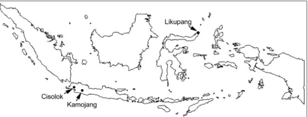
Environmental samples were collected from geothermal areas (Cisolok, Kamojang, and Likupang) in Indonesia (Fig. 1). The environmental temperature and pH of each sample was 70oC and pH 7.5 for Cisolok, 80oC and pH 4.0 for Kamojang, and 80oC and pH 7.0 for Likupang. After collecting the samples, genomic DNA was directly extract- ed from each environmental sample using SDS-based DNA extraction method as previously described [29]. Partial 16S rRNA gene was amplified using a set of bacterial specific universal primers; 27F (5’-AGAGTTTGATCCTGGCTCAG- 3’) and 1492R (5’-GGTTACCTTGTTACGACTT-3’). The PCR condition included initial denaturing at 94oC for 1 min followed by 30 cycles of 94oC for 1 min, 55oC for 30 sec, 72oC for 1 min, and a final extension step of 72oC for 5 min. The amplified PCR products of expected size were extracted and purified from agarose gel with QIAquick Gel Extraction kit (Qiagen, CA, USA). The purified DNA fragments were cloned into the pGEM-T vector (Promega, WI, USA) and transformed into E. coli TOP10 cells to construct the 16S rRNA gene libraries. After sequencing each 16S rRNA gene from the libraries, too short sequences of less than 350 bp in length were excluded for the further analysis. The resulting sequences were compared to 16S rRNA gene sequence from the nonredundant sequence database by using BLASTN program at National Centre of Biotechnological Information (NCBI) to determine their
approximate taxonomic identifications and investigate the microbial diversity.
Analysis of Microbial Diversity in Cisolok
Cisolok located in West Java has the extreme environ- ment with high temperature and high sulfate concentration [12]. Based on 155 representative 16S rRNA gene sequences, two most major abundant phyla were shown in Cisolok;
Proteobacteria (38.7%) with major class, β-Proteobacteria (27.1%) and Cyanobacteria (11.0%) with major genus, Thermosynechococcus (10.3%) (Table 1 and Fig. 2). Un- classified bacteria with no phylum affiliation also ac- counted for 26.4% of all 16S rRNA gene sequences. Con- cerning the evolution of tolerance, Cyanobacteria have been established in diverse environmental habitats showing wide ranges in temperature, salinity, pH, and irradiance [23]. As a good example, Cyanobacteria community con- taining Synechococcus in hot spring flows with marked temperature gradients was developed. Similarly, the diversity analysis of Cisolok showed that Thermosynechococcus was distributed with 10.3%. The 16S rRNA gene sequencing in Cisolok also exhibited the presence of chemolithotrophic thermophiles including the sulfate reducer Thermodesulfo- bacteria and hydrogen-oxidizing Aquificae. The occurrence of such microorganisms is consistent with the environment of Cisolok where sulfate-rich hot spring is, suggesting the lithotrophic base for primary productivity in Cisolok through sulfate reduction and hydrogen oxidation [14].
Sample collection from geothermal hot stream in Cisolok expected the resulting sequences to be majorly derived from thermophiles. Accordingly, some divisions were detected including Deinococcus-Thermus, Thermodesulfobacteria,
Fig. 1. Map of Indonesia showing the sampling sites (colosed circles) for analysis of microbial diversity in this study. Environmental samples were collected from geothermal hot stream at Cisolok (6o57’23”S, 106o28’46”E), volcano area at Kamojang (7o07’30”S, 107o47’60”E), West Java, and thermal vent under the sea water at Likupang (1o41’35”N, 125o00’47”E), north Sulawesi, Indonesia.
Aquificae, and Dictyoglomi which could be cultured from the geothermal habitats. However, several other microbial divisions characterized by mesophiles were also detected, including Proteobacteria, Firmicutes, and Plantomycetes, suggesting the possible contamination from groundwater flow into the hot stream in Cisolok [14].
Analysis of Microbial Diversity in Kamojang
Kamojang is one of the geothermal areas in West Java, Indonesia and is around the environment with an extremely low pH, high sulfate concentration, and high temperature [1]. After sequencing 171 representatives, the two most abundant microbial orders were Acidithiobacillales (46.2%) belonged to γ-Proteobacteria, followed by Aquificales (12.9%), whereas unclassified bacteria also accounted for a high portion (21.6%) (Table 1 and Fig. 2). The major distribution of Acidithiobacillales is responsible for the extreme condi- tions including acidic environment and high concentrations of sulfates and ferric irons, being supported by the previous report showing that the iron- and sulfur-oxidizing Acidi- thiobacillus ferrooxidans and sulfur-oxidizing A. thiooxidans are present in these extreme conditions [20]. Concerning the iron cycle, A. ferrooxidans utilizes the reduced ferrous iron as an energy source, producing the oxidized ferric iron which is relatively stable at acidic pH in the presence of elemental sulfur [6]. Therefore, the abundance of Acidithio- bacillales seems to play an important role in the generation and maintenance of the extremely acidic conditions of the Table 1. Frequency and phylogenetic affiliation of 16S rRNA
gene libraries from the geothermal areas of Cisolok, Kamojang, and Likupang in Indonesia.
Division
Sampling area Number of clones (%) Cisolok Kamojang Likupang Actinobacteria
Acidimicrobidae 1 (0.6%)
Aquificae
Aquificales 1 (0.6%) 22 (12.9%)
Bacteroidetes/Chlorobi group Bacteroidetes
Sphingobacteria 3 (1.9%) 7 (6.7%)
Flavobacteria 5 (4.8%)
Unclassified Bacteroidetes 1 (0.6%) 17 (16.2%)
Chlorobi 3 (1.9%)
Environmental samples 2 (1.3%) Chloroflexi
Chloroflexales 3 (1.9%)
Unclassified Chloroflexi 1 (0.6%) Cyanobacteria
Thermosynechococcus 16 (10.3%) Unclassified Cyanobacteria 1 (0.6%) Deferribacteres
Defferribacterales 2 (1.3%) Deinococcus-Thermus
Thernales 4 (2.6%)
Dictyoglomi
Dictyoglomales 1 (0.6%)
Firmicutes Bacilli
Bacillales 4 (2.6%) 1 (0.6%) 1 (1.0%)
Unclassified Bacilli 1 (1.0%)
Clostridia
Clostridiales 5 (3.2%) 2 (1.9%)
Unclassified Firmicutes 4 (3.8%)
Fusobacteria
Fusobacteriales 2 (1.3%)
Nitrospirae
Nitrospirales 1 (0.6%)
Planctomycetes
Planctomycetales 2 (1.3%) Proteobacteria
α-Proteobacteria 1 (0.6%) 3 (2.9%)
Caulobacterales 1 (0.6%)
Rhodobacterales 1 (0.6%) 1 (1.0%)
Rhizobiales 2 (1.3%) 1 (1.0%)
Unclassified α-Proteobacteria 3 (1.9%) β-Proteobacteria
Burkholderiales 15 (9.7%) 6 (3.5%) Hydrogenophilales 6 (3.9%) 4 (2.3%)
Nitrosomonadales 1 (0.6%)
Rhodocyclales 7 (4.5%) 1 (0.6%) 1 (1.0%) Unclassified β-Proteobacteria 14 (9.0%) 7 (4.1%) 1 (1.0%)
Table 1. Continued.
Division
Sampling area Number of clones (%) Cisolok Kamojang Likupang
γ-Proteobacteria 1 (1.0%)
Acidithiobacillales 79 (46.2%)
Alteromonadales 16 (15.2%)
Oceanospirillales 6 (5.7%)
Pseudomonadales 2 (1.3%) 8 (4.7%)
Thiotrichales 7 (6.7%)
Unclassified γ-Proteobacteria 1 (0.6%) 1 (0.6%) 5 (4.8%) Environmental samples 1 (0.6%)
δ-Proteobacteria 7 (4.5%) 1 (0.6%) 2 (1.9%) Unclassified δ-Proteobacteria 3 (2.9%) Thermodesulfobacteria
Thermodesulfobacteriales 3 (1.9%)
Unclassified Bacteria 41 (26.5%) 37 (21.6%) 21 (20.0%) Total clones 155 (100%)171 (100%)105 (100%)
ecosystem in Kamojang area.
The explanation of extremely acidic condition based on the distribution of Acidithiobacillales could be strongly supported by the previous study on microbial diversity of acidic hot spring (Kawah Hujan B) in Kamojang area [1].
The study described that the majority of the sequences was closely related to Crenarchaeota frequently found in extremely acidic environments [19] and capable to utilize elemental sulfur as energy source [5].
Analysis of Microbial Diversity in Likupang
Total of 105 representative clones from thermal vent under the sea water at Likupang, north Sulawesi, Indonesia were sequenced and analyzed, resulting that the highest proportion was γ-Proteobacteria (33.3%), followed by Bacteroidetes (27.6%) and unclassified bacteria group (20%). The minor groups were also affiliated to Firmicutes (7.6%) and α-Proteobacteria (4.8%) (Table 1 and Fig. 2).
In fact, these populations could be common characteristics exhibited in the microbial communities of marine environ- ments with hydrothermal vents. For example, the microbial diversity analysis of hydrothermal vent (Black point) in the Southern Tyrrhenian Sea, Italy, showed γ-Proteobacteria and Firmicutes to be the dominant populations [22]. In γ- Proteobacteria group, Alteromonadales were accounted with a high proportion (15.2%), followed by Thiotrichales (6.7%) and Oceanospirillales (5.7%). Alteromonadales, which is prevalent in different oceanic regions, but more frequently in deep sea [4], is primarily composed of heterotrophic marine bacteria playing a significant role in the biogeochemical cycle of carbon, nitrogen, and sulfur [16]. Thiotrichales group including Thiomicrospira, Thiothrix, and Beggiatoa has been identified to be chemolithotrophic sulfur-oxidizing bacteria group [26]. Considering the previous studies that these sulfur-oxidizing bacteria were frequently observed in the hydrothermal vents and wide range of marine habitats, these microbial communities could be typical components in the thermal vent under the sea water at Likupang where they can utilize the reduced sulfur compounds as electron donors [21].
Bacteroidetes phylum has been known to occur ubiquitously in marine habitats and their colonies known to exhibit specific color pigmentations due to the presence of flexirubin-type pigments [18]. Our diversity analysis of Likupang also showed that Bacteroidetes group had the second proportion (27.6%) where Sphingobacteria and Flavobacteria classes accounted with 6.7% and 4.8%
proportion, respectively.
In summary, we analyzed and compared the microbial communities of several geothermal areas in Indonesia via culture-independent approach with 16S rRNA gene sequencing (Fig. 2). Regardless of sampling areas, there was a large portion of unclassified bacteria (20.0-26.5%), representing the possible existence of a novel sub-class or even higher class in these geothermal areas [27]. However, the fact that 16S rRNA gene sequence-based analysis cannot completely represent the environmental habitats Fig. 2. Distribution of the predominant phyla (A) and detailed
Proteobacteria (B) identified from 16S rRNA gene libraries.
Four classes (α, β, γ, and δ-Proteobacteria) belonged to Proteo- bacteria phylum were used in the case of Proteobacteria. The combined distribution of minor phyla affiliated below 7% of total sequenced clones (except Proteobacteria) was represented to be
“Others”.
with just classifying the microbial distribution roughly, will encourage to further studies on microbial diversity with fluorescence in-situ hybridization analysis (FISH) as well as other specific genes such as rpoB and gyrB as well as fluorescence in-situ hybridization [9]. Nevertheless, the detail microbial diversity analysis from each sampling area indicates that the majority of libraries appear to be plausible community groups corresponding to each environment of sampling area. From the point that there have been just few studies reporting the microbial community of geothermal areas in Indonesia [1, 3], the present study could contribute to expand the knowledge of the microbial diversity and provide a basic ecosystem of geothermal areas in Indonesia.
Additionally, this study could support the big challenge looking for novel biomaterials and functional enzymes for industrial applications [17, 24].
REFERENCES
1. Aditiawati, P., H. Yohandini, F. Madayanti, and Akhmaloka.
2009. Microbial diversity of acidic hot spring (Kawah Hujan B) in geothermal field of Kamojang area, west Java-Indone- sia. Open Microbiol. J. 3: 58-66.
2. Amann, R. I., W. Ludwig, and K. H. Schleifer. 1995. Phylo- genetic identification and in situ detection of individual microbial cells without cultivation. Microbiol. Rev. 59: 143- 169.
3. Aminin, A. L. N., F. M. Warganegara, P. Aditiawati, and Akhmaloka. 2008. Culture-independent and culture-depen- dent approaches on microbial community analysis at Gedongsongo (GS-2) hot spring. Int. J. Integr. Biol. 2: 145- 152.
4. Biers, E. J., S. Sun, and E. C. Howard. 2009. Prokaryotic genomes and diversity in surface ocean waters: interrogating the global ocean sampling metagenome. Appl. Environ.
Microbiol. 75: 2221-2229.
5. Bintrim, S. B., T. J. Donohue, J. Handelsman, G. P. Roberts, and R. M. Goodman. 1997. Molecular phylogency of Archaea from soil. Proc. Natl. Acad. Sci. USA 94: 277-282.
6. Brock, T. D. and J. Gustafson. 1976. Ferric iron reduction by sulfur- and iron-oxidizing bacteria. Appl. Environ. Micro- biol. 32: 567-571.
7. Burns, D. G., H. M. Camakaris, P. H. Janssen, and M. L.
Dyall-Smith. 2004. Combined use of cultivation-dependent and cultivation-independent methods indicates that members of most haloarchaeal groups in an Australian crystallizer pond are cultivable. Appl. Environ. Microbiol. 70: 5258- 5265.
8. Dahllöf, I., H. Baillie, and S. Kjelleberg. 2000. rpoB-Based microbial community analysis avoids limitations inherent in 16S rRNA gene intraspecies heterogeneity. Appl. Environ.
Microbiol. 66: 3376-3380.
9. Friedrich, A. B., H. Merkert, T. Fendert, J. Hacker, P.
Proksch, and U. Hentschel. 1999. Microbial diversity in the marine sponge Aplysina cavernicola (formerly Verongia cav- ernicola) analyzed by fluorescence in situ hybridization (FISH). Mar. Biol. 134: 461-470.
10. Ghosh, D., B. Bal, V. K. Kashyap, and S. Pal. 2003. Molec- ular phylogenetic exploration of bacterial diversity in a Bakreshwar (India) hot spring and culture of Shewanella- related thermophiles. Appl. Environ. Microbiol. 69: 4332- 4336.
11. Goh, K. M., Y. S. Chua, R. N. Z. R. A. Rahman, R. Chan, and R. Illias. 2011. A comparison of conventional and mini- primer PCR to elucidate bacteria diversity in Malaysia Ulu Slim hot spring using 16S rDNA clone library. Rom. Bio- technol. Lett. 16: 6247-6255.
12. Herdianita, N. R. and B. Priadi. 2008. Arsenic and mercury concentrations at several geothermal systems in West Java, Indonesia. ITB J. Sci. 40A: 1-14.
13. Hobel, C. F. V., V. T. Marteinsson, S. Hauksdóttir, Ó. Frid- jónsson, S. Skírnisdóttir, G. Ó. Hreggvidsson, and J. K.
Kristjánsson. 2004. Use of low nutrient enrichments to access novel amylase genes in silent diversity of thermo- philes. World J. Microbiol. Biotechnol. 20: 801-809.
14. Hugenholtz, P., C. Itulle, K. L. Hershberger, and N. R. Pace.
1998. Novel division level bacterial diversity in a Yellow- stone hot spring. J. Bacteriol. 180: 366-376.
15. Janssen, P. H. 2006. Identifying the dominant soil bacteria taxa in libraries of 16S rRNA and 16S rRNA genes. Appl.
Environ. Microbiol. 72: 1719-1728.
16. Karl, D. M. 2002. Microbiological oceanography – hidden in a sea of microbes. Nature 415: 590-591.
17. Kim, J. N., M. J. Seo, E. A. Cho, S. J. Lee, S. B. Kim, C. I.
Cheigh, and Y. R. Pyun. 2005. Screening and characteriza- tion of an esterase from a metagenomic library. J. Microbiol.
Biotechnol. 15: 1067-1072.
18. Kirchman, D. L. 2002. The ecology of Cytophaga-Flavobac- teria in aquatic environments. FEMS Microbiol. Ecol. 39:
91-100.
19. Kozubal, M., R. W. Macur, S. Korf, W. P. Taylor, G. G. Ack- erman, A. Nagy, and W. P. Inskeep. 2008. Isolation and dis- tribution of a novel iron-oxidizing crenarchaeon from acidic geothermal springs in Yellowstone national park. Appl. Envi- ron. Microbiol. 74: 942-949.
20. López-Archilla, A. I., I. Marín, and R. Amils. 2001. Micro- bial community composition and ecology of an acidic aquatic environment: the Tino River, Spain. Microb. Eol. 41:
20-35.
21. Maugeri, T. L., V. Lentini, C. Gulgliandolo, F. Italiano, S.
Cousin, and E. Stackebrandt. 2009. Microbial communities at two shallow hydrothermal vents off Panarea Island (Edo- lian Islands, Italy). Extremophiles 13: 199-212.
22. Maugeri, T. L., V. Lentini, C. Gugliandolo, S. Cousin, and E.
Stackebrandt. 2010. Microbial diversity at a hot, shallow-sea hydrothermal vent in the Southern Tyrrhenian sea (Italy).
Geomicrobiol. J. 27: 380-390.
23. Miller, S. R. and R. W. Castenholz. 2000. Evolution of ther- motolerance in hot spring Cyanobacteria of the genus Syn- echococcus. Appl. Environ. Microbiol. 66: 4222-4229.
24. Oren, A. 2002. Diversity of halophilic microorganisms:
Environments, phylogency, physiology, and applications. J.
Ind. Microbiol. Biotechnol. 28: 56-63.
25. Park, S. J., C. H. Kang, and S. K. Rhee. 2006. Characteriza- tion of the microbial diversity in a Korean solar saltern by 16S rRNA gene analysis. J. Microbiol. Biotechnol. 16: 1640- 1645.
26. Ruby, E. G., C. O. Wirsen, and H. W. Jannasch. 1981. Chem- olithotrophic sulfur-oxidizing bacteria from the Galapagos rift hydrothermal vents. Appl. Environ. Microbiol. 42: 317-
324.
27. Wu, C., F. Yang, R. Gao, Z. Huang, B. Xu, Y. Dong, T.
Hong, and X. Tang. 2010. Study of fecal bacterial diversity in Yunnan snub-nosed monkey (Rhinopithecus bieti) using phylogenetic analysis of cloned 16S rRNA gene sequences.
Afr. J. Biotechnol. 9: 6278-6289.
28. Yin, H., L. Cao, M. Xie, Q. Chen, G. Qiu, J. Zhou, L. Wu, D.
Wang, and X. Liu. 2008. Bacterial diversity based on 16S rRNA and gyrB genes at Yinshan mine, China. Syst. Appl.
Microbiol. 31: 302-311.
29. Zhou, J., M. A. Bruns, and J. M. Tiedje. 1996. DNA recov- ery from soils of diverse composition. Appl. Environ. Micro- biol. 62: 316-322.
국문초록
16S rRNA 분석을 통한 인도네시아의 Cisolok, Kamojang, Likupang 지열지대 내 미생물 다양성 분석
서명지1,2*·김정녀3·변유량3
1한국식품연구원 발효기능연구단
2과학기술연합대학원대학교 식품생명공학
3연세대학교 생명공학과
인도네시아 지열지대의 미생물 다양성을 16S rRNA 염기서열 분석을 통해 조사하였다. 전체적으로 어떠한 phylum 계통군에도 포함되지 않는 unclassified bacteria가 20.0-26.5% 존재하였으며 sampling 지역에 상관없이 Proteobacteria 가 우점 phylum 계통군으로 나타났다. Cisolok 주변의 지열 지역을 조사한 결과 β-Proteobacteria (27.1%)와 Cyanobacteria (11.0%)가 높은 비율을 차지한 반면 Kamojang의 화산 주변 지역의 경우에는 γ-Proteobacteria (51.5%) 그리고 Aquificales (12.9%)가 우점 계통군으로 나타났다. 또한 Likupang 열수구의 경우에는 γ-Proteobacteria (33.3%) 와 Bacteroidetes (27.6%)가 높은 비율로 나타났다. 본 연구를 통해 인도네시아 각 지열지대에 분포하는 미생물 군집은 각 지역의 환경적인 특징 (극한 지열 및 해양 서식지)과 밀접한 연관성이 있음을 알 수 있었다.