46(4) : 283∼ 288 (2015)
283
들깨 잎 추출물에서 분리한 Isoegomaketone(IK)의 항 비만 효능
소양강1,2·조윤호1·남보미1·이승영1·김진백1·강시용1·정혜광2·진창현1*
1
한국원자력연구원,2
충남대학교 약학대학Anti-Obesity Effect of Isoegomaketone Isolated from Perilla frutescens (L.) Britt. cv. Leaves.
Yangkang So1,2, Yun Ho Jo1, Bo Mi Nam1, Seung Young Lee1, Jin-Baek Kim1, Si-Yong, Kang1, Hye Gwang Jeong2, and Chang Hyun Jin1*
1
Korea Atomic Energy Research Insititute, Jeongeup, Jeollabuk 580-185, Korea2
Colleage of Phamacy, Chungnam Natinal University, Daejeon, Chungnam 305-764, KoreaAbstract − In this study, we investigated anti-obesity effect of isoegomaketone (IK) isolated from leaves extract of Perilla frute- scens (L.) Britt. cv. We verified differentiation and lipid accumulation by Oil Red O staining in 3T3-L1 cells after IK treatment with differentiation media. IK inhibited mRNA expression of adipocyte specific genes that were related with differentiation of 3T3-L1 cells. We confirmed the effects of IK on body weight and visceral fat mass in obese mice. Mice were randomly divided into three groups; normal diet group (ND), high-fat diet group (HFD) and high-fat diet with IK group (HFD-IK). The obesity mice were induced by feeding the 45% high-fat diet to the C57BL/6J mice during 4 weeks. After HFD-IK was orally admin- istered 10 mg/kg of IK. As a result, the body weight of HFD and HFD-IK was increased 2.4 times and 1.7 times of ND, respec- tively. Also visceral fat mass of HFD was increased 24 times but in the case of HFD-IK was increased to 13 times in comparison with ND. Taken together, our findings suggest that IK reduced differentiation and adiogenesis in 3T3-L1 cells, decreased the body weight and visceral fat mass in obesity mice. These results suggest that IK may have a potential benefit as anti-obesity material.
Key words − Isoegomaketone, Anti-obesity, 3T3-L1 cells, C57BL/6J, High-fat diet
비만은 서양화되고 있는 식습관의 문제로 체내에서 요구 하는 에너지의 소비량보다 더 많은 에너지를 섭취함으로써 지방 조직이 축적되어 외형적 변화를 야기시키는 대표적인 질병으로 알려져 있다. 그러나 비만은 단순한 질병으로 정 의 내리기에는 어려운 문제로 보고 있다.
1)
그 이유는 비만 이 환경, 심리, 유전, 행동들을 비롯한 생리학적 외부 요인 들에 의해서도 영향을 받기 때문이다. 이와 같은 요인들로 인하여 암을 비롯한 당뇨병, 고지혈증, 고혈압, 관절염, 동 맥경화, 지방간 등의 원인이 되는 대사성 질환을 야기 시키 게 된다.2,3)
또한, 비만 인구율이 전세계적으로 지속적인 증 가 추세를 보이고 있어 비만은 심각한 사회 문제로 관심이 집중되고 있다.4)
비만을 치료하기 위한 연구는 이전부터 수 행되어왔다. 특히, 화학적 치료제들의 효능이 뛰어나 대중적으로 사용되었지만, 심각한 부작용들이 밝혀지면서
5-7)
천 연물 소재로 제조된 치료제에 대한 관심이 높아지고 있다.이와 같은 연구들은 대개 전통의학이나 민간처방을 바탕으 로 그 효능과 안정성이 널리 알려진 천연물 소재들 중에서 선별하여 진행되고, 관련된 연구들이 많이 수행되고 있다.
일본, 중국, 우리나라와 동아시아가 원산지이며 식용으로 잎이 널리 섭취되고 있는 들깨(Perilla frutescens (L.) Britt.
cv.)의 경우 민간에서 오한, 발열, 감기 및 구토 등의 증상을 완화시키기 위해 사용하였다.
8)
이전 연구들을 통해서 들깨 잎은 다양한 방법으로 추출되어 불포화지방산을 비롯한 필 수지방산, 플라보노이드(flavonoids) 등과 지용성 비타민에 속하는 비타민 A, D, E들의 함량이 높게 함유되어 있다고 보고되어있다. 이를 바탕으로 생리활성 연구를 통해 항암, 항염증, 항산화 및 항알레르기 등과 같은 다양한 효능이 검 증되었다.9)
그 중 isoegomaketone(IK) 성분의 경우 perillaketone(PK)
*교신저자(E-mail):[email protected] (Tel): +82-63-570-3162
성분과 함께 들깨 추출물의 주요한 성분 중의 하나로 들깨 에서 나는 특유한 향을 내는 방향성 성분이다.
10)
IK는 항암 을 비롯한 항염증, apoptosis 등에 효과를 나타내는 것으로 밝혀졌다. 특히 동물실험을 통해 IK의 뛰어난 항염증 효능 이 증명되었으며, 그 기작에 대한 연구도 보고된 바 있다.11,12)
항염증 생리활성은 비만을 억제하는 기작에도 관련 되어 있다는 연구가 있다. 지방세포들이 모여 구성되는 결합조직 인 지방조직이 면역세포 중에 하나로 포식, 염증 유도, 세 포 활성화, 세포 제거 등을 수행하는 대식세포와 연관되어 있기 때문이다. 대식세포는 Th1 cytokine인 IFN-γ에 의한 classical activation(M1)과 Th2 cytokine인 IL-4, IL-13에 의 한 alternative activation(M2)로 존재한다. 비만으로 인한 지 방조직의 염증화는 지방조직 안으로 M2 대식세포의 침투 가 증가하여 tumor necrosis factor-alpha(TNFα), nitric oxide synthase(iNOS), interleukin-6(IL-6) 등의 발현을 높이 기 때문이다. 따라서 항염증에 효능을 보이는 물질이 지방 조직의 염증화를 개선하여 비만으로 질병이 발전함을 억제 하는 효능을 보여준다.13)
하지만 아직까지 IK의 뛰어나 항 염증 효능에도 불구하고 IK의 항비만 효능과 관련된 연구 는 미비한 실정이다.따라서 본 연구에서는 항비만 효능에 대한 IK의 생리활 성을 알아보기 위해 지방세포 분화에 관련한 유전자들의 감 소 및 고지방 식이를 통해 비만을 유도한 마우스 동물모델 을 이용하여 체중변화 여부를 검증하고자 하였다.
재료 및 방법
식물재료 − 본 실험에서 사용된 축엽자소 잎은 한국원자 력연구원 첨단방사선연구소에서 축엽자소 종자에 200 Gy 감마선을 처리한 후 안정화 시킨 돌연변이 품종이다. IK의 함량이 기존의 야생 품종에 비하여 높은 이 품종은 성장한 잎만을 수확하여 음건조 식물의 잎을 음건조하여 분쇄 후 진공 상태로 보관하여 사용하였다. 이 식물에 대한 감정은 한국원자력연구원 첨단방사선연구소 방사선육종연구팀 김 진백 박사님을 통하였다.
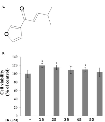
실험재료 − 축엽자소의 성장한 잎만을 수확하여 음건조 후 파쇄하여 120 g의 시료를 얻었다. 이후 헥산 추출 한 뒤 감압 농축하여 건조를 통해 12 g 추출물을 확보하였고, 추 출물을 컬럼크로마토그라피를 이용하여 10 mg의 순도 99%
의 IK 를 분리하였다(Fig. 1A).
14)
시약 및 재료 − Dulbecco’s Modified Eagle’s Medium(DMEM), 100 units/mL Pen/Strep/Glutamine(P/S/
G), 100 mM MEM Sodium Pyruvate and Bovine Serum Albumin(BSA)들은 Invitrogen Corporation's GIBCO(Carlsbad, CA, USA)에서 구입하였다. Fetal Bovine Serum(FBS)는 Hyclone(Logan, UT)회사 제품을 사용하였다. 3-isobuty-1- methylxanthine(IBMX), dexamethasone(DEX), insulin와 Oil Red O 염색시약은 Sigma Chemical Co.(St. Louis, MO, USA)에서 구입하였다. Cell viability assay kit는 대일 랩(Korea)에서 구입하였으며, Cdk2와 PPARγ-specific antibodies는 Cell signaling Tec(Beverly, MA)에서 구입하였 다. β-tubulin은 Santa Cruz Biotechnology(Santa Cruz, CA)에서 구입하였고, goat anti-rabbit IgG HRP-conjugated antibodies는 Invitrogen(Carlsbad, CA)회사에서 구입하였다.
세포 배양 − 3T3-L1 지방전구세포는 10% BSA, P/S/G와 Sodium Pyruvate 를 함유한 DMEM 배지에서 70~80% 배 양 후 계대하여 사용하였다.
독성 실험 − 3T3-L1 지방전구세포를 96 well plate에 2×10⁵ cells/mL로 계대하여 24시간 동안 배양하였다. IK를 15, 25, 35, 45, 50μM 농도별로 처리하여 이후 24시간을 배 양하였다. 세포 독성은 cell viability kit를 이용하여 측정하 였다.
지방세포 분화 − 3T3-L1 지방전구세포를 6 well plate에 3×10⁵ cells/mL로 계대 후 48시간 동안 배양하였다. 지방세 포 분화를 위해 DMEM(10% FBS, P/S/G와 Sodium Pyruvate), 0.5 mM IBMX, 1 M Dex, 1 mg/mL insulin을 포함하는 MDI(methylxanthine, dexamethasone과 insulin을 포함하는 배지) 로 바꿔서 72시간 동안 배양하였다. 이 후 48시간 동안 1 mg/mL insulin을 포함하는 DMEM(10%
FBS, P/S/G, Sodium Pyruvate)배지에 세포를 배양한 후 같 Table І. Primer sequences used in real-time PCR
Primers Forward Reverse
C/EBPα GGTGCGCAAGAGCCGAGATAAAG AGTTCACGGCTCAGCTGTTCCAC
PPARγ CCAGAGTCTGCTGATCTGCG GCCACCTCTTTGCTCTGCTC
SREBP1 GCGCCATGGACGAGCTG TTGGCACCTGGGCTGCT
aP2 CTCACCTGGAAGACAGCTCCTCCTCG TTCCATCCAGGCCTCTTCCTTTGGCTC
GLUT4 CGAGCTGGACGACGGACACTC AGACATAGCTCATGGCTGGAACCCG
LPL CAGAGTTTGACCGCCTTCC AATTTGCTTTCGATGTCTGAGAA
ACSL1 AAAGATGGCTGGTTACACACG CGATAATCTTCAAGGTGCCATT
β-actin TGAGAGGGAAATCGTGCGTGAC GCTCGTTGCCAATAGTGATGACC
은 배지에 48시간 동안 한번 더 배양하여 9일 동안 지방전 구세포를 지방세포로 분화시켰다.
Oil Red O 염색 − 세포 계대에서 지방세포 분화까지 9 일동안의 실험이 끝난 후 10% formalin으로 세포를 고정하 였다. Oil Red O 염색시약으로 1시간 동안 지방방울들을 염 색하여 세척 후 현미경으로 확인하였다.
Real-time PCR − 지방전구세포를 지방세포로 분화시키 는 과정 중 분화배지에 IK 50 μM을 처리하였고, 9일의 분 화 과정이 끝난 후 QIAGEN 제품을 이용하여 RNA를 추 출 후 대조군과 비교하였다(Table І).
마우스 실험 − 5주령의 수컷 C57BL/6J 마우스를 중앙실 험동물㈜로부터 구입하였다. 1주일 동안 사육실에서 순화를 시킨 후 마우스를 정상식이 대조군(ND, normal diet), 45%
고지방식이 대조군(HFD, high-fat diet)과 45% 고지방식이- IK 실험군(HFD-IK, high-fat diet with IK)으로 나누어 총 8 주간 사육을 진행하였다. 처음 4주 동안은 고지방식이사료 섭취를 통해 비만 동물 모델로 유도하였고, 이후 4주간 HFD- IK는 고지방식이 사료와 함께 PBS용액(1×PBS with 0.5%
Tween20)에 녹인 IK 10 mg/kg을 1일 1회 경구투여하였다.
마우스의 체중과 식이섭취량은 1일 1회 측정하였으며 8주 간의 사육기간이 끝이 난 후에 혈액과 지방을 추출하여 실 험에 사용하였다.
통계 처리 − 실험결과는 평균±표준편차로 표현하였다. 두 실험군 간의 유의성 검정은 Student’s t-test를 이용하여 p<0.05 수준의 경우 통계적 유의성이 있는 것으로 판정하였다.
결과 및 고찰
3T3-L1 지방전구세포에서 IK의 독성 영향 − IK가 3T3- L1 지방전구세포에서 독성에 미치는 영향을 실험하였다. 24
시간 계대배양 후 IK를 15, 25, 35, 45, 50 μM 농도별 처리 후 24시간이 지난 다음 cell viability assay kit를 이용하여 독성을 측정한 결과 독성은 나타나지 않았다(Fig. 1B).
3T3-L1 지방전구세포 분화과정 중 중성지방 억제 − 분 화 과정 중의 IK 처리가 지방세포 내의 지방방울들의 감소 Fig. 1. Effect of IK on cell viability in 3T3-L1 cells. (A) Structure of IK. (B) 3T3-L1 cells were treated with various cencentrations of IK for 24 h. Cell viability was determined with the EZ-cytox assay kit. Results were expressed as % of control absorbance. Each value represents the mean±SD. (n=5)
*p<0.05 vs. control.
Fig. 2. Effect of IK on adipocyte differentiation in 3T3-L1 cells. 3T3-L1 cells were treated with various cencentrations of IK in dif- ferentiation medium during adipogenesis from 0-9 days. Lipid droplets were stained in 3T3-L1 cells using Oil Red O solution.
에 영향을 미치는 지를 확인하기 위해 Oil Red O 염색시약 을 통해서 지방방울들을 염색하여 확인하였다. IK를 15, 25, 35, 45, 50μM로 처리한 후 농도 의존적으로 지방방울들이 감소되는지 현미경을 통해 관찰하였다(Fig. 2). 이러한 지방 방울들의 변화는 IK로 인해 지방세포 분화를 비롯한 지방 합성 및 저장을 억제하는 작용이 기여한 결과로 보여진다.
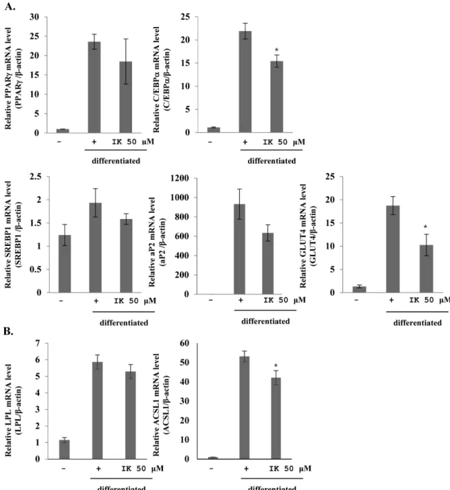
3T3-L1 지방전구세포 분화에 IK가 미치는 영향 − 3T3- L1 지방전구세포가 지방세포로의 분화에 IK가 미치는 영향
을 관찰하기 위해 adipogenesis 관련 유전자들의 발현 감소 를 확인하였다. IK를 50 μM 농도로 분화배지에 처리하여 실험을 진행하였다. 지방분화 실험이 끝난 다음 세포에서 mRNA를 분리하고 real-time PCR을 통해서 지방합성 및 지 방축적과 관련이 있는 유전자들의 mRNA 발현량을 확인하 였다. 그 결과 지방전구세포가 지방세포로의 분화와 지방 합성을 과속화 시킬 수 있는 중요한 활성 유전자들
15-18)
CCAAT/enhancer binding protein alpha(C/EBPα),Fig. 3. Inhibitory effects of IK on adipogenesis mRNA expression in 3T3-L1. Expression level of adipocyte specific genes(C/EBPα, PPARγ, SREBP1, aP2, GLUT4, LPL and ACSL1) were measured by real-time PCR. 3T3-L1 cells were treated IK 50 μM with dif- ferentiation medium. mRNA expression level of genes were normalized with b-actin. Each value represents the mean±SD. (n=3)
*p<0.05 vs. positive.
Peroxisome proliferator-activated receptor gamma(PPARγ), adipocyte-specific lipid binding protein(aP2), sterol regulatory element-binding protein 1(SREBP1), glucose transporter 4(GLUT4), Lipoprotein lipase(LPL)와 acyl Co- A synthetase long chain family member(ACSL1)의 발현이 감소됨이 관찰되었다(Fig. 3). C/EBPα와 PPARγ는 지방전 구세포가 지방세포로의 분화 단계에서 발현되어지며, 동시 에 발현되었을 때 지방세포 특이 유전자들의 mRNA의 전 사 활성화를 유도하고 SREBP1의 경우는 PPARγ의 활성을 증가시키는 역할로서 지방세포로의 분화에 영향을 미친다.
따라서 IK를 분화과정 중에 처리함에 따라 이들 유전자들 의 감소를 유도하여 지방세포로의 분화를 억제시킴을 나타 내었다. C/EBPα, PPARγ와 SREBP1의 발현으로 aP2, GLUT, ACSL1, LPL 등의 지방세포 특이 유전자들이 활성 화된다. aP2와 ACSL1은 지방합성에 관여하고, GLUT4는 분화 및 축적에 관여하는 유전자로 알려져 있다. 따라서 IK 는 C/EBPα, PPARγ와 SREBP1 유전자들을 감소시킴으로 써 이들 유전자들로 인해서 활성화 되는 aP2, GLUT, ACSL1, LPL 유전자들 또한 감소하게 만들어 지방세포분화 를 비롯한 지방 합성과 축적을 억제함을 나타내었다. 이를 통해 IK에 의한 지방세포에서의 특이 유전자들의 발현 억 제가 지방방울들의 수와 크기 감소에 영향을 미치는 것으 로 사료된다.
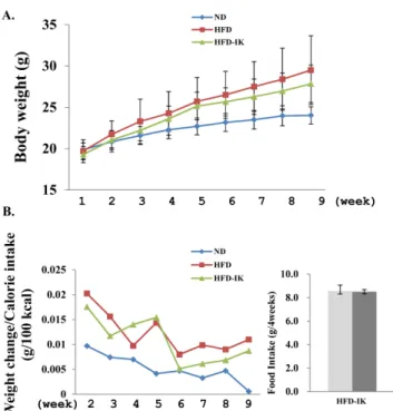
체중 및 조직 무게에 미치는 영향 − 고지방식이로 비만 이 유도된 C57BL/6J 수컷 마우스를 통해 IK가 체중에 미 치는 영향을 조사하고자 10 mg/kg씩 실험동물군에 경구투 여하고 그 변화를 확인하였다. 1주 순화기간을 제외한 총 8 주 동안 실험동물군들의 체중 변화 및 사료섭취량을 1일 1 회 측정하였다. 4주간의 45% 고지방식이사료 섭취를 통해 ND를 제외한 실험군들을 비만동물모델로 유도하였다. 그 결과 ND에 비하여 HFD과 HFD-IK의 체중은 2배 가까운 증가를 보여주었다. 이후 진행된 4주 동안, 이전 4주 동안 진행했던 식이사료를 계속 섭취하면서 ND와 HFD에게는 PBS용액을 경구투여하고 HFD-IK는 IK를 녹인 PBS용액을 경구투여하였다. 실험 결과 HFD는 ND와 비교하여 체중이 2.4배 증가한 반면 HFD-IK는 1.7배 정도가 증가됨을 보여 주었다(Fig. 4A). 체중변화 결과는 weight change/Calorie intake(g/100 kcal)로 계산하였는데
19)
HFD-IK는 경구투여 전 까지 HFD에 비해서 높은 체중변화를 보여주었지만 IK를 경구투여하고 나서는 다소 감소함을 확인하였다(Fig. 4B).또한, 비만모델로 유도시킨 기간 동안의 사료 섭취량과 IK 를 경구투여하는 기간동안의 사료 섭취량에는 유의적인 차 이를 확인할 수 없었다(Fig. 4B). 이는 HFD-IK에서 비만동 물모델의 체중 감소가 열량섭취 차이에 영향을 받아 유도 된 결과가 아닌 것으로 사료되며, IK로 인한 식욕억제가 나 타나지 않음을 보여준다. 이후 해부를 통해서 간장 주변의
내장지방 감소를 관찰하여 보았다. HFD를 ND와 비교하여 보았을 때 24배 정도 증가한 것에 비해 HFD-IK는 13배 정 도의 증가함이 관찰되었다(Fig. 5). 따라서 비만 동물 모델 실험을 통해 IK는 식욕 감퇴를 일으키지 않으며, 체중 및 내장지방 감소를 유도하고 이는 IK가 항비만 효능이 있음 을 나타낸다. 하지만 추후 이에 관한 메커니즘과 항비만에 대하여 연구가 진행되어야 한다.
Fig. 4. Effect of IK on the mice weight. Male C57BL/6J mice were fed a normal or 45% high-fat diet without IK or with IK treatment during 8 weeks. (A) Body weight of mice, (B) changes of body weight and averages of food intake for 4 weeks. Each value represents the mean±SD. (n=7), ND: nor- mal diet mice, HFD: high-fat diet mice, HFD-IK: high-fat diet with IK mice, gray: induced obesity animal model period, dark gray: orally IK administered period.
Fig. 5. Effect of IK on the perirenal fat pad. Each value rep- resents the mean±SD. (n=3) *p<0.05 HFD vs. HFD-IK.
결 론
본 연구는 지방전구세포에 IK를 처리함으로써 지방세포 의 분화, 합성 및 지방 축적에 관련한 유전자들이 감소하는 것을 확인할 수 있었으며, 고지방식이로 유도된 비만 동물 모델을 이용한 실험을 통해 HFD-IK에서 체중 및 내장 지 방이 감소됨을 확인하였다. 이러한 IK의 항비만 효능은 확 연하게 두드러지는 결과를 보여주지는 않았으나 차후 IK가 항비만 개선 및 예방을 위한 천연물 소재로 이용될 개발 가 능성을 보여준다.
사 사
이 논문은 2015년도 한국원자력연구원의 연구개발 프로 그램과 정부(미래창조과학부)의 재원으로 한국연구재단의 지원을 받아 수행된 연구임(방사선기술개발사업, No.
2012M2A2A6010575).
인용문헌
1. McGee, D. L. (2005) Body mass index and mortality:A meta- analysis based on person-leveldata from twenty-six obser- vational studies. Ann. Epidemiol. 15: 377-398.
2. Peter, G. K. (2000) Obesity as a medical problem. Nature 404: 635-643.
3. Lementowski, P. W. and Zelicof, S. B. (2008) Obesity and osteoarthritis. Am. J. Orthop. 37: 148-51.
4. Yiying, Z., Ricardo, P., Margherita, M., Marisa, B., Lori, L.
and Jeffrey, M. F. (1994) Positional cloning of the mouse obese gene and its human homologue. Nature 372: 425-432.
5. Johansson, K., Neovius, K., DeSantis, S. M., Rössner, S. and Neovius, M. (2009) Discontinuation due to adverse events in randomized trials of orlistat, sibutramine and rimonabant: a meta-analysis. Obes. Rev. 10: 564-575.
6. Tewari, A., Ali, A., O’Donnell, A. and Butt, M. S. (2009) Weight loss and 2,4-dinitrophenol poisoning. Br. J. Anaesth.
102: 566-567.
7. Heidi, M., Connolly, M. D., Jack, L., Crary, M. D., Michael, D., McGoon, M. D., Donald, D., Hensrud, M. D., M.P.H., Brooks, S. Edwards, M. D., William, D., Edwards, M. D. and Hartzell, V. Schaff, M. D. (1997) Valvular heart disease asso- ciated with fenfluramine-phentermine. N. Engl. J. Med. 337:
581-588.
8. Peng, Y., Ye, J. and Kong, J. (2005) Determination of phe- nolic compounds in Perilla frutescens L. by capillary elec- trophoresis with electrochemical detection. J. Agric. Food Chem. 53: 8141-8147.
9. Lee, J. I., Han, E. D., Lee, S. T. and Park, H. W. (1986) Study on the evaluation of oil quality and the differences of fatty acid composition between varieties in perilla (Perilla frute- scens Britton var. japonica Hara). Korean J. Breed 18: 228- 233.
10. Madoka, H., Ichiho, I., Toru, Y., Robert, P. A. and Gisho, H.
(2004) cDNA isolation and functional expression of myrcene synthase from Perilla frutescensn. Biol. Pharm. Bull. 27:
1979-1985.
11. Jin, C. H., Lee, H. J., Park, Y. D., Choi, D. S., Kim, D. S., Kang, S. Y., Seo, K. I. and Jeong, I. Y. (2010) Isoegoma- ketone inhibits lipopolysaccharide-induced nitric oxide pro- duction in RAW 264.7 macrophages through the heme oxygenase-1 induction and inhibition of the interferon-beta- STAT-1 pathway. J. Agric. Food Chem. 58: 860-867.
12. Cho, B. O., Jin, C. H., Park, Y. D., Ryu, H. W., Byun, M. W., Seo, K. I. and Jeong, I. Y. (2011) Isoegomaketone induces apoptosis through caspase-dependent and caspase-indepen- dent pathways in human DLD1 cells. Biosci. Biotechnol. Bio- chem. 75: 1306-1311.
13. Heilbronn, L. K. and Campbell, L. V. (2008) Adipose tissue macrophages, low grade inflammation and insulin resistance in human obesity. Curr. Pharm. Des. 14: 1255-1230.
14. MacDougald, O. A. and Lane, M. D. (1995) Transcriptional regulation of gene expression during adipocyte differentia- tion. Annu. Rev. Biochem. 64: 345-373.
15. Latasa, M. J., Moon, Y. S., Kim, K. H. and Sul, H. S. (2000) Nutritional regulation of the fatty acid synthase promoter in vivo: sterol regulatory element binding protein functions through an upstream region containing a sterol regulatory ele- ment. Proc. Natl. Acad. Sci. U. S. A. 97: 10619-10624.
16. Rosen. E. D. (2005) The transcriptional basis of adipocyte development. Prostaglandins Leukot. Essent. Fatty Acids 73:
31-34.
17. Kong, C. S., Kim, J. A., Eom, T. K. and Kim, S. K. (2010) Phosphorylated glucosamine inhibits adipogenesis in 3T3-L1 adipocytes. J. Nutr. Biochem. 21: 438-443.
18. Bulló, M., García-Lorda, P., Peinado-Onsurbe, J., Hernández, M., Del Castillo, D., Argilés, J. M. and Salas-Salvadó, J.
(2002) TNFalpha expression of subcutaneous adipose tissue in obese and morbid obese females: relationship to adipocyte LPL activity and leptin synthesis. Int. J. Obes. Relat. Metab.
Disord. 26: 652-658.
19. Park, H. J., Lee, E. J., Kim, J., Kim, J. Y., Kwon, O. and Kim, M. K. (2009) Effect of Leucine Intake on Body Weight Reduction in Rats Fed High Fat Diet. Korean J. Nutr. 42:
714-722.
(2015. 10. 22 접수; 2015. 11. 19 심사; 2015. 12. 1 게재확정)