호박즙, 옥수수수염차, 팥차 및 혼합물이 식이유도 비만동물모델에서 체중과 항산화 활성에 미치는 영향
박재희․이은지․박은주 경남대학교 식품영양생명학과
Effect of Pumpkin, Corn Silk, Adzuki Bean, and Their Mixture on Weight Control and Antioxidant Activities in High Fat Diet-Induced Obesity Rats
Jae-Hee Park, Eunji Lee, and Eunju Park
Department of Food, Nutrition and Biotechnology, Kyungnam University
ABSTRACT Pumpkin juice (PJ), corn silk tea (CT), and adzuki bean tea (AT) have long been used for treatment of obesity in Korea. This study investigated the efficacy of PJ, CT, AT, and their mixture (PCA) on alteration of body weight and antioxidant metabolism in high-fat diet (HFD)-induced obese rats. After being fed HFD for 4 weeks, SD rats were divided into six groups fed a normal diet (ND), HFD, HFD+PJ [250 mg/kg body weight (BW)], HFD+CT (250 mg/kg BW), HFD+AT (250 mg/kg BW), and HFD+PCA (PJ : CT : AT=1:1:1, 250 mg/kg BW) for another 9 weeks. HFD consumption resulted in total lipid, triglyceride, and total cholesterol accumulation in adipose tissue, which was reduced by administration of PJ, CT, AT, or PCA. The plasma oxygen radical absorbance capacity value and hepatic glutathione peroxidase activity significantly increased compared to the HFD group. The liver thiobarbituric acid reactive substances was significantly lower in the PCA group than the HFD group. HFD-induced DNA damage in hepatocytes, as measured by comet assay, decreased in the PJ, AT, and PCA-supplemented groups. The PCA group exerted a superior antigenotoxic effect compared to other treatments. PCA recovered the concentration of plasma adipo- nectin, which was reduced by HFD. Adipocyte surface area (%) was significantly higher in the HFD group than the ND group, significantly lower in the PJ and PCA groups than the HFD group, and not significantly different compared with the ND group. Based on the results, supplementation of PJ, CT, AT, and PCA exhibited lipid-lowering effects in adipocytes of HFD-induced obese rats. Furthermore, the PCA group exhibited superior antioxidant activity in all treated groups. This study suggests that a mixed beverage consisting of PJ, CT, and AT may be a significant source of natural antioxidants, which might be helpful in preventing obesity and progress of various oxidative stresses induced by HFD.
Key words: diet-induced obese, pumpkin, corn silk, adzuki bean, physiological activity
Received 23 May 2016; Accepted 20 June 2016
Corresponding author: Eunju Park, Department of Food, Nutrition and Biotechnology, Kyungnam University, Changwon, Gyeongnam 51767, Korea
E-mail: [email protected], Phone: +82-55-249-2218
서 론
비만은 현재 전 세계적으로 심각한 건강문제로 떠오르고 있으며, 우리나라 또한 국민건강영양조사 결과 비만 유병률 이 1998년 26.0%에서 2014년 31.5%로 크게 증가한 것으 로 나타났다(1). 비만은 생활습관상의 원인 또는 유전적 원 인에 의한 체내 지방 축적이 과잉된 상태로 높은 산화적 스 트레스와 항산화 대사의 불균형은 만성적인 산화적 스트레 스의 상태를 나타내게 되며(2,3), 이는 심혈관계 질환이나 당뇨, 호흡기 질환, 골관절염 같은 질병을 유발하게 한다.
따라서 비만을 치료하려는 방법에 많은 관심이 모여지고 있
으나(4), 비만을 치료하려는 방법들 중 약물 요법들에서 부 작용이 상당수 보고되고 있어, 최근에는 식품 및 천연물에서 체중조절에 효과적인 기능성 소재들을 찾아내고 이들의 작 용기전을 밝히는 연구가 활발히 진행되고 있다(5-8).
민간요법은 예로부터 민간에서 전해져 오는 질병 치료를 목적으로 시행되어온 방법들로, 한의학이나 현대의학의 근 본이 생활 속에서 시작되고 있다는 점에서 의료 행위의 원조 라 할 수 있다(9). 전통적으로 체중감소를 위해 행해지는 민 간요법으로는 호박즙이 가장 많이 음용되고 있으며 그다음 으로는 옥수수수염차, 팥 삶은 물이 음용되고 있다. 늙은 호 박(
Cucurbita moschata
Duch.)은 민간요법에서 부종에 약 효가 좋다고 하여 해독용으로 널리 사용되고 있으며(10), 옥수수수염(corn silk)은 벼과에 속하는 옥수수(Zea mays
Linne)의 수염으로 고혈압, 혈당 강하, 이뇨작용 등에 효능 이 있다고 보고되고 있다(11,12). 옥수수수염이 포함된 생 약복합물은 3T3-L1의 지방축적 억제율을 농도 의존적으로증가시켰으며, 지방축적에 관여하는 효소 glycerol-3-phos- phate dehydrogenase(GPDH)의 활성을 억제시켰다. 또 한, 고지방식이로 유도한 비만동물 모델에서 옥수수수염 함 유 생약복합물의 체중과 식이효율 감소에 의한 항비만 효과 가 보고되었으며(13),
in vitro
수준에서 옥수수수염의 항산 화 활성이 보고되었다(14,15). 팥(Vigna angularis
var.nipponensis)에는 사포닌이 풍부하게 함유되어 있고, 사포 닌은 소변을 원활하게 배출하는 이뇨 효과가 있으며 부기를 빼 주는 데 도움이 되는 것으로 보고되고 있다(16). 최근에 는 단일 소재보다는 천연 소재 혼합 섭취로 항비만 효과를 확인한 연구(13,17)들이 보고되는 추세로,
in vivo
수준에서 체중감소에 도움이 되는 것으로 보고된 호박즙, 옥수수수염 차, 팥차 추출물 혼합 시 그 효과를 살펴보는 것이 의미가 있을 것으로 생각된다.비만 및 대사증후군 발병 기전 연구 및 치료제 개발을 위 한 동물 모델로는 spontaneous mutant, transgenic mu- tant, knockout(또는 knock in) 등의 유전자 변이동물모델 이 주이다. 그동안 비만 연구에서 주로 사용되어온 ob/ob 또는 db/db 마우스는 C57 Bl/6J 마우스를 대상으로 렙틴 또는 렙틴 수용체 변이를 유발시킨 종으로서 leptin resist- ance 및 제2형 당뇨병 연구에 적합한 모델이나, 이러한 단일 유전자 변이에 의한 비만이 매우 드물게 나타난다(식욕 억제 에 관여하는 MC4-R 유전자 변이의 경우 전체 비만의 약 4%)는 점을 감안하면 인체 비만 연구 적용에 있어 한계가 있다. 그러므로 비만이 사회적 관심과 문제로 대두하고 비만 에 의해 유발되는 질병의 치료에 관심이 집중됨에 따라 동물 을 이용한 다양한 식이성 비만모델(dietary obesity mod- els) 개발이 진행되고 있다(18). 그리고 여러 연구를 통해 고지방식이(high-fat diet, HFD)를 통한 비만모델은 체중 조절이나 비만기전 연구에 적합한 모델로 알려져 있다(19).
따라서 본 연구에서는 SD rat에 4주 동안 고지방식이를 급여하여 식이유도 비만동물모델로 만든 후 민간요법으로 체중감량을 위해 음용되어온 호박즙, 옥수수수염차, 팥차를 9주 동안 음료의 형태로 공급하여 이 추출물들의 항산화 활 성과 체중감소에 어떠한 영향을 미치는지 비교해 보고자 하 였으며, 이들 세 가지 음료를 혼합 섭취 시 그 상승효과를 살펴보고자 하였다.
재료 및 방법
시료 준비
본 실험에 사용된 호박, 옥수수수염, 팥 추출물들은 일반 적으로 음용하는 형태와 같은 조건으로 제조하여 실험에 사 용하였다. 호박 추출물은 2012년 가을에 수확된 늙은 호박 (4~5 kg/개)을 구입하여 꼭지를 제거하고 수세 정선한 후 일정한 크기로 절단하여 오쿠(OC-8300R, Gyeonggi, Ko- rea)를 사용하여 원액으로 제조하였다. 옥수수수염은 강원
도에서 2012년에 생산되어 건조된 것을 구입하여 사용하였 다. 옥수수수염차는 옥수수수염 20 g에 1 L의 물을 가하고 30분 동안 침출하여 옥수수수염을 건져낸 후 추출물만을 시료로 사용하였다. 팥은 창원시 농협에서 2011년에 생산 된 것을 구입하여 사용하였다. 팥차는 건조된 팥 50 g에 1 L의 물을 가하고 30분 동안 열탕처리 하여 팥을 건져낸 후 추출물만을 시료로 사용하였다. 각 추출물은 동결 건조 후 분말화하여 사용하였다. 이때 추출수율은 호박즙 1.5%, 옥 수수수염차 2.4%, 팥차 1%로 계산되었다.
실험동물 사육
5주령의 Sprague-Dawley 수컷 흰쥐 42마리를 1주일간 동물 사육실 환경에 적응시킨 후 고지방식이로 비만을 4주 간 유도한 뒤 6그룹으로 나누어 9주간 사육하였다. AIN-76 기본식이를 기준으로 정상대조군(normal diet, ND)은 기본 식이에 물을 함께 섭취하였고, 고지방식이섭취군(high-fat diet, HFD)은 lard 15%를 첨가한 고지방식이에 물을 함께 섭취하였다. 고지방식이(HFD)+호박즙(pumpkin juice, PJ) 섭취군은 호박즙 분말(250 mg/kg BW)이 첨가된 물을 함께 섭취하였고, 고지방식이(HFD)+옥수수수염차(corn silk tea, CT)섭취군은 옥수수수염차 분말(250 mg/kg BW)이 첨가 된 물을 함께 섭취하였다. 고지방식이(HFD)+팥차(adzuki bean tea, AT)섭취군은 팥차 분말(250 mg/kg BW)이 첨가된 물을 함께 섭취하였고, 고지방식이(HFD)+혼합음료(PCA) 섭취군은 호박즙 분말 : 옥수수수염차 분말 : 팥차 분말을 동 량의 비율로 혼합하여 250 mg/kg BW의 농도로 섭취하였 다. 이때 각 분말은 실험동물들의 하루 평균 물 섭취량인 20 mL에 혼합하여 공급하였고, 각 추출물이 혼합된 음료를 모두 섭취한 것을 확인한 후 증류수를 자유식(
ad libitum
)으 로 공급하였다. 실험동물은 경남대학교 동물사육실에서 stainless steel wire cage에 한 마리씩 분리하여 사육하였 고, 해당 식이를 증류수 또는 음료와 함께 자유로이 섭취하 도록 하였다. 사육기간 동안 사육실의 온도는 25±5°C, 습도 는 50%를 유지하였으며 명암은 12시간(07:00~19:00)을 주기로 자동 조절하였다. 식이섭취량과 음료섭취량은 매일 기록하였고, 일주일 간격으로 체중을 측정하였다. 본 동물실 험의 연구계획서는 경남대학교 동물실험 윤리위원회의 심 의 및 승인을 거쳐 수행되었다(KUIAC-13-08).혈액분리 및 장기 채취
13주간의 사육이 끝난 후 12시간 공복을 유도한 흰쥐를 가볍게 마취시킨 다음 대동맥에서 전혈을 취하여 lithium- heparinic polystyrene tube에 담아 3,000 rpm에서 30분 간 원심분리 한 후 혈장을 취하였다. 혈액을 취한 후 간과 지방조직을 적출하여 차가운 0.9% 생리식염수로 세척하여 표면의 혈액을 제거하고 여과지로 물기를 닦아 액체 질소에 급속 냉동시켜 분석 전까지 -80°C에 보관하였다.
혈장, 간 및 지방조직의 지질 농도 측정
혈장 총콜레스테롤과 HDL-콜레스테롤 농도는 Allain 등 (20)의 방법에 기초를 둔 진단 kit(Choongwae Pharm Corp., Seoul, Korea)을 이용하여 비색법으로 측정하였다. 중성지 방 농도는 lipase-glycerol phosphate oxidase 방법(21)에 기초를 둔 진단 kit(Choongwae Pharm Corp.)을 이용하여 비색법으로 측정하였다. LDL-콜레스테롤은 Friedewald 등(22)에 의한 계산법[총콜레스테롤-(HDL-콜레스테롤-
중성지방/5)]으로 산출하였다.
간과 지방조직의 총지질 함량은 Folch 등(23)의 방법으 로 분석하였다. 지방조직의 총콜레스테롤과 중성지방 농도 는 진단 kit(Choongwae Pharm Corp.)을 이용하여 비색법 으로 측정하였다.
지방조직의 형태학적 관찰
부고환지방의 조직을 10% neutral buffered formalin을 사용하여 고정 후 탈수 및 포매 과정을 거쳐 파라핀 블럭을 제작하였다. 그리고 두께 4 μM의 관상 절편으로 제작한 후 xylene으로 파라핀을 제거하고, hematoxylin과 eosin으로 염색하여 광학현미경(Nikon, Tokyo, Japan)으로 관찰하였 다. 부고환지방의 표면적의 측정은 Image J software(Na- tional Institute of Mental Health, Bethesda, MD, USA)를 사용하여 측정하였다.
아디포넥틴 분석
혈장 아디포넥틴은 rat adiponectin ELISA kit(Assay Pro, St. Charles, MO, USA)을 이용하여 측정하였다.
혈장 oxygen radical absorbance capacity(ORAC) 측정
2,2'-Azobis(2-amidinopropane) dihydrochloride(AAPH) 를 peroxyl radical generator로 사용하여 최종농도는 20 nM이 되도록 시료에 처리하였으며, Ou 등(24)의 방법에 따 라 형광 표준용액 fluorescein의 최종농도는 40 nM이 되도 록 처리하였다. Control standard로 1 μM의 Trolox를 사용 하였으며, 각 시료는 FLUOstar OPTIMA micro-plate reader(BMG Labtech, Ortenberg, Germany)에 의해 ex- citation wavelength 485 nm, emission wavelength 535 nm에서 150 cycle 동안 측정되었다. ORAC value는 각 시 료의 형광 값 감소 곡선 아래 부분의 총면적을 산출하여 1 μM Trolox equivalent(TE)로 나타내었고, 최소 3번 반복 하여 실험하였다.적혈구, 간 항산화 효소 활성 측정
Glutathione-peroxidase(GSH-Px)의 활성은 간 조직을 250 mM potassium phosphate buffer(pH 7.0) 1 mL와 함께 균질화한 후, 10,000×
g
에서 20분간 원심분리 하였다.사이토졸 층을 포함하는 25 μL의 상층액을 10 mM EDTA, 10 mM NaN3, 10 mM GSH, 2 mM NADPH 그리고 1 unit
의 glutathione reductase와 함께 실온에서 5분 동안 방치 한 후, 이 혼합물에 2.5 mM H2O2 25 μL를 더해 반응을 측정 한 것으로 H2O2 감소율은 spectrophotometer를 이용하여 340 nm에서 70초간 측정되었으며 조직의 단백질은 BCA protein assay로 정량하였다.
간 조직의 catalase 활성은 간을 균질화한 다음 600×
g
에 서 10분간 원심분리 하였다. 이때 얻은 상층액을 10,000×g
에서 20분 동안 원심분리 하여 얻은 pellet에 1× RBC를 처리하여 10분 동안 아이스에 방치한 후 다시 10,000×g
에 서 20분간 원심분리 하여 pellet을 50 mM Na-K phos- phate buffer(pH 7.0)로 세척한 다음 실험에 사용하였다.실험방법은 Na-K phosphate buffer 600 μL와 시료 50 μL 를 섞어 30 mM hydrogen peroxide 300 μL를 첨가한 후 UV/VIS spectrophotometer로 240 nm에서 30초 동안 hydrogen peroxide의 감소량을 측정하였다. 조직의 단백 질 정량은 BCA protein assay(Pierce Biotechnology, Rockford, IL, USA)를 이용하였으며, 활성도는 단백질 1 mg이 시간당 생성시킨 hydrogen peroxide를 nmole로 나 타내었다.
간 지질과산화물 측정
간 0.1 g을 1.15% KCl 용액에 넣고 균질화한 시료 및 standard 400 μL와 TBARS 용액[0.8% TBA : 20% acetic acid(pH 3.5) : 8.1% SDS=150:150:20(v/v)] 1.6 mL를 혼 합한 후, oil bath를 이용하여 95°C에서 1시간 동안 반응시 켰다. 반응이 끝난 후 차가운 ice water에 10분간 냉각시키 고, 증류수 1 mL와 n-butanol : pyridine solution=15:1 혼 합용액 5 mL를 첨가하여 충분히 혼합한 다음 4,000 rpm으 로 15분간 원심분리 하였다. 원심분리 후 상층액 200 μL를 취해 540 nm에서 흡광도를 측정하였다. 간의 protein 함량 을 BCA protein assay로 측정한 후 최종값은 μM/mg pro- tein으로 나타내었다.
간세포 DNA 손상 측정
간세포의 DNA 손상도는 해부 당일 collagenase(150 units/1 g liver)가 함유된 10 mL의 Hank's Buffered Salt Solution(HBSS) 용액이 담긴 flask에 옮겨 37°C shaking incubator에서 10분간 보관한 후 40×
g
에서 5분간 원심분 리 하였다. 상층액만 취해 700×g
에서 다시 한 번 10분간 원심분리 한 후 바닥에 모인 세포를 취해 150 μL의 1%LMA와 섞은 다음, 1% normal melting agarose gel(NMA) 을 미리 입혀둔 슬라이드 위에 현탁액이 골고루 분산되도록 분주하여 cover glass로 덮어 4°C 냉장고에 보관하였다.
4°C 냉장고에서 젤이 굳으면 cover glass를 벗기고 그 위에 다시 1% LMA 용액 75 μL를 분주하여 다시 냉장고에 보관 하였다. 슬라이드의 산화적 스트레스 처리는 200 μM H2O2
용액을 5분간 처리한 후 PBS로 세척하였다. Cell lysis를 위해 미리 차갑게 준비해둔 alkali lysis buffer(2.5 M NaCl,
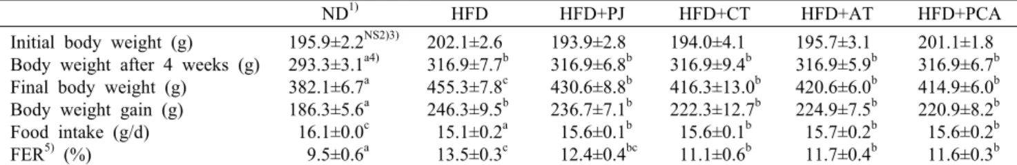
Table 1. Effect of pumpkin juice (PJ), corn silk tea (CT), adzuki bean tea (AT), and the mixture of PJ, CT, and AT (PCA) on
the body weight change, energy intake, food intake, and food efficiency ratio in diet-induced obese ratsND1) HFD HFD+PJ HFD+CT HFD+AT HFD+PCA
Initial body weight (g) Body weight after 4 weeks (g) Final body weight (g)
Body weight gain (g) Food intake (g/d) FER5) (%)
195.9±2.2NS2)3) 293.3±3.1a4) 382.1±6.7a 186.3±5.6a 16.1±0.0c 9.5±0.6a
202.1±2.6 316.9±7.7b 455.3±7.8c 246.3±9.5b 15.1±0.2a 13.5±0.3c
193.9±2.8 316.9±6.8b 430.6±8.8b 236.7±7.1b 15.6±0.1b 12.4±0.4bc
194.0±4.1 316.9±9.4b 416.3±13.0b 222.3±12.7b 15.6±0.1b 11.1±0.6b
195.7±3.1 316.9±5.9b 420.6±6.0b 224.9±7.5b 15.7±0.2b 11.7±0.4b
201.1±1.8 316.9±6.7b 414.9±6.0b 220.9±8.2b 15.6±0.2b 11.6±0.3b
1)ND, normal diet; HFD, high-fat diet; HFD+PJ, HFD supplemented with pumpkin juice; HFD+CT, HFD supplemented with corn silk tea; HFD+AT, HFD supplemented with adzuki bean; HFD+PCA, HFD supplemented with the mixture of PJ, CT, and AT.
2)Values are the mean±SE for 7 animals in each group.
3)NS: no significant difference.
4)Values with different letters in the same row are significantly different at P<0.05 by Duncan's multiple range test.
5)FER: food efficiency ratio.
Table 2. Effect of pumpkin juice (PJ), corn silk tea (CT), adzuki bean tea (AT), and the mixture of PJ, CT, and AT (PCA) on
adipose tissue weight in diet-induced obese rats (g/kg BW)ND1) HFD HFD+PJ HFD+CT HFD+AT HFD+PCA
Subcutaneous Epididymal Retro-peritoneal Abdominal
Brown adipose tissue Total adipose tissue
8.1±0.4NS2)3) 11.9±0.6a4) 2.7±0.3NS 4.7±0.4NS 0.8±0.1NS 28.1±1.3a
10.4±1.0 17.0±1.1c 3.5±0.3 5.8±0.5 0.8±0.1 39.1±2.1c
10.1±0.4 15.6±0.9bc 3.1±0.5 6.1±0.6 0.9±0.1 36.0±1.7bc
9.5±1.2 16.4±1.0c 3.7±0.2 6.5±0.7 0.8±0.1 37.6±2.8bc
9.9±0.9 16.8±0.8c 3.5±0.4 6.6±0.7 0.9±0.1 38.6±2.5bc
10.5±1.0 13.5±1.2ab 3.4±0.4 5.7±0.7 1.0±0.1 31.5±2.9ab
1)ND, normal diet; HFD, high-fat diet; HFD+PJ, HFD supplemented with pumpkin juice; HFD+CT, HFD supplemented with corn silk tea; HFD+AT, HFD supplemented with adzuki bean; HFD+PCA, HFD supplemented with the mixture of PJ, CT, and AT.
2)Values are the mean±SE for 7 animals in each group.
3)NS: no significant difference.
4)Values with different letters in the same row are significantly different at P<0.05 by Duncan's multiple range test.
100 mM EDTA, 10 mM Tris)에 1% Triton X-100을 섞은 후 슬라이드를 담가 4°C의 암실조건에 1시간 동안 침지시켰 다. Lysis가 끝난 슬라이드는 전기영동 수조에 배열하여 차 가운 electrophoresis buffer(300 mM NaOH, 10 mM Na2EDTA)를 채워 20분간 unwinding 후, 25 V/300±3 mA 의 전압을 걸어 20분간 전기영동을 실시하였다. 전기영동이 끝나고 0.4 M Tris buffer(pH 7.5)로 충분히 세척하고 ethidium bromide로 핵을 염색하여 형광 현미경(LEICA DMLB, Wetzlar, Germany)의 CCD camera(Nikon)를 통 해 보내진 세포핵 이미지를 comet image analyzing sys- tem(Komet, version 5.0, Kinetic Imaging, Liverpool, UK)이 설치된 컴퓨터로 분석하였다.
통계처리
모든 데이터의 통계처리는 SPSS/Windows 18.0(IBM, Chicago, IL, USA)을 이용하여 분석하였고, 결과는 평균±
표준오차로 나타내었다. 각 항목은 일원배치 분산분석(one- way ANOVA)을 시행하여, Duncan's multiple range test 로 신뢰 수준은
P
<0.05에서 평균값들에 대해 그룹 간의 유 의성 차이를 검증하였다.결과 및 고찰
체중증가량, 식이섭취량 및 식이섭취효율
초기체중, 최종체중, 체중증가량, 식이섭취량 및 식이섭 취효율은 Table 1에 나타내었다. 초기체중에서는 그룹 간 유의성이 나타나지 않았으나, 최종체중에서는 ND군보다 HFD군의 체중이 19.2% 유의적 증가를 보여 고지방식이 섭 취로 인한 비만 유도를 확인할 수 있었다. 고지방식이 섭취 군들 간의 최종 체중 비교 시 HFD군에 비해 음료섭취그룹이 6~9% 유의적으로 감소하였다. 이는 HFD군보다 음료섭취 군들(HFD+CT, HFD+AT, HFD+PCA)의 식이섭취효율이 유의적으로 낮은 데서 기인한 것으로 생각한다.
지방조직 무게의 변화
실험동물의 지방조직 무게의 변화를 Table 2에 나타내었 다. 부고환지방조직 무게는 HFD군이 ND군보다 유의적으 로 증가하였고, HFD군에 비해 HFD+PCA군은 유의적으로 감소하였으며, 호박즙, 옥수수수염차, 팥차 개별 섭취군은 감소하는 경향만 나타내었다. 다른 부위의 지방조직 무게는 그룹 간 유의적인 차이를 나타내지 않았다. 전체 지방조직 무게 또한 HFD군이 ND군보다 유의적으로 높았으며, HFD +PCA군은 유의적으로 감소하는 결과를 나타내었는데 이 는 부고환지방조직 무게가 반영된 결과로 생각된다. 호박즙,
ab abc bc
a c
a
0 50 100 150 200 250
ND HFD PJ CT AT PCA
Adipocyte surface area (% of ND) .
ND HFD HFD+PJ HFD+CT HFD+AT HFD+PCA
Fig. 1. Epididymal adipocytes surface area of obese rats fed the
experimental diets for 9 weeks. ND, normal diet; HFD, high-fat diet; HFD+PJ, HFD supplemented with pumpkin juice; HFD+CT, HFD supplemented with corn silk tea; HFD+AT, HFD sup- plemented with adzuki bean; HFD+PCA, HFD supplemented with the mixture of PJ, CT, and AT. Mean surface area for sub- cutaneous adipocytes was measured using Image J software.
Each bar represents the mean±SE (n=7 per group). Bars with different letters are significantly different at P<0.05 by Duncan's multiple range test.
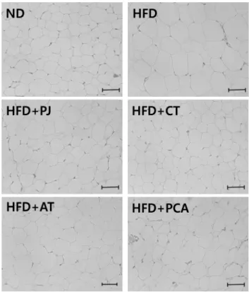
Fig. 2. Histological analysis of epididymal adipose tissue of rats
fed the experimental diets for 9 weeks. ND, normal diet; HFD, high-fat diet; HFD+PJ, HFD supplemented with pumpkin juice;HFD+CT, HFD supplemented with corn silk tea; HFD+AT, HFD supplemented with adzuki bean; HFD+PCA, HFD supplemented with the mixture of PJ, CT, and AT. All sections were stained with hematoxylin and eosin; magnification, ×100. Magnifica- tion bar=100 μm.
옥수수수염차, 팥차 개별 섭취군 역시 부고환지방조직 무게 의 영향으로 전체 지방조직 무게가 감소하는 경향만 보였다.
비만은 체지방의 증가, 특히 복강 내에 위치한 지방조직의 증가가 건강상의 위해요인으로 작용한다고 보고하여(25) 호박즙, 옥수수수염차, 팥차 혼합음료가 체중뿐만 아니라 체 지방 감소에도 영향을 미치는 것으로 생각된다. Do 등(26) 의 연구에서 늙은 호박 물 추출물(0.5~5 mg/mL)을 3T3- L1 세포에 처리하여 지방 축적이 억제되는 것을 농도 의존 적으로 확인하였지만, 그 유효 성분에 대해서는 보고하고 있지 않았다. 사포닌은 리파아제 활성을 낮추어 섭취된 지방 이 소화관에서 흡수되는 것을 저해함으로써 지방조직의 무 게가 증가하는 것을 억제하는 것으로 보고되고 있는데(16), 팥에서는 adzuki saponin Ⅰ~Ⅷ 종이 보고되고 있으며(27), 옥수수수염에도 사포닌이 함유된 것으로 보고되었다(11, 12). 따라서 본 연구에서는 고지방식이에 의한 지방조직 무 게 증가에 대해 각각의 시료 유효성분의 개별 효과보다는 이들 성분의 상승작용이 부고환지방과 총지방 무게 감소에 효과적이었던 것으로 생각된다.
지방조직의 형태학적 분석과 지방세포의 표면적
비만은 에너지 섭취와 소비 간의 불균형으로 인해 과도하 게 체지방이 축적되는 현상으로 지방세포의 수와 크기가 증 가하게 된다. 따라서 지방세포 크기의 측정은 항비만 효능을 입증할 수 있는 효과적인 방법으로 잘 알려져 있다(28). 지 방조직에서 그룹 간의 유의적 차이를 보였던 부고환지방세 포 표면적을 비교해보면 HFD군은 ND군보다 유의적으로 지방세포 표면적이 증가하였으며, HFD+PJ군(-78%)과 HFD +PCA군(-65%)의 지방세포 표면적은 HFD군에 비해 유의 적으로 감소하였으며, ND군과 유의적 차이를 나타내지 않
았다(Fig. 1). HFD+PJ군에서 지방세포 표면적 감소는 지방 세포에서 중성지방 농도가 현저하게 감소에 의한 것으로 생 각한다. Rhee 등(29)의 연구에서도 고지방식이로 유도된 비 만 쥐에 녹차 카테킨 공급 시 부고환지방의 크기가 감소하였 는데, 이는 중성지방의 합성이 감소하여 체지방 축적이 감소 하므로 인해 지방세포의 hypertrophy를 억제한 것으로 보 고하였다. Maeda 등(30)은 아디포넥틴 농도가 내장 지방과 음의 상관관계가 있음을 보고하여, HFD+PCA군의 부고환 지방조직 세포 표면적 감소는 증가한 아디포넥틴에 의한 것 으로 생각된다. 부고환지방조직의 형태학적 분석은 Fig. 2 에 나타내었다.
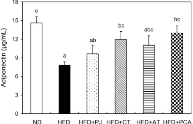
아디포넥틴에 미치는 영향
지방세포에서 분비되는 아디포넥틴은 체내 지방 축적 및 식욕을 조절하고 염증을 감소시키며 인슐린 민감도를 촉진 하고, 비만인에서 혈청 아디포넥틴의 수치가 정상인의 혈청 아디포넥틴보다 감소하는 것으로 보고되고 있다(31,32). 아 디포넥틴은 ND군에 비해 HFD군에서 유의적으로 감소하였 고, HFD군에 비해 HFD+CT군과 HFD+PCA군이 유의적 으로 증가하였다(Fig. 3). 항산화제는 산화적 스트레스를 낮 추어 아디포넥틴 발현을 조절하는 것으로 보고되고 있으며
Table 3. Effect of pumpkin juice (PJ), corn silk tea (CT), adzuki bean tea (AT), and the mixture of PJ, CT, and AT (PCA) on
plasma and hepatic tissue lipid profilesND1) HFD HFD+PJ HFD+CT HFD+AT HFD+PCA
Plasma lipid profile (mg/dL) Triglyceride
Total cholesterol HDL-cholesterol LDL-cholesterol
Hepatic tissue lipid profile (mg/g) Total lipid
Triglyceride Total cholesterol
68.8±4.6NS2)3) 157.9±3.6NS 37.1±1.6NS 134.5±3.7NS 19.3±1.5b4) 0.56±0.03NS 0.62±0.04NS
67.4±3.7 152.8±10.4 31.9±2.4 134.4±4.0 18.0±1.3ab 0.60±0.04 0.68±0.05
64.3±3.2 164.2±4.4 35.7±1.3 141.4±4.0 19.9±0.9b 0.58±0.02 0.71±0.06
64.3±2.3 166.4±5.7 34.8±1.6 144.5±4.7 16.0±1.3a 0.57±0.03 0.63±0.04
65.1±3.3 167.5±4.3 36.6±2.0 143.9±3.7 16.2±0.9a 0.54±0.01 0.58±0.02
61.8±3.1 160.1±4.6 34.3±2.1 138.2±3.6 17.3±0.8ab 0.55±0.01 0.58±0.02
1)ND, normal diet; HFD, high-fat diet; HFD+PJ, HFD supplemented with pumpkin juice; HFD+CT, HFD supplemented with corn silk tea; HFD+AT, HFD supplemented with adzuki bean; HFD+PCA, HFD supplemented with the mixture of PJ, CT, and AT.
2)Values are the mean±SE for 7 animals in each group.
3)NS: no significant difference.
4)Values with different letters in the same row are significantly different at P<0.05 by Duncan's multiple range test.
bc bc abc
ab
a c
0 3 6 9 12 15 18
ND HFD PJ CT AT PCA
Adiponectin (μg/mL) .
ND HFD HFD+PJ HFD+CT HFD+AT HFD+PCA
Fig. 3. Effect of pumpkin juice (PJ), corn silk tea (CT), adzuki
bean tea (AT), and the mixture of PJ, CT, and AT (PCA) on adipocytokine levels in plasma. ND, normal diet; HFD, high-fat diet; HFD+PJ, HFD supplemented with pumpkin juice; HFD+CT, HFD supplemented with corn silk tea; HFD+AT, HFD sup- plemented with adzuki bean; HFD+PCA, HFD supplemented with the mixture of PJ, CT, and AT. Each bar represents the mean±SE (n=7 per group). Bars with different letters are sig- nificantly different at P<0.05 by Duncan's multiple range test.
(33,34) 포도씨 procyanidins(35), catechins(36), γ-ory- zanol(37) 등의 항산화제들이 아디포넥틴 발현을 증가시킨 연구 결과들이 보고되었다. 따라서 HFD+PCA군에서 호박 즙, 옥수수수염차, 팥차의 항산화 성분들에 의해 고지방식이 에 의한 산화적 스트레스가 감소되어 아디포넥틴 농도가 증 가한 것으로 생각된다.
혈장, 간 조직, 부고환지방조직 지질 분석
혈장과 간 조직의 지질 분석 시 모든 그룹에서 유의적인 차이를 나타내지 않았다(Table 3). Kim 등(17)의 결과에서 도 고지방식이군(AIN-76 식이+20.5% lard 첨가)의 혈액 총콜레스테롤, 중성지질, HDL-콜레스테롤은 정상식이군과 유의적 차이가 나타나지 않았는데 그 이유에 대해서는 언급 하고 있지 않았다. 고지방식이로 유도한 비만 흰쥐에 함초 분말의 항비만 효과를 탐색한 Kim 등(38)의 연구에서도 혈
액, 간, 분변의 지질 농도가 고지방식이 섭취에 의해서는 유 의하게 증가하였으나 함초 분말 섭취에 따른 차이는 나타나 지 않아, 이는 함초 동결건조 분말 섭취가 장내 지방의 흡수 나 혈중 지질 농도에 영향을 미치지 않는 것으로 보고하였 다. 그러나 함초 분말 섭취에 의해서 부고환지방조직의 지방 축적 억제 효과는 확인하였다.
반면 부고환지방조직에서 총지질 함량, 중성지방, 총콜레 스테롤 농도는 HFD군이 ND군보다 유의적으로 높았으며, 음료섭취군은 HFD군에 비해 유의적으로 낮게 나타났다 (Fig. 4). 이는 늙은호박 물 추출물(500 mg/kg)이 고지방식 이(콜레스테롤 15%, sodium cholate 1%, 옥수수유 84%) 로 유도된 동물모델에서 혈중 콜레스테롤과 중성지질을 낮 추는 효과를 보고한 Lim과 Choi(10)의 연구 결과와 유사하 였다. Bae 등(39)은 인삼의 사포닌이 간세포 내 콜레스테롤 과 지방산의 생합성 및 분해를 촉진시키는 한편 콜레스테롤 대사를 항진시키는 것으로 보고하고 있으며, 또한 콜레스테 롤 장내흡수 억제 효과로 인해 체중, 부고환지방조직 무게 감소, 지방성분 축적이 억제된 것으로 보고하였다. 따라서 본 연구 결과에서도 늙은 호박 내 유효성분과 옥수수수염과 팥에 들어있는 사포닌의 작용으로 지방조직 내 지질 농도가 감소한 것으로 생각한다. 그리고 지방조직의 무게와 지질 농도에 유의적 효과를 보인 늙은 호박의 생리활성 성분과 그 작용 기전에 관하여는 추후 체계적인 연구가 필요할 것으 로 생각된다.
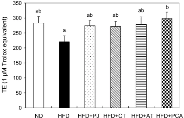
혈장 ORAC 값에 미치는 영향
혈장 ORAC assay는 혈장에서 peroxy radical 소거능을 측정하는 방법으로, 그 결과는 Fig. 5에 나타내었다. ORAC 활성은 정상식이군보다 고지방식이 섭취에 의해 유의적으 로 감소하지는 않았지만, 고지방식이와 호박즙, 옥수수수염 차, 팥차 혼합물 섭취 시 고지방식이 섭취군보다 ORAC 활성 이 유의적으로 증가하였다. HFD+PCA군을 제외한 나머지 음료섭취군은 HFD군에 비해 증가하는 경향만 보였다. 고지
abc bc c
ab d
a
0 300 600 900 1200 1500
ND HFD PJ CT AT PCA
Total lipid (mg/g) .
ND HFD HFD HFD HFD HFD +PJ +CT +AT +PCA
bc c bc a d
ab
0 1 2 3 4 5 6
ND HFD PJ CT AT PCA
Triglyceride (mg/g) .
ND HFD HFD HFD HFD HFD +PJ +CT +AT +PCA
b b ab b
c
a
0 1 2 3 4 5 6 7
ND HFD PJ CT AT PCA
Total cholesterol (mg/g) .
ND HFD HFD HFD HFD HFD +PJ +CT +AT +PCA
Fig. 4. Effect of pumpkin juice (PJ), corn silk tea (CT), adzuki bean tea (AT), and the mixture of PJ, CT, and AT (PCA) on
epididymal adipose tissue lipid profiles. ND, normal diet; HFD, high-fat diet; HFD+PJ, HFD supplemented with pumpkin juice;HFD+CT, HFD supplemented with corn silk tea; HFD+AT, HFD supplemented with adzuki bean; HFD+PCA, HFD supplemented with the mixture of PJ, CT, and AT. Each bar represents the mean±SE (n=7 per group). Bars with different letters are significantly different at P<0.05 by Duncan's multiple range test.
ab b ab ab
a ab
0 50 100 150 200 250 300 350
ND HFD PJ CT AT PCA
TE (1 μM Trolox equivalent) .
ND HFD HFD+PJ HFD+CT HFD+AT HFD+PCA
Fig. 5. Effect of pumpkin juice (PJ), corn silk tea (CT), adzuki
bean tea (AT), and the mixture of PJ, CT, and AT (PCA) on oxygen radical absorbance capacity (ORAC) and total radical trapping capacity (TRAP) in plasma. ND, normal diet; HFD, high-fat diet; HFD+PJ, HFD supplemented with pumpkin juice;HFD+CT, HFD supplemented with corn silk tea; HFD+AT, HFD supplemented with adzuki bean; HFD+PCA, HFD supplemented with the mixture of PJ, CT, and AT. Each bar represents the mean±SE (n=7 per group). Bars with different letters are sig- nificantly different at P<0.05 by Duncan's multiple range test.
a ab ab ab
b ab
0 0.4 0.8 1.2
ND HFD PJ CT AT PCA
TBARS (μM/mg protein) .
ND HFD HFD+PJ HFD+CT HFD+AT HFD+PCA
Fig. 6. Effect of pumpkin juice (PJ), corn silk tea (CT), adzuki
bean tea (AT), and the mixture of PJ, CT, and AT (PCA) on TBARS in liver. ND, normal diet; HFD, high-fat diet; HFD+PJ, HFD supplemented with pumpkin juice; HFD+CT, HFD supple- mented with corn silk tea; HFD+AT, HFD supplemented with adzuki bean; HFD+PCA, HFD supplemented with the mixture of PJ, CT, and AT. Each bar represents the mean±SE (n=7 per group). Bars with different letters are significantly different atP<0.05 by Duncan's multiple range test.
방 식이에 의해 라디칼 생성이 증가한 것을 확인할 수 있었 으며, 호박즙, 옥수수수염차, 팥차를 각각 섭취하는 것보다 세 가지를 혼합하여 섭취하였을 때 라디칼 소거 효과가 증가 하는 것을 확인할 수 있었다. 늙은 호박의 경우 카로틴 색소 는 비타민 A의 전구체로서 뿐만 아니라 활성산소의 소거제 (40)로 작용함으로써 항산화 작용을 발휘하는 기능성 성분 으로 보고되고 있으며, lutein, α-carotene, β-carotene이 30% 정도 함유되어 있다(41). 옥수수수염 유래 플라보노이 드로는 maysin, apimaysin, methoxymaysin 등이 있으며 이 중 maysin은 옥수수수염에 가장 많이 함유된 대표적인 기능성 물질로 종양 세포주에 대한 세포독성 효과 및 라디칼 소거 활성 등이 보고되어 있다(42,43). 팥의 색소는 antho- cyanin계의 cyanidin으로 알려져 있으며(44), 이들 색소는 항산화(45) 및 항종양 효과(46)를 나타내는 것으로 보고되 고 있다.
간 지질과산화물 형성에 미치는 영향
지질과산화물은 활성산소종 등 산화적 스트레스에 의해 체내 각종 지질성분으로부터 생성된다. 생성된 지질과산화 물은 세포막의 투과성을 변형시켜 DNA, 탄수화물, 단백질, 지질의 손상을 유발하여 노화, 고혈압, 동맥경화, 심장병 등 각종 질병을 일으킬 수 있다(47). 간에서의 지질과산화물을 분석한 결과를 살펴보면 HFD군이 ND군보다 증가하는 경 향을 보였고, HFD+PCA군은 HFD군보다 유의적으로 감소 하였다(Fig. 6). 홍삼 성분이 지질과산화에 미치는 효과를 살펴본 Sung 등(48)은 홍삼에 함유된 사포닌을 비롯하여 정유성분, 폴리아세틸렌, 페놀성분, 배당체 및 산성펩타이드 등(49)의 생리활성 성분이 지질과산화 억제 효과가 있다고 보고 있다.
따라서 HFD+PCA군의 지질과산화 정도가 HFD군에 비 해 억제된 이유는 옥수수수염과 팥에 함유된 사포닌과 플라 보노이드, 안토시아닌과 늙은 호박의 카로틴 간의 상승작용 으로 인한 것으로 생각한다.
Table 4. Effect of pumpkin juice (PJ), corn silk tea (CT), adzuki bean tea (AT), and the mixture of PJ, CT, and AT (PCA) on
erythrocyte and hepatic antioxidant enzyme activitiesND1) HFD HFD+PJ HFD+CT HFD+AT HFD+PCA
GSH-Px (μM/mg protein) Catalase (mM/mg protein)
286.6±35.9a2)3) 1.1±0.2NS
237.1±9.2a 1.0±0.1
246.9±16.3a 0.9±0.1
468.4±78.0b 1.0±0.2
339.1±36.8ab 1.0±0.2
680.2±77.1c 1.3±0.1
1)ND, normal diet; HFD, high-fat diet; HFD+PJ, HFD supplemented with pumpkin juice; HFD+CT, HFD supplemented with corn silk tea; HFD+AT, HFD supplemented with adzuki bean; HFD+PCA, HFD supplemented with the mixture of PJ, CT, and AT.
2)Values are the mean±SE for 7 animals in each group.
3)Values with different letters in the same row are significantly different at P<0.05 by Duncan's multiple range test.
4)NS: no significant difference.
b c cd c
a d
0 20 40 60 80 100
ND HFD PJ CT AT PCA
Tail DNA (%) .
ND HFD HFD+PJ HFD+CT HFD+AT HFD+PCA
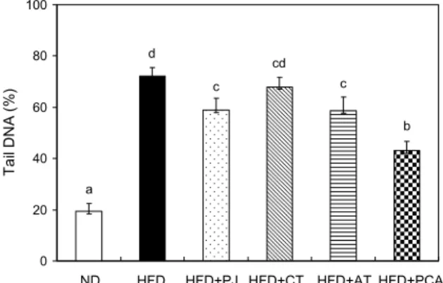
Fig. 7. Antigenotoxic effect of pumpkin juice (PJ), corn silk
tea (CT), adzuki bean tea (AT), and the mixture of PJ, CT, and AT (PCA) on high-fat diet induced DNA damage in hepatocytes.ND, normal diet; HFD, HFD+PJ, HFD supplemented with pumpkin juice; HFD+CT, HFD supplemented with corn silk tea;
HFD+AT, HFD supplemented with adzuki bean; HFD+PCA, HFD supplemented with the mixture of PJ, CT, and AT. Each bar represents the mean±SE (n=7 per group). Bars with different letters are significantly different at P<0.05 by Duncan's multiple range test.
간 조직 항산화 효소 활성에 미치는 영향
비만은 지질과산화물의 형성을 촉진해 체내 항산화 시스 템의 불균형을 일으키는 과정과 관련이 있다(50,51). 지방 이 축적될 경우 몸 전체에서 산화스트레스가 증가하는 양상 을 보이는데, 비만 쥐의 경우 지방조직에서 활성산소종의 생산이 선택적으로 많이 이루어진다. 생체 내에는 이러한 산화적 손상을 예방하거나 복구하는 체계로 superoxide dismutase(SOD), glutathione(GSH), glutathione perox- idase(GSH-Px), glutathione S-transferase(GST), cata- lase(CAT) 등이 있다(52). 비만으로 인한 산화스트레스는 항산화 효소 활성을 감소시켜 항산화 방어 시스템을 저하시 킨다고 알려진바 활성산소종과 항산화 효소와의 항상성의 불균형은 DNA, 단백질 및 지질의 산화 변형을 통하여 노화 를 촉진하며 심혈관 질환, 염증성 질환 및 암과 같은 질환을 유발하게 된다(53,54). 간 항산화 효소 활성을 분석한 결과 는 Table 4에 나타내었다. 간 조직 GSH-Px는 HFD군과 ND군 간의 유의적 차이는 없었으나 고콜레스테롤 식이섭취 군에서 감소하는 경향을 나타내었고, 옥수수수염차 섭취군 과 호박즙, 옥수수수염차, 팥차 혼합물 섭취군은 고콜레스테 롤 식이섭취군보다 GSH-Px 활성이 유의적으로 증가했으 며, 이들 내 함유된 항산화 성분의 상승작용에 의한 결과로 생각된다. GSH-Px는 GSH와 과산화수소로부터 GSSG와 물, 알코올을 생성하는 반응을 촉매하여 생체 내 과산화수소 나 지질과산화물을 제거하는 작용을 한다(55). 간 조직 cat- alase는 모든 그룹에서 유의적인 결과를 나타내지 못하였 다.
간세포 DNA 손상에 미치는 영향
Comet assay는 소형 전기영동 겔 방법으로 인체에 유해 한 독성물질에 의한 DNA 손상 정도를 간단하고 빠르게 측 정하여 유전 독성의 발생 여부를 알아내는 좋은 도구로 이용 되고 있다(56). 간세포 DNA 손상은 알칼리 환경의 전기영 동을 이용한 comet assay를 수행한 후 DNA 손상 정도를 세포의 파괴된 파편의 길이로 측정하였다. 고지방식이로 비 만을 유도한 동물의 간세포에 H2O2 200 μM 농도를 처리하 여 내재적인 DNA 손상 정도를 관찰하였고, 이에 따른 간세 포의 DNA 손상에 대한 음료 섭취 효과는 Fig. 7에 제시하였 다. 본 실험에서 ND군에 비해 HFD군은 유의적으로 DNA 손상이 증가하였다. 이는 고지방식이에 의한 활성산소의 증
가로 체내 지질과산화물이 과다 생성되면서 DNA 손상이 많이 유발되는 것으로 생각한다. HFD군에 비해 HFD+PJ 군, HFD+AT군, HFD+PCA군은 DNA 손상이 유의적으로 감소하였으며, 그 효과는 HFD+PCA군에서 가장 높게 나타 났다. 이 결과로 호박즙, 팥차 각각의 항산화 성분들이 고지 방 식이로 유도된 DNA 손상 회복 효능을 가진다는 것을 확인할 수 있었으며, 그 효과는 호박즙, 옥수수수염차, 팥차 의 혼합물에 함유된 항산화 성분들의 상승작용에 의해 증가 한 것으로 생각된다.
요 약
4주간 식이로 유도된 비만 동물에서 호박즙(250 mg/kg BW), 옥수수수염차(250 mg/kg BW), 팥차(250 mg/kg BW)와 이 들 혼합음료(호박즙 분말 : 옥수수수염차 분말 : 팥차 분말, 1:1:1, 250 mg/kg BW)를 9주간 투여하여 그 효능을 알아본 결과 호박즙, 옥수수수염차, 팥차 개별 섭취 시 체중감소 효 과는 보였으나 그 기전은 본 연구에서 규명하지 못하였다.
그러나 혼합음료 섭취군에서는 지방조직 무게의 유의적 감 소로 인한 체중 저하 효능을 확인할 수 있었다. 부고환지방
조직의 표면적은 고지방식이섭취군(HFD)군에 비해 고지방 식이+혼합음료섭취군(HFD+PCA)군에서 유의적으로 감소 하였으며, HFD+PCA군의 아디포넥틴은 HFD군에 비해 유 의적으로 증가하였다. 음료섭취군들 중 고지방식이+혼합음 료섭취군(HFD+PCA)은 고지방식이섭취군(HFD)보다 ORAC value가 증가하였고, GSH-Px의 간 항산화 효소에서도 HFD군보다 HFD+PCA군이 유의적으로 증가하였다. 또한, HFD+PCA군은 고지방식이에 의한 지질과산화물 생성을 유의적으로 감소시켰으며, 비만으로 인한 산화적 스트레스 에 따른 DNA 손상을 현저히 낮추었다. 따라서 산화적 스트 레스에 의해 유도되는 비만 치료에 호박즙, 옥수수수염차, 팥차를 각각 섭취하기보다는 이들 세 가지를 동일한 비율로 혼합 섭취 시 각각의 항산화 효능의 상승작용에 의한 항비만 효과가 현저하게 나타낼 것으로 생각한다.
감사의 글
이 논문은 2011년도 정부(미래창조과학부)의 재원으로 한국 연구재단의 지원을 받아 수행된 기초연구사업임(No. NRF- 2011-0014277).
REFERENCES
1. Korean Statistical Information Service. Obesity. http://kosis.
kr/statisticsList/statisticsList_01List.jsp?vwcd=MT_ZTITLE
&parentId=D#SubCont (accessed Jan 2016).
2. Ferretti G, Bacchetti T, Moroni C, Savino S, Liuzzi A, Bal- zola F, Bicchiega V. 2005. Paraoxonase activity in high- density lipoproteins: a comparison between healthy and obese females. J Clin Endocrinol Metab 90: 1728-1733.
3. Higdon JV, Frei B. 2003. Obesity and oxidative stress: a direct link to CVD?. Arterioscler Thromb Vasc Biol 23: 365- 367.
4. Antipatis VJ, Gill TP. 2001. Obesity as a global problem.
In International Text Book of Obesity. Björntorp P, ed. John Wiley & Sons Inc., Chichester, West Sussex, UK. p 3-22.
5. Seo YH. 2005. Patent trend of anti-obesity supplementary food. Food World 8: 116-122.
6. Han LK, Kimura Y, Okuda H. 2005. Anti-obesity effects of natural products. Stud Nat Prod Chem 30: 79-110.
7. Moro CO, Basile G. 2000. Obesity and medicinal plants.
Fitoterapia 71: S73-S82.
8. Rayalam S, Della-Fera MA, Baile CA. 2008. Phytochemicals and regulation of the adipocyte life cycle. J Nutr Biochem 19: 717-726.
9. Ku BH. 1987. Korea folk medicine. Parkchul Press Inc., Seoul, Korea. p 90.
10. Lim JP, Choi H. 2001. Effects of the water extract from
Cucurbita maxima Duchesne on inflammation and hyper-
lipidemia in rats. Korean J Med Crop Sci 9: 280-283.11. Velazquez DV, Xavier HS, Batista JE, de Castro-Chaves C.
2005. Zea mays L. extracts modify glomerular function and potassium urinary excretion in conscious rats. Phytomedi-
cine 12: 363-369.
12. Rau O, Wurglics M, Dingermann T, Abdel-Tawan M, Schu- bert-Zsilavecz M. 2006. Screening of herbal extracts for ac- tivation of the human peroxisome proliferator-activated re-
ceptor. Pharmazie 61: 952-956.
13. Chin HS, Pack KJ, Pack SH, Kim JK. 2009. The effects of herbal extract mixture on anti-obesity. J Korean Soc Food
Sci Nutr 38: 32-38.
14. Maksimović Z, Malencić D, Kovacević N. 2005. Polyphenol contents and antioxidant activity of Maydis stigma extracts.
Bioresour Technol 96: 873-877.
15. Maksimović ZA, Kovacević N. 2003. Preliminary assay on the antioxidant activity of Maydis stigma extracts. Fitoter-
apia 74: 144-147.
16. Han LK, Kimura Y, Okuda H. 2003. Anti-obesity effects of tea saponins. 7th International Symposium on Green Tea, Seoul, Korea. p 65-72.
17. Kim HS, Kim TW, Kim DJ, Hwang HJ, Lee HJ, Choe M.
2007. Effects of natural plants supplementation on adipocyte size of the epididymal fat pads in rats. J Korean Soc Food
Sci Nutr 36: 419-423.
18. Vasselli JR, Weindruch R, Heymsfield SB, Pi-Sunyer FX, Boozer CN, Yi N, Wang C, Pietrobelli A, Allison DB. 2005.
Intentional weight loss reduces mortality rate in a rodent model of dietary obesity. Obes Res 13: 693-702.
19. Cho JH, Lee NJ, Hong SH, Kim DK, Shin S, Park JH, Kang JK, Kim YB, Hwang SY. 2005. Effect of Hwalgidan SJ-201 on obesity induced by high-fat diet in Zucker rats. Lab Anim
Res 21: 158-163.
20. Allain CC, Poon LS, Chan CS, Richmond W, Fu PC. 1974.
Enzymatic determination of total serum cholesterol. Clin
Chem 20: 470-475.
21. McGowan MW, Artiss JD, Strandbergh DR, Zak B. 1983.
A peroxidase-coupled method for the colorimetric determi- nation of serum triglycerides. Clin Chem 29: 538-542.
22. Friedewald WT, Levy RI, Fredrickson DS. 1972. Estimation of the concentration of low-density lipoprotein cholesterol in plasma, without use of the preparative ultracentrifuge.
Clin Chem 18: 499-502.
23. Folch J, Lees M, Sloane Stanley GH. 1957. A simple meth- od for the isolation and purification of total lipides from animal tissues. J Biol Chem 226: 497-509.
24. Ou B, Hampsch-Woodill M, Prior RL. 2001. Development and validation of an improved oxygen radical absorbance capacity assay using fluorescein as the fluorescent probe.
J Agric Food Chem 49: 4619-4626.
25. Björntorp P. 1988. The associations between obesity, adi- pose tissue distribution and disease. Acta Med Scand Suppl 723: 121-134.
26. Do GP, Lee HJ, Do JR, Kim HK. 2012. Antiobesity effect of the Cucubita moschata Duch extracts in 3T3-L1 adipo- cyets. Korean J Food Preserv 19: 138-143.
27. Ha TJ, Lee BW, Park KH, Jeong SH, Kim HT, Ko JM, Baek IY, Lee JH. 2014. Rapid characterisation and compar- ison of saponin profiles in the seeds of Korean Leguminous species using ultra performance liquid chromatography with photodiode array detector and electrospray ionisation/mass spectrometry (UPLC-PDA-ESI/MS) analysis. Food Chem 146: 270-277.
28. Moon GA, Choi SM, Kim SH, Kim SS, Kang JY, Yoon Y. 2003. Human and animal study on the natural food for obesity and metabolic syndrome risk factors. J Korean Soc
Food Sci Nutr 32: 1394-1400.
29. Rhee SJ, Kim KR, Kim HT, Hong JH. 2007. Effects of cat- echin on lipid composition and adipose tissue in obese rats fed high fat diet. J Korean Soc Food Sci Nutr 36: 540-547.
30. Maeda N, Takahashi M, Funahashi T, Kihara S, Nishizawa H, Kishida K, Nagaretani H, Matsuda M, Komuro R, Ouchi
N, Kuriyama H, Hotta K, Nakamura T, Shimomura I, Matsuzawa Y. 2001. PPARγ ligands increase expression and plasma concentrations of adiponectin, an adipose-derived pro- tein. Diabetes 50: 2094-2099.
31. Goldfine AB, Kahn CR. 2003. Adiponectin: linking the fat cell to insulin sensitivity. Lancet 362: 1431-1432.
32. Gil JH, Lee JA, Kim JY, Hong YM. 2008. Leptin, adipo- nectin, interleukin-6 and tumor necrosis factor-α in obese adolescents. Korean J Pediatr 51: 597-603.
33. Furukawa S, Fujita T, Shimabukuro M, Iwaki M, Yamada Y, Nakajima Y, Nakayama O, Makishima M, Matsuda M, Shimomura I. 2004. Increased oxidative stress in obesity and its impact on metabolic syndrome. J Clin Invest 114:
1752-1761.
34. Subauste AR, Burant CF. 2007. Role of FoxO1 in FFA-in- duced oxidative stress in adipocytes. Am J Physiol Endocri-
nol Metab 293: E159-E164.
35. Terra X, Montagut G, Bustos M, Llopiz N, Ardèvol A, Bladé C. Fernández-Larrea J, Pujadas G, Salvadó J, Arola L, Blay M. 2009. Grape-seed procyanidins prevent low-grade in- flammation by modulating cytokine expression in rats fed a high-fat diet. J Nutr Biochem 20: 210-218.
36. Cho SY, Park PJ, Shin HJ, Kim YK, Shin DW, Shin ES, Lee HH, Lee BG, Baik JH, Lee TR. 2007. (-)-Catechin sup- presses expression of Kruppel-like factor 7 and increases expression and secretion of adiponectin protein in 3T3-L1 cells. Am J Physiol Endocrinol Metab 292: E1166-E1172.
37. Justo ML, Rodriguez-Rodriguez R, Claro CM, Alvarez de Sotomayor M, Parrado J, Herrera MD. 2013. Water-soluble rice bran enzymatic extract attenuates dyslipidemia, hyper- tension and insulin resistance in obese Zucker rats. Eur J
Nutr 52: 787-797.
38. Kim MJ, Jun HY, Kim JH. 2015. Anti-obesity effect of Ko- rean Hamcho (Salicornia herbacea L.) powder on high-fat diet-induced obese rats. J Nutr Health 48: 123-132.
39. Bae MJ, Sung TS, Choi C. 1990. Effect of ginseng fraction components on plasma, adipose and feces steroids in obese rat induced by a high fat diet. Korean J Ginseng Sci 14:
404-415.
40. Seddon JM, Ajani UA, Sperduto RD, Hiller R, Blair N, Burton TC, Farber MD, Gragoudas ES, Haller J, Miller DT, Yannuzzi LA, Willett W. 1994. Dietary carotenoids, vita- mins A, C and E advanced age-related macular degenera- tion. JAMA 272: 1413-1420.
41. Kim SR, Ha TY, Song HN, Kim YS, Park YK. 2005. Com- parison of nutritional composition and antioxidative activity for Kabocha squash and pumpkin. Korean J Food Sci Tech-
nol 37: 171-177.
42. Lee EA, Byrne PF, McMullen MD, Snook ME, Wiseman BR, Widstrom NW, Coe EH. 1998. Genetic mechanisms un- derlying apimaysin and maysin synthesis and corn earworm antibiosis in maize (Zea mays L.). Genetics 149: 1997-2006.
43. Kim SL, Snook ME, Lee JO. 2003. Radical scavenging ac- tivity and cytotoxicity of maysin (C-glycosylflavone) iso- lated from silks of Zea mays L.. Korean J Crop Sci 48:
392-396.
44. Yoshida K, Sato Y, Okuno R, Kameda K, Isobe M, Kondo T. 1996. Structural analysis and measurement of anthocya- nins from colored seed coats of Vigna, Phaseolus, and Gly-
cine legumes. Biosci Biotech Biochem 60: 589-593.
45. Ariga T, Koshiyama I, Fukushima D. 1988. Antioxidative properties of procyanidins B-1 and B-3 from azuki beans in aqueous systems. Agric Biol Chem 52: 2717-2722.
46. Koide T, Hashimoto Y, Kamei H, Kojima T, Hasegawa M, Terabe K. 1997. Antitumor effect of anthocyanin fractions extracted from red soybeans and red beans in vitro and in
vivo. Cancer Biother Radiopharm 12: 277-280.
47. Jovanovic SV, Simic MG. 2000. Antioxidants in nutrition.
Ann N Y Acad Sci 899: 326-334.
48. Sung KS, Chun C, Kwon YH, Chang CC. 2000. Effects of red ginseng component administration on glutathione and lipid peroxidation levels in mice liver. J Ginseng Res 24:
176-182.
49. Lee KJ. 2012. Characteristics of physico-chemical proper- ties and analysis of functional components in soymilk with red ginseng extract. PhD Dissertation. Chosun University, Gwangju, Korea.
50. Carantoni M, Abbasi F, Warmerdam F, Klebanov M, Wang PW, Chen YD, Azhar S, Reaven GM. 1998. Relationship between insulin resistance and partially oxidized LDL par- ticles in healthy, nondiabetic volunteers. Arterioscler Thromb
Vasc Biol 18: 762-767.
51. Hirashima O, Kawano H, Motoyama T, Hirai N, Ohgushi M, Kugiyama K, Ogawa H, Yasue H. 2000. Improvement of endothelial function and insulin sensitivity with vitamin C in patients with coronary spastic angina: possible role of reactive oxygen species. J Am Coll Cardiol 35: 1860-1866.
52. Halliwill B. 1996. Antioxidant in human health and disease.
Annu Rev Nutr 16: 33-50.
53. Matsuda M, Shimomura I. 2013. Increased oxidative stress in obesity: Implications for metabolic syndrome, diabetes, hypertension, dyslipidemia, atherosclerosis, and cancer. Obes
Res Clin Pract 7: e330-e341.
54. Trakshel GM, Maines MD. 1988. Characterization of gluta- thione S-transferases in rat kidney. Alteration of composi- tion by cis-platinum. Biochem J 252: 127-136.
55. Rotruck, JT, Pope AL, Ganther HE, Swanson AB, Hafeman DG, Hoekstra WG. 1973. Selenium: biochemical role as a component of glutathione peroxidase. Science 179: 588-590.
56. Kim MJ, Chu WM, Park EJ. 2012. Antioxidant and anti- genotoxic effects of Shiitake mushrooms affected by differ- ent drying methods. J Korean Soc Food Sci Nutr 41: 1041- 1048.