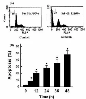
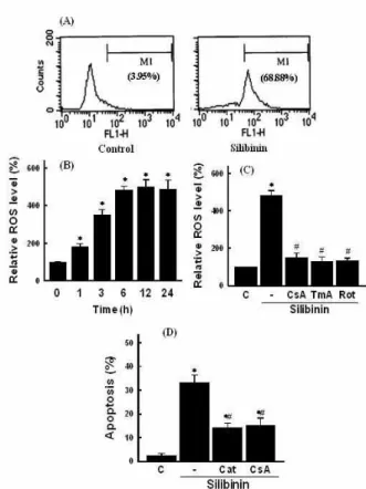
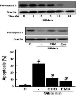
Silibinin Induces Apoptotic Cell Death Via ROS-dependent Mitochondrial Pathway in Human Glioma Cells
7
0
0
전체 글
(2)
(3)
(4)
(5)
(6)
(7)
수치
관련 문서
Detection of changes in mitochondrial membrane potential and release of mitochondrial cytochrome C Cells were treated with 5, 10, 20, 40 μmol/L curcumin (reference
Salinomycin Induces Apoptosis in PC-3 Human Prostate Cancer Cells via G1 Cell Cycle Arrest and Ca 2+ -Dependent Mitochondrial Pathway.. Kwang youn Kim, Sun nyoung
Reactive oxygen species mediate caspase activation and apoptosis induced by lipoic acid in human lung epi- thelial cancer cells through Bcl-2
Although the current results indicate that CCH1-induced apoptosis is accompanied by mitochondrial cytochrome c release and subsequent activation of caspase cascade
Dimethoxycurcumin, a Structural Analogue of Curcumin, Induces Apoptosis in Human Renal Carcinoma Caki Cells Through the Production of Reactive Oxygen Species, the Release
In addition, the HO-1 inducer cobalt protoporphyrin (CoPP) and the metabolic product of HO-1 bilirubin mimicked diazoxide to inhibit sFlt-1 release and reactive oxygen species
Juniperus chinensis extract induces apoptosis via reaction oxygen species (ROS) generation in human pancreatic cancer cell lines.. Boram Go 1,3 · Song-I Han 2 · Jungwhoi Lee 2
Oleanolic acid induced autophagic cell death in hepatocellular carcinoma cells via PI3K⁄Akt⁄mTOR and ROS-dependent pathway.. Yang Shi 1 , Qingwei Song 1 , Dianhe Hu 1 , Xiaohu Zhuang
