• 검색 결과가 없습니다.

PCR-based Assay for the Specific Detection of Pseudomonas syringae pv. tagetis using an AFLP-derived Marker

N/A
N/A
Protected

Academic year: 2021

Share "PCR-based Assay for the Specific Detection of Pseudomonas syringae pv. tagetis using an AFLP-derived Marker"

Copied!
5
0
0

로드 중.... (전체 텍스트 보기)

전체 글

(1)

Research Article Open Access http://dx.doi.org/10.5423/RPD.2015.21.1.001

Research in Plant Disease

©The Korean Society of Plant Pathology pISSN 1598-2262, eISSN 2233-9191

This is an open access article distributed under the terms of the Creative Commons Attribution License (http://creativecommons.org/licenses/by/2.0), which permits unrestricted use, distribution, and reproduction in any medium, provided the original work is properly cited.

PCR-based Assay for the Specific Detection of Pseudomonas syringae pv. tagetis using an AFLP-derived Marker

Eun-Sung Song 1 , Song-Yi Kim 2 , Soo-Cheon Chae 2 , Jeong-Gu Kim 1 , Heejung Cho 1 , Seunghwan Kim 1 and Byoung-Moo Lee 1 *

1

National Academy of Agricultural Science, Rural Development Administration, Jeonju 560-500, Korea

2

Department of Horticulture, Kong-Ju National University, Yesan 340-802, Korea

A PCR method has been developed for the pathovar-specific detection of Pseudomonas syringae pv. tagetis, which is the causal agent of bacterial leaf spots and apical chlorosis of several species within the Compositae family. One primer set, PSTF and PSTR, was designed using a genomic locus derived from an amplified fragment length polymorphism (AFLP) fragment produced a 554-bp amplicon from 4 isolates of P. syringae pv. tagetis.

In DNA dot-blot analysis with the PCR product as probe, a positive signal was identified in only 4 isolates of P.

syringae pv. tagetis. These results suggest that this PCR-based assay will be a useful method for the detection and identification of P. syringae pv. tagetis.

Keywords : AFLP, Detection, PCR, Pseudomonas syringae pv. tagetis

Introduction

Pseudomonas syringae pv. tagetis was first described in Denmark as a pathogen that affects marigold production (Hellmers, 1955). It is now known as a phytopathogenic bacte- rium that is the causal agent of bacterial leaf spots and apical chlorosis in several species within the family Compositae:

African marigold (Tagetes erecta L.), sunflower (Helianthus annuus L.), common ragweed (Ambrosia artemisiifolia L.), Jerusalem artichoke (Helianthus. tuberosus L.), dandelion (Taraxacum officinale Weber), compass plant (Silphium perfoliatum L.) and another sunflower species (Helianthus sal- icifolius A. Diter) (Gulya et al., 1981; Hellmers, 1955; Rhodehamel and Durbin, 1985; Rhodehamel and Durbin, 1989a; Shane and Baumer, 1984; Styer and Durbin, 1982).

P. syringae pv. tagetis produces a toxin (tagetitoxin) in host leaves that is then translocated to emerging leaves, where it inhibits RNA polymerase III, thereby preventing chloroplast bio- genesis and resulting in apical chlorosis (Mathews and Durbin, 1990; Steinberg et al., 1990). The pathogens are divided into three classes based on their capability to produce tagetitoxin:

class 1 and 2 strains produce tagetitoxin in plants; class 3 strains do not produce the toxin (Rhodehamel and Durbin, 1989b).

There are many reports that specific detection methods for phytotoxin-producing P. syringae pathovars have been devel- oped based on genes required for their production (Bereswill et al., 1994; Lydon and Patterson, 2001; Schaad et al., 1995;

Sorensen et al., 1998). Recently, a PCR protocol to distinguish P.

syringae pv. tagetis from other P. syringae pathovars and closely related species was developed based on genes required for tag- etitoxin production (Kong et al., 2004). However, this approach is unable to distinguish the bacterium from other Pseudomonas isolates at the pathovar level. Furthermore, Pseudomonas spe- cies other than P. syringae pv. tagetis have been reported to induce apical chlorosis in Canada thistle and pea (Suzuki et al., 2003; Zhang et al., 2002). Therefore, a PCR-based assay that is able to unambiguously distinguish P. syringae pv. tagetis from P. syringae pv. helianthi and other apical chlorosis-inducing Pseudomonas species is needed.

In this study, we report the development of a pathovar-spe- cific marker derived from the AFLP technique for detecting and distinguishing P. syringae pv. tagetis from other pathovars and species of Pseudomonas and Xanthomonas. The specificity of the PCR-based assay using pathovar-specific primers was vali- dated by testing 47 isolates collected from various geographical

*Corresponding author Tel : +82-63-238-4565 Fax: +82-63-238-4554 E-mail: [email protected]

Received December 1, 2014

Revised February 13, 2015

Accepted March 2, 2015

(2)

regions and host plants.

Material and Methods

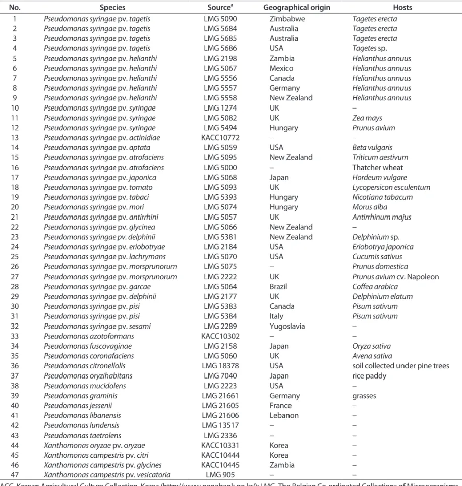
Bacterial strains and DNA isolation. The bacterial strains that are listed in Table 1 were obtained from the Korean Ag- ricultural Culture Collection (KACC) in Suwon, Korea, and the

Belgian Co-ordinated Collections of Micro-organisms (BCCM) in Brussels, Belgium. The genomic DNA was isolated as described previously (Song et al., 2014).

AFLP PCR analysis. The AFLP assay was performed using a previously described method (Song et al., 2014), with minor modification. Genomic DNA (approximately 300 ng) was

Table 1. List of bacterial strains used in this study

No. Species Source

a

Geographical origin Hosts

1 Pseudomonas syringae pv. tagetis LMG 5090 Zimbabwe Tagetes erecta

2 Pseudomonas syringae pv. tagetis LMG 5684 Australia Tagetes erecta

3 Pseudomonas syringae pv. tagetis LMG 5685 Australia Tagetes erecta

4 Pseudomonas syringae pv. tagetis LMG 5686 USA Tagetes sp.

5 Pseudomonas syringae pv. helianthi LMG 2198 Zambia Helianthus annuus

6 Pseudomonas syringae pv. helianthi LMG 5067 Mexico Helianthus annuus

7 Pseudomonas syringae pv. helianthi LMG 5556 Canada Helianthus annuus

8 Pseudomonas syringae pv. helianthi LMG 5557 Germany Helianthus annuus

9 Pseudomonas syringae pv. helianthi LMG 5558 New Zealand Helianthus annuus

10 Pseudomonas syringae pv. syringae LMG 1274 UK -

11 Pseudomonas syringae pv. syringae LMG 5082 UK Zea mays

12 Pseudomonas syringae pv. syringae LMG 5494 Hungary Prunus avium

13 Pseudomonas syringae pv. actinidiae KACC10772 - -

14 Pseudomonas syringae pv. aptata LMG 5059 USA Beta vulgaris

15 Pseudomonas syringae pv. atrofaciens LMG 5095 New Zealand Triticum aestivum

16 Pseudomonas syringae pv. atrofaciens LMG 5000 - Thatcher wheat

17 Pseudomonas syringae pv. japonica LMG 5068 Japan Hordeum vulgare

18 Pseudomonas syringae pv. tomato LMG 5093 UK Lycopersicon esculentum

19 Pseudomonas syringae pv. tabaci LMG 5393 Hungary Nicotiana tabacum

20 Pseudomonas syringae pv. mori LMG 5074 Hungary Morus alba

21 Pseudomonas syringae pv. antirrhini LMG 5057 UK Antirrhinum majus

22 Pseudomonas syringae pv. glycinea LMG 5066 New Zealand -

23 Pseudomonas syringae pv. delphinii LMG 5381 New Zealand Delphinium sp.

24 Pseudomonas syringae pv. eriobotryae LMG 2184 USA Eriobotrya japonica

25 Pseudomonas syringae pv. lachrymans LMG 5070 USA Cucumis sativus

26 Pseudomonas syringae pv. morsprunorum LMG 5075 - Prunus domestica

27 Pseudomonas syringae pv. morsprunorum LMG 2222 UK Prunus avium cv. Napoleon

28 Pseudomonas syringae pv. garcae LMG 5064 Brazil Coffea arabica

29 Pseudomonas syringae pv. delphinii LMG 2177 UK Delphinium elatum

30 Pseudomonas syringae pv. pisi LMG 5383 Canada Pisum sativum

31 Pseudomonas syringae pv. pisi LMG 5384 Italy Pisum sativum

32 Pseudomonas syringae pv. sesami LMG 2289 Yugoslavia -

33 Pseudomonas azotoformans KACC10302 - -

34 Pseudomonas fuscovaginae LMG 2158 Japan Oryza sativa

35 Pseudomonas coronafaciens LMG 5060 UK Avena sativa

36 Pseudomonas citronellolis LMG 18378 USA soil collected under pine trees

37 Pseudomonas oryzihabitans LMG 7040 Japan rice paddy

38 Pseudomonas mucidolens LMG 2223 USA -

39 Pseudomonas graminis LMG 21661 Germany grasses

40 Pseudomonas jessenii LMG 21605 France -

41 Pseudomonas libanensis LMG 21606 Lebanon -

42 Pseudomonas lundensis LMG 13517 - -

43 Pseudomonas taetrolens LMG 2336 - -

44 Xanthomonas oryzae pv. oryzae KACC10331 Korea -

45 Xanthomonas campestris pv. citri KACC10444 Korea -

46 Xanthomonas campestris pv. glycines KACC10445 Zambia -

47 Xanthomonas campestris pv. vesicatoria LMG 905 - -

a

KACC, Korean Agricultural Culture Collection, Korea (http://www.genebank.go.kr/); LMG, The Belgian Co-ordinated Collections of Microorganisms

(BCCM), Belgium; ‘-’ unknown.

(3)

digested with EcoRI and MseI enzymes and was then ligated to the ends of the restricted DNA fragments with EcoRI adaptor and MseI adaptor (Table 2). A pre-selective PCR reaction was performed with the AccuPower PCR Premix (Bioneer, Daejeon, Korea) in a 25 ml reaction mixture containing 1 ml of DNA (50 ng/

ml), 10 pmol of Eco0 (5’-GACTGCGTACCAATTC-3’), and 10 pmol of Ms0 (5’-GATGAGTCCTGAGTAA-3’). The pre-selective PCR and AFLP PCR amplification were conducted as described previously (Song et al., 2014). The amplified products were resolved in a 1.2% agarose gel with a 1-kb DNA ladder (TNT Research, Seoul, Korea) as a reference, stained with ethidium bromide, and visu- alized on a UV transilluminator.

Primer design and PCR amplification. The specific DNA fragment was eluted as described previously (Song et al., 2014).

DNA was directly used in the ligation reaction with a pGEM-T Easy Vector (Promega, Madison, WI, USA) and was then trans- ferred into competent DH5a (RBC Bioscience, Taipei, Taiwan) cells according to the supplier’s instructions. The sequencing re- action was performed with an ABI Prism 3730 DNA Sequencer

(Life Technologies, Carlsbad, CA, USA). After trimming the vec- tor sequence, one pair of primers was designed based on the obtained sequence.

The specificity of the designed primers was evaluated against P. syringae pv. tagetis and other Pseudomonas and Xanthomon- as species. The PCR reaction was performed with premixed polymerase (Taq PreMix; TNT Research, Seoul, Korea) in a 20 ml reaction mixture containing 1 ml of DNA (50 ng/ml), 10 pmol of PSTF (5’-AATGAGCTGAAATTCAACGG-3’), and 10 pmol of PSTR (5’-CGACCTGGATATAAGTTGCC-3’). The PCR amplification was performed with a T100 Thermal Cycler (Bio-Rad, Hercules, CA, USA) under the following conditions: initial denaturation (5 min at 96°C), 25 cycles (15 s at 96°C; 15 s at 62°C; and 30 s at 72°C), and a final extension (5 min at 72°C). Subsequently, 5 ml of each reaction mixture was resolved in a 1.2% agarose gel, stained with ethidium bromide, and visualized on a UV transilluminator.

DNA dot-blot analysis. A DNA dot-blot analysis was per- formed using a previously described method (Kang et al., 2007), with some modifications. A total volume of 5 ml of genomic

Fig. 1. Agarose gel electrophoresis of PCR amplicons amplified from Pseudomonas syringae pv. tagetis isolates using the pathovar-specific PSTF/

PSTR primer set. Lane M: size marker (1-kb ladder); lanes 1-47: Pseudomonas and Xanthomonas isolates (numbers 1-47, respectively, in Table 1).

Table 2. Oligonucleotide adaptors and primers used for AFLP analysis

No. Primer name Sequence (5’ - 3’) No. Primer name Sequence (5’ - 3’)

1 EcoRI-Adaptor Forward CTCGTAGACTGCGTACC 1

MseI-Adaptor Forward TACTCAGGACTCAT

2 Reverse AATTGGTACGCAGTCTAC 2 Reverse GACGATGAGTCCTGAG

3 EcoRI + 0 Eco0 GACTGCGTACCAATTC 3 MseI + 0 Ms0 GATGAGTCCTGAGTAA

4

EcoRI + 3

Eco1 GACTGCGTACCAATTCAGG 4

MseI + 3

Ms1 GATGAGTCCTGAGTAACAT

5 Eco2 GACTGCGTACCAATTCACG 5 Ms2 GATGAGTCCTGAGTAACTT

6 Eco3 GACTGCGTACCAATTCAAC 6 Ms3 GATGAGTCCTGAGTAACAC

7 Eco4 GACTGCGTACCAATTCACA 7 Ms4 GATGAGTCCTGAGTAACTA

8 Eco5 GACTGCGTACCAATTCACC 8 Ms5 GATGAGTCCTGAGTAACAG

9 Eco6 GACTGCGTACCAATTCAGC 9 Ms6 GATGAGTCCTGAGTAACTC

10 Eco7 GACTGCGTACCAATTCACT 10 Ms7 GATGAGTCCTGAGTAACAA

11 Eco8 GACTGCGTACCAATTCAAG 11 Ms8 GATGAGTCCTGAGTAACTG

12 Eco9 GACTGCGTACCAATTCAGA 12 Ms9 GATGAGTCCTGAGTAACCA

13 Eco10 GACTGCGTACCAATTCAGT 13 Ms10 GATGAGTCCTGAGTAACGA

(4)

DNA (approximately 250 ng) was spotted onto an Amersham Hybond-N+ nylon membrane (GE Healthcare, Little Chalfont, UK), which was then air-dried and baked at 80°C for 2 h. The PCR product from P. syringae pv. tagetis LMG 5090 was labeled with [a-

32

P] dCTP using a random primer method according to the manufacturer’s instructions (Ladderman Labeling Kit, Takara Bio, Otsu, Japan). The pre-hybridization and hybridiza- tion were conducted as described by Sambrook and Russell (2001). The membrane was exposed to an imaging screen (Fuji, Tokyo, Japan) for 3 h, and captured radiation was visualized us- ing a Personal Molecular Imager system (Bio-Rad).

Results

Specificity of an AFLP-derived marker. In order to develop the pathovar-specific marker, 100 AFLP primer combinations (EcoRI + 3 / MseI + 3) were tested with 4 isolates of P. syringae pv. tagetis and 12 isolates of P. syringae pathovars, including the closely related P. syringae pv. helianthi (data not shown). From this, a specific 594-bp amplicon for P. syringae pv. tagetis was cloned and sequenced. The sequence was analyzed for similar- ity with sequences in the National Center for Biotechnology Information (NCBI) GenBank database. BLASTN results showed that no significant similarity was found. Based on this sequence, the PSTF/PSTR primer set was designed to amplify a 554-bp amplicon.

The specificity of the designed primers was evaluated by test- ing all isolates shown in Table 1. The PCR product was amplified from only 4 isolates of P. syringae pv. tagetis from among 47 isolates of other pathovars and species of Pseudomonas and Xanthomonas (Fig. 1). These results indicate that the pathovar- specific primers are highly specific for detecting this pathogen.

DNA dot-blot analysis. To confirm whether the entire 554- bp amplicon was unique to P. syringae pv. tagetis, the amplicon was used as a probe against genomic DNA extracted from P.

syringae pv. tagetis and other Pseudomonas and Xanthomonas isolates shown in Table 1. Positive signals were found in only 4 isolates of P. syringae pv. tagetis from among 47 isolates of other pathovars and species of Pseudomonas and Xanthomonas, including the closely related P. syringae pv. helianthi and other apical chlorosis-inducing Pseudomonas species (Fig. 2). This result revealed that this amplicon is highly conserved in P.

syringae pv. tagetis and does not share considerable homology with other bacteria.

Discussion

The plant pathogen P. syringae pv. tagetis causes apical chlo- rosis and bacterial leaf spots in various Asteraceae, including the weeds common ragweed and dandelion (Gulya et al., 1981;

Hellmers, 1955; Rhodehamel and Durbin, 1985; Rhodehamel and Durbin, 1989a; Shane and Baumer, 1984; Styer and Durbin, 1982). Since the isolation of this pathogen from weeds display- ing apical chlorosis, it has been evaluated as a biological agent to control Canada thistle in soybean and woollyleaf bursage in cotton (Gronwald et al., 2002; Sheikh et al., 2001). Apical chlo- rosis-inducing Pseudomonas species other than this pathogen have also been reported in Canada thistle and pea (Suzuki et al., 2003; Zhang et al., 2002). However, pathovar-specific prim- ers, which could be used for identifying a particular P. syringae pv. tagetis, are still lacking. Therefore, we utilized the AFLP technique to identify a specific polymorphic amplicons for P.

syringae pv. tagetis. Polymorphic band produced only from this pathogen was cloned and sequenced. The sequence was used Fig. 2. DNA dot-blot analysis using PCR amplicon with PSTF and PSTR from Pseudomonas syringae pv. tagetis LMG 5090. Lanes 1-4: P. syringae pv.

tagetis; lanes 5-47: corresponding to isolates numbered in Table 1.

(5)

to design pathovar-specific primers that precisely distinguished P. syringae pv. tagetis from other pathovars and species of Pseu- domonas and Xanthomonas (Fig. 1).

Previously, Kong et al. (2004) described a PCR method for the identification of P. syringae pv. tagetis based on genes required for tagetitoxin production, but was unable to differentiate between P. syringae pv. tagetis and P. syringae pv. helianthi or P. syringae pv. atrofaciens. In contrast, the PCR technique described in this study was able to unambiguously differentiated 4 isolates of P. syringae pv. tagetis from among other Pseudo- monas and Xanthomonas isolates, including both P. syringae pathovars (Fig. 1). Furthermore, a DNA dot-blot analysis using the PCR product as a probe showed a positive signal for all the P. syringae pv. tagetis (Fig. 2), confirming that the entire 554-bp amplicon was highly conserved in this pathogen. This fragment was analyzed by a BLASTN search and showed no significant matches with known nucleotide sequences. BLASTX results re- vealed that the sequences showed relatively low similarity (32%) to the hypothetical protein from Paenibacillus sp. WLY78. These results suggest that the specificity of the primers for P. syringae pv. tagetis described in the present study is due to the unique- ness of the DNA sequence within the amplified region.

In conclusion, the results presented herein indicate that this PCR-based assay could be a reliable and useful method for the specific detection of P. syringae pv. tagetis strains.

Acknowledgements

This study was supported by the 2014 Post-doctoral Fellow- ship Program (Project No. PJ0100852014) of the National Acad- emy of Agricultural Science, Rural Development Administration, Republic of Korea.

References

Bereswill, S., Bugert, P., Volksch, B., Ullrich, M., Bender, C. L. and Geider, K. 1994. Identification and relatedness of coronatine- producing Pseudomonas syringae pathovars by PCR analysis and sequence determination of amplification products. Appl.

Environ. Microbiol. 60: 2924-2930.

Gronwald, J. W., Plaisance, K. L., Ide, D. A. and Wyse, D. L. 2002. As- sessment of Pseudomonas syringae pv. tagetis as a biocontrol agent for Canada thistle. Weed Sci. 50: 397-404.

Gulya, T. J., Urs, R. and Banttari, E. E. 1981. Apical chlorosis of sun- flower caused by Pseudomonas syringae pv. tagetis. Plant Dis. 66:

598-600.

Hellmers, E. 1955. Bacterial leaf spot of African marigold (Tagetes erecta) caused by Pseudomonas tagetis sp. n. Acta Agric. Scand. 5:

185-200.

Kang, M. J., Lee, M. H., Shim, J. K., Seo, S. T., Shrestha, R., Cho, M. S., Hahn, J. H. and Park, D. S. 2007. PCR-based specific detection of

Ralstonia solanacearum by amplification of cytochrome c1 signal peptide sequences. J. Microbiol. Biotechnol. 17: 1765-1771.

Kong, H., Patterson, C. D., Zhang, W., Takikawa, Y., Suzuki, A. and Lydon, J. 2004. A PCR protocol for the identification of Pseudomonas syringae pv. tagetis based on genes required for tagetitoxin pro- duction. Biol. Control. 30: 83-89.

Lydon, J. and Patterson, C. D. 2001. Detection of tabtoxin-producing strains of Pseudomonas syringae by PCR. Lett. Appl. Microbiol. 32:

166-170.

Mathews, D. E. and Durbin, R. D. 1990. Tagetitoxin inhibits RNA synthesis directed by RNA polymerases from chloroplasts and Escherichia coli. J. Biol. Chem. 265: 493-498.

Rhodehamel, N. H. and Durbin, R. D. 1985. Host range of strains of Pseudomonas syrinage pv. tagetis. Plant Dis. 69: 589-591.

Rhodehamel, N. H. and Durbin, R. D. 1989a. Two new hosts of Pseu- domonas syringae pv. tagetis. Plant Dis. 73: 368. (Abstract) Rhodehamel, N. H. and Durbin, R. D. 1989b. Toxin production by

strains of Pseudomonas syringae pv. tagetis. Physiol. Mol. Plant Pathol. 35: 301-311.

Sambrook, J. and Russell, D. W. 2001. Molecular cloning: a labora- tory manual, 3rd ed. Cold Spring Harbor Laboratory Press, Cold Spring Harbor, New York.

Schaad, N. W., Cheong, S. S., Tamaki, S., Hatziloukas, E. and Panopoulos, N. J. 1995. A combined biological and enzymatic amplification (BIO-PCR) technique to detect Pseudomonas syringae pv. pha- seolicola in bean seed extracts. Phytopathology 85: 243-248.

Shane, W. W. and Baumer, J. S. 1984. Apical chlorosis and leaf spot of Jerusalem artichoke incited by Pseudomonas syringae pv.

tagetis. Plant Dis. 68: 257-260.

Sheikh, T., Wheeler, T. A., Dotray, P. A. and Zak, J. C. 2001. Biological control of woollyleaf bursage (Ambrosia grayi) with Pseudomo- nas syringae pv. tagetis. Weed Technol. 15: 375-381.

Song, E. S., Kim, S. Y., Noh, T. H., Cho, H. J., Chae, S. C. and Lee, B. M.

2014. PCR-based assay for rapid and specific detection of the new Xanthomonas oryzae pv. oryzae K3a race using an AFLP- derived marker. J. Microbiol. Biotechnol. 24: 732-739.

Sorensen, K. N., Kim, K. H. and Takemoto, J. Y. 1998. PCR detection of cyclic lipodepsinonapeptide-producing Pseudomonas syringae pv. syringae and similarity of strains. Appl. Environ. Microbiol. 64:

226-230.

Steinberg, T. H., Mathews, D. E., Durbin, R. D. and Burgess, R. R. 1990.

Tagetitoxin: a new inhibitor of eukaryotic transcription by RNA polymerase III. J. Biol. Chem. 265: 499-505.

Styer, D. J. and Durbin, R. D. 1982. Common ragweed: a new host of Pseudomonas syringae pv. tagetis. Plant Dis. 66: 71. (Abstract) Suzuki, A., Togawa, M., Ohta, K. and Takikawa, Y. 2003. Occurrence

of white top of pea caused by a new strain of Pseudomonas sy- ringae pv. pisi. Plant Dis. 87: 1404-1410.

Zhang, W., Sulz, M., Mykietiak, T., Cole, D., Andreiuk, R., Tewari, J.

P. and Harker, N. 2002. First report on Canada thistle disease caused by a bacterium in Alberta. Can. J. Plant Pathol. 24: 507.

(Abstract)

수치

Table 1. List of bacterial strains used in this study
Fig. 1. Agarose gel electrophoresis of PCR amplicons amplified from Pseudomonas syringae pv

참조

관련 문서