- 69 -
서론
지방종이란, 지방조직으로 이루어진 양성 종양이다.
간충직에서 유래하는 가장 흔한 연부조직 종양으로서, 인구의 약 2%에서 발생하나, 입턱얼굴 부위에서는 비교 적 드물게 발생한다.1,2) 지방종은 치료가 필요하다면 수 술적으로 절제한다. 대개의 경우 지방종의 절제는 기술 적으로 어렵지 않으나, 해부학적으로 까다로운 곳에 위
치하거나 크기가 큰 경우에는 완전 절제가 쉽지 않을 수 있다. 우리는 여기서 관자의 근육 밑층부터 시작하여 관골궁 아래를 지나며 볼의 근육 밑층까지 이어진 거대 지방종에 대한 증례를 소개하고자 한다.
증례
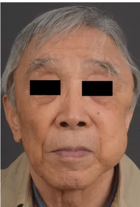
82세 남성이 좌측 관자 부위와 좌측 볼에 지속적으로 크기가 증가하는 2개의 종괴를 주소로 내원하였다. 관자 부위의 종괴는 2년 전, 볼의 종괴는 1년 전부터 인지하였 다고 한다(Fig. 1). 이학적 검사 상 두 종괴는 모두 부드럽 고 유동적이었다. 볼의 종괴는 약 10 × 8 cm, 관자 부위의 종괴는 약 5 × 5 cm 정도 크기였다. 환자 얼굴의 감각은 대칭적이고 온전하였으며, 안면신경마비의 증거는 보이 지 않았다. 전산화단층촬영 영상에서는 두 개의 종괴가 아닌, 방사선 투과성을 띤 하나의 종괴가 좌측 관자의 대한두경부종양학회지, 제36권 제2호, 2020. pp.69-72
Korean Journal of Head & Neck Oncology, Vol.36, No.2
https://doi.org/10.21593/kjhno/2020.36.2.69 ISSN 1229-5183(Print) / ISSN 2586-2553(Online)
증례 보고: 2개의 종괴를 모방하는 볼 지방 덩이에서 유래한 거대 지방종
김태운⋅김상화+
서울대학교병원 성형외과
Case Report: A Giant Lipoma from the Buccal Fat Pad Resembling Two Different Masses
Taewoon Kim, MD, Sang Wha Kim, MD, PhD+
Department of Plastic and Reconstructive Surgery, Seoul National University Hospital
= Abstract =
We report a surgically challenging case of a large lipoma located from the temple, across the zygomatic region to the buccal area, resembling two different masses.
An 82-year-old man presented with two persistently growing soft masses at his left temple and cheek. A com- puted tomographic scan revealed a single large radiolucent mass extending from the submuscular layer of the left temple crossing beneath the zygomatic arch to the buccal region, rather than two individual masses. Excision was performed through upper gingivobuccal and temporal incisions. The mass was dissected through both incisions, cut in half, and extracted from both sides.
No complications were observed. The biopsy result was consistent with a lipoma. Four months later, the scars were inconspicuous, and the patient was satisfied.
Considering the size, shape, and location, this is a rare and intriguing case. The bi-directional approach allowed for successful total excision without any complications, leaving inconspicuous scars.
Key Words : Lipoma⋅Neoplasms⋅Cheek
Received Revised Accepted
: August 25, 2020 : October 15, 2020 : October 19, 2020
+Corresponding author: Sang Wha Kim, M.D., Ph.D.
Department of plastic and reconstructive surgery, college of Medicine, Seoul National University, Seoul National University Hospital, 101 Daehak-ro, Jongno-gu, Seoul, 110-744, Seoul, Korea
Tel: +82-2-2072-2374, Fax: +82-2-3675-7792 E-mail: [email protected]
- 70 -
근육 밑층부터 시작하여 관골궁 아래를 지나며 볼의 근 육 밑층까지 이어진 것으로 드러났다(Fig. 2A, 2B, 2C).
신체 검진 소견 및 방사선 영상 상 방사선 투과성인 특성 이 지방종을 시사하였다.1)
종괴의 크기와 자라는 속도는 수술적 제거의 적응증에 합당하였고 환자 또한 제거를 강력히 희망하였다. 입안 절개가 계획되었고, 외부 절개도 필요할 수 있음을 사전 에 설명하고 환자의 동의를 구하였다.
수술은 전신 마취 하에서 진행되었다. 이하선관에 주 의하며 좌상측 잇몸볼 절개가 가해졌고, 저작근 아래로 박리하여 지방종의 볼 부분을 노출시켰다. 가능한 만큼 박리를 시행하고 추출을 시도하였으나, 종괴 전체를 빼 내기에는 불가능하여 관자 부위에 추가 절개를 가하였 다. 여기를 통해 관자근과 깊은관자근막 사이를 박리하 면서 종괴를 주변 조직과 분리시켰다. 세심한 박리 끝에 종괴를 반으로 가르고, 양측에서 추출하였다. 음압 배액 관을 2군데 모두 삽입하였고, 봉합을 실시하였다(Fig.
3A, 3B).
환자는 수술 후 4일째에 배액관 2개 모두 제거 후 퇴원
A B C
Fig. 2. Pre-operative computed tomographic scan. A pre-operative computed tomographic scan revealed a single large radio- lucent mass extending from the submuscular layer of the left temple, crossing the zygomatic region beneath the zygomatic arch and lateral to the lateral maxillary wall, to the submuscular layer of the buccal region, rather than two individual masses.
A
B
Fig. 3. Immediate post-operative photo. The immediate post-operative photo of the patient just after surgical excision. Figure 3B shows the extent of the gingivobuccal incision.
Fig. 1. Pre-operative photo. A pre-operative photo of the 82- year-old man who presented with two persistently growing large masses at the left temple and left cheek.
- 71 - 하였다. 혈종, 장액종, 벌어짐, 감염 등 합병증은 일절 관찰되지 않았고, 안면신경의 모든 가지는 온전하였다.
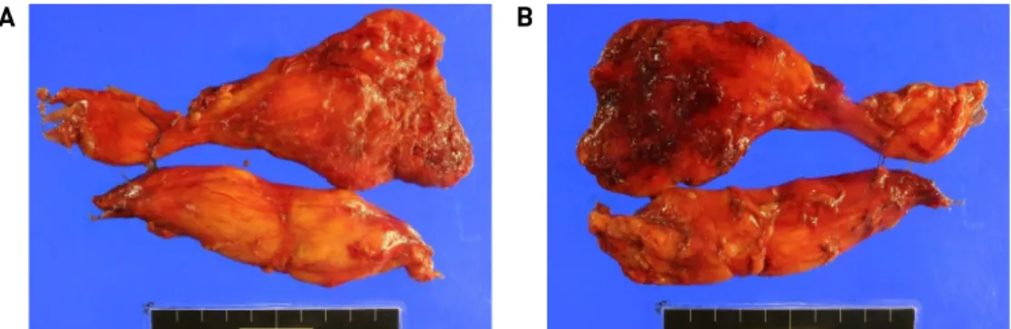
조직검사 결과 육종 성분이 없는 양성 지방종에 합당하 였고, 크기는 16.5 × 9.0 × 1.2 cm로 측정되었다(Fig. 4A, 4B). 4개월 경과 후 반흔은 눈에 거의 띄지 않았고, 환자 는 결과에 만족하였다(Fig. 5).
고찰
지방종은 대개의 경우 증상이 없으며, 경과관찰이 가 능하다. 수술적 절제의 적응증으로는 크기(직경 5 cm 이 상), 통증 혹은 불편감, 크기의 빠른 증가 속도, 암 등과의
감별 진단, 미용적 필요성 등이 있다3). 크기가 빠르게 증 가하는 경우에는 악성 변화가 있을 수 있어 제거를 요한 다. 본 증례에서의 지방종은 크기가 16.5 × 9.0 × 1.2 cm로 컸던 점, 그리고 2년 안에 빠르게 크게가 증가했다는 점 이 수술적 적응증에 해당하였다. 또한, 환자가 미용적인 이유로 절제를 원하여 방문했다는 점도 수술적 적응증이 되겠으며, 이것이 환자가 본과를 방문한 주된 이유임을 고려하여 저자들은 최대한 눈에 띄는 반흔을 남기지 않 는 접근 방법을 고민하였다.
지방종의 크기, 모양, 위치를 고려하였을 때 본 증례는 희귀하고 독특한 증례이다. 이 환자의 지방종의 위치는 볼 지방 덩이의 위치와 정확하게 일치하였다. 종괴의 앞 부분은 관골궁 아래에서 협근과 상악골 위로, 그리고 저 작근과 큰광대근 아래로 뻗어나갔다. 종괴의 관자 부분 은 관골궁 아래에서부터 위쪽으로 뻗어나가면서 깊은관 자근막과 관자근 사이의 층에 위치하였다4,5). 볼 지방 덩 이에서 유래한 지방종에 대한 문헌은 꽤 찾아볼 수 있었 으나, 본 증례에서와 같이 2개의 종괴처럼 나타났던 경우 는 드물었다. Kakudo et al.은 본 증례와 유사한 2개의 증례를 발표하였고, “Dumbbell-formed lipomas under the zygomatic arch”라 칭하였다6). 이 문헌에서 첫번째 증례 에서는 볼 부위와 관자 부위에 2개의 외부 절개를 가하여 제거하였고, 두번째 증례에서는 귀앞에서부터 아래턱밑 까지 이어지는 외부 절개를 이용하였다.
볼 지방 덩이에서 유래한 지방종의 제거를 위해 문헌 에서 외과의들은 관자 부위 절개, 아래턱밑 절개, 볼 절개 등 다양한 외부 절개를 이용하였다5-8). 다른 이들은 구강 내 절개를 이용하였다9-11). Fig. 3B에 나와있는 것처럼, 저자들의 구강내 절개는 안면부 골절 수술 시 이용하는 잇몸볼 절개에서 이하선관 출구 바로 옆까지 이어졌다.
구강 내 잇몸볼 절개를 이용할 시에는 이하선관 출구를 피하여 절개를 하고, 적절한 층을 찾아서 박리하는 것이 중요하다. 볼 지방덩이와 앞부분과 옆부분은 안면신경 A
B
Fig. 4. Specimen photo. The biopsy result was consistent with a lipoma without sarcoma component, and the size was measured 16.5 x 9.0 x 1.2 cm.
Fig. 5. 4-month post-operative photo. Four months after the operation, the patient’s scar was inconspicuous, and the pa- tient was satisfied with the results.
- 72 - 의 볼가지와 광대가지가 지나가므로, 이 부분에서의 박 리는 특별히 주의를 하며 진행해야 한다. 또한, 볼 지방덩 이의 볼 돌기(buccal process)를 지나는 이하선관 역시 염 두에 두고 박리해야 한다. 이러한 구조물들의 손상을 방 지하기 위해서는 시야가 잘 확보될 수 있도록 지혈을 해가며 진행하는 것이 좋다. 정상적인 볼지방덩이와 달 리 지방종은 특유의 매끈하고 반짝거리는 성질이 있어 적절한 층을 찾아 박리만 했다면 종괴는 쉽게 구분이 가능하다.
저자들이 알고 있는 한, 외부 절개와 구강내 절개를 동시에 사용한 것은 본 증례가 처음이다. 이러한 쌍방향 성 절개는 신경이나 혈관 손상 없이 눈에 띄지 않는 반흔 을 남기며 지방종을 완전히 절제할 수 있도록 해주었다.
저자들은 이러한 쌍방향성 절개가 앞으로 유사한 증례에 서 유용하게 사용될 수 있을 것이라고 생각한다.
References
1) Kransdorf MJ, Murphey MD. Imaging of Soft Tissue Tumors.
New York: Lippincott Williams & Wilkins; 2006. p. 81.
2) Kransdorf MJ. Benign soft-tissue tumors in a large referral pop- ulation: distribution of specific diagnoses by age, sex, and
location. Am J Roentgenol. 1995;164:395-402.
3) Boyer M, Monette S, Nhuyen A, Zipp T, Nimunkar AJ. A review of techniques and procedures for lipoma treatment. Clinical Dermatology. 2015;3:105-112.
4) Bruce D, Jackson IT, Halim A, Triplett WW, Ferreira M.
Anatomy of the Buccal Fat Pad and Its Clinical Significance.
Plast Reconstr Surg. 1989;83:257-262.
5) Chia CY, Rovaris DA, Fontana R. Giant lipoma of the buccal fat pad: case report and literature review. Revista Brasileira de cir- urgia plastica. 2016;31:112-117.
6) Kakudo N, Kusumoto K, Takemoto T, Tanaka Y, Kurokawa I, Ogawa Y. Dumbbell-formed lipomas under the zygomatic arch. J Plast Reconstr Aesthet Surg. 2008;61:107-110.
7) Trento GS, Stringhini DJ, Rebellato NLB, Scariot R. Extra-Oral Excision of a Buccal Fat Pad Lipoma. J Craniofac Surg. 2017;
28:e226-e227.
8) Kim JW, Kang SJ. Pediatric Lipoma of the Buccal Fat Pad. J Craniofac Surg. 2012;23:1934-1935.
9) de Wijn RS, van der Heijden EP, Kon M. On lipoma of the buccal fat pad: Report of two cases and review of the literature. J Plast Reconstr Aesthet Surg. 2009;62:28-35.
10) Brucoli M, Arcuri F, Borello G, Benech A. Surgical Technique of the Transoral Approach to Remove a Lipoma of the Buccal Fat Pad. J Craniofac Surg. 2011;22:2415-2418.
11) Arora BK. Large buccal fat pad lipoma: A rare case report. Int J Case Rep Imag. 2019;10:1-5.