• 검색 결과가 없습니다.

Rhabdoid tumor in the gluteal region of a Shit-tzu dog

N/A
N/A
Protected

Academic year: 2021

Share "Rhabdoid tumor in the gluteal region of a Shit-tzu dog"

Copied!
3
0
0

로드 중.... (전체 텍스트 보기)

전체 글

(1)

大韓獸醫學會誌

(2009)

49

卷第

4

號 Korean J Vet Res

(2009) 49(4) : 361~363

361

<증례보고>

Rhabdoid tumor in the gluteal region of a Shit-tzu dog

Jae-Yong Chung

1

, Sun-Hee Do

2,

*

1Daegu Polytechnic College University, Daegu 706-711, Korea

2College of Veterinary Medicine, Konkuk University, Seoul 143-701, Korea

(Accepted: December 1, 2009)

Abstract : Rhabdoid tumor is an aggressive neoplasm of animals and human. It is similar with rhabdomyosarcoma histopathologically. But cellular origin of this neoplasm showed no striated muscle origin by immunohistological and ultrastructural studies. Castrated male Shit-tzu dog, 6 years old, had a mass in the left gluteal region near to the tail. The mass was examined histopathologically and immunohistologically. Histopathologically, the tumor was consisted of large polygonal cell with abundant eosinophilic cytoplasm. The nuclei in some cells were marked eccentrically located. Immunohisto- chemically, many neoplastic cells were positive for vimentin. These findings were similar to histopathological and immunohistological features of human rhabdoid tumor and few rhabdoid tumors in animals.

Keywords : dog, malignant, rhabdoid tumor, soft tissue, vimentin

Introduction

Rhabdoid tumor (RT) is highly aggressive neoplasm of unknown cellular origin in human, usually occurring in the kidney and central nervous system of infants or children. RT is originally described as the variant of Wilms’ tumor during the evaluation of National Wilms’ Tumor Study results [2]. And it is primarily a kidney tumor that occurs mainly in children [1]. Later RT was reported in many other tissues including skin, heart, thymus, liver, soft tissue, central, and peripheral nervous system [6]. It was similar with rhabdomyo- sarcoma under the light microscope by histopatho- logically. But cellular origin of this neoplasm was revealed no striated muscle origin by immunohisto- logical and ultrastructural studies. The exact pathogenesis of RT is unknown. So it was called to “rhabdoid” [10].

In animals, RT was reported in the gastric wall of an aged orangutan [8], the brain of a dog [9], the orbit of a horse [4], and cutaneous tissue in a cat [5]. The diagnosis of RT is based on the characteristics of histopathological, ultrastructural, and immunohistoche- mical features [9]. Histopathologically, RT is typically composed of large polygonal to globoid eosinophilic

cells with globular intracytoplasmic inclusions. Nuclei are vesicular, often with marginated chromatin, and contain large distinct nucleoi. Ultrastructurally, the cytoplasmic inclusion is composed of discrete aggregates of intermediate filaments, arranged in whorls. And cytoplasmic inclusion which is aggregates of the intermediate filaments react consistently with vimentin antibody in immunohistochemistry. Less frequently, RT reacts to cytokeratin, epithelial membrane antigen, glial fibrillary acidic protein, or neuron-specific enolase, respectively by the cases and occurred region [4, 5, 8, 9].

Case report

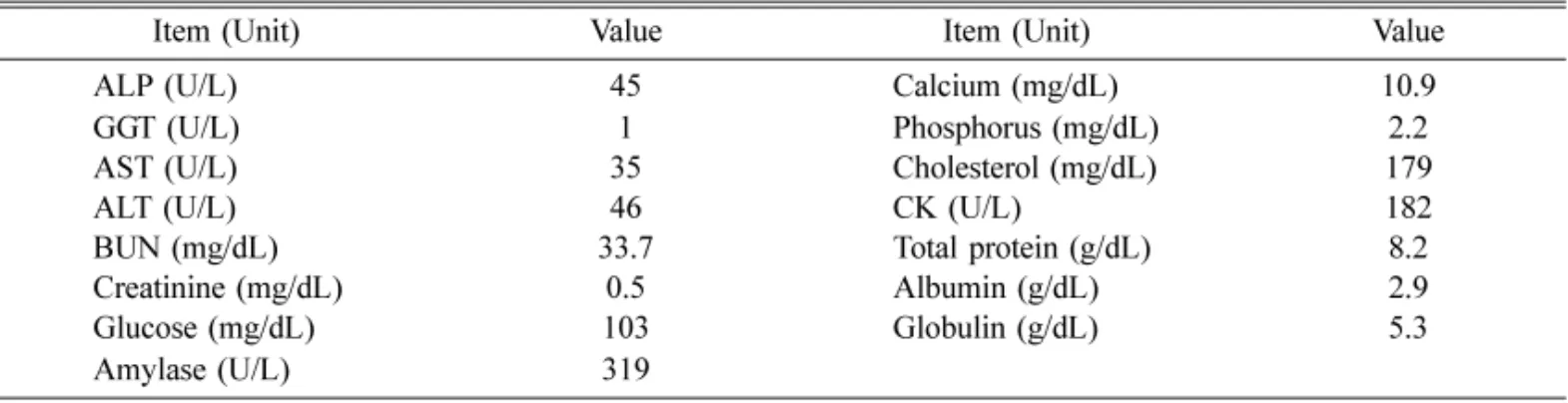
A six years old castrated male Shit-tzu dog brought to a local veterinary clinic with subcutaneous proliferative mass in the left gluteal region near to the tail. The mass was firm and poorly circumscribed with 4 × 3.5 × 4.5 cm in size. The excised gluteal mass was slightly lobulated and white to gray colored mucoid discharges on the cut section. The dog was vaccinated according to routine vaccination program and had no medical history relating the tumor. No abnormality was founded in physical and radiographic examination.

*Corresponding author: Sun-Hee Do

College of Veterinary Medicine, Konkuk University, Seoul 143-701, Korea

[Tel: +82-2-450-3706 Fax: +82-2-450-3037, E-mail: [email protected]]

(2)

362 Jae-Yong Chung, Sun-Hee Do

Complete blood count and serum chemistry results were within normal limits except blood urea nitrogen and phosphorus (Table 1). But they were slight elevation compared with reference range. A surgical excision was performed due to physical discomfort.

The specimen was examined histopathologically and immunohistologically. It was fixed with 10% neutral- buffered formalin, and paraffin-embedded tissues were sectioned at 4

µ

m-thicks and stained with hematoxylin and eosin. After heating, deparaffinization in xylene, rehydration in graded alcohols, blockage of endogenous- peroxidase. Mouse monoclonal antibodies against

α

- SMA (Dako, USA), vimentin (Dako, USA) and desmin (Dako, USA) were used as primary antibodies.

In the histopathological examination, the mass consisted of large polygonal to round cell with abundant cytoplasms showing high cellularity and vascularity (Fig. 1A). The neoplastic cells formed cellular sheet and/or nest infiltrated in adipose tissue.

Tumor cells had abundant eosinophilic cytoplasms and prominent round to oval vesicular nuclei as well as round hyaline-like inclusions and eccentric crescent- shaped nuclei in some of them (Fig. 1B). The neoplastic cells showed moderate pleomorphism. In addition, tumoric cells had positive immunoreactivity for vimentin (Fig. 1C) except

α

-SMA and desmin. The expression of vimentin was differential diagnostic method of other tumors which were similar to histopathological appearance. All the microscopic features were similar to the previous results of the RT occurred in human and animals.

Discussion

RT is a very aggressive and metastasized highly in human and animal cases. In aged orangutan case, large gastric masses were present around the cardia and in Table 1. Serum chemistry results of the case

Item (Unit) Value Item (Unit) Value

ALP (U/L) 45 Calcium (mg/dL) 10.9

GGT (U/L) 1 Phosphorus (mg/dL) 2.2

AST (U/L) 35 Cholesterol (mg/dL) 179

ALT (U/L) 46 CK (U/L) 182

BUN (mg/dL) 33.7 Total protein (g/dL) 8.2

Creatinine (mg/dL) 0.5 Albumin (g/dL) 2.9

Glucose (mg/dL) 103 Globulin (g/dL) 5.3

Amylase (U/L) 319

Fig. 1. (A) Neoplastic cells were admixed with adipose

tissue and collagenous stroma. (B) Tumoric cells revealed

characteristics of rhabdoid cells having eosinophilic

cytoplasmic inclusions and pleomorphic and large

eccentrically-placed nuclei with vesicular chromatin. (C)

Strong positive immunoreactivity for vimentin were

observed and some neoplastic cells exhibiting eccentric

nuclei and intracytoplasmic globular inclusions (arrows). A

(scale bar = 100

µ

m) and B (scale bar = 50

µ

m): H&E

stain, C (scale bar = 50

µ

m): ABC method.

(3)

Rhabdoid tumor in a dog 363 the corpus, which is metastasis to the liver, pancreas,

and right ovary [8]. RT that occurred in the horse was invasive as well. It appeared to take only approximately 3 months to affect the entire orbit and metastasize to the lymph nodes of the head and neck as well as the surrounding tissue [4]. In contrast to previous studies, our case revealed localized tumoric mass in subcutaneous gluteal region and no metastasis in the physical and radiographic examinations.

In human, renal RT and extra-renal RT containing chest wall, thymus, heart, liver, pelvis, uterus, vulva, prostate, urinary bladder, soft tissue, skin, and paravertebral region have been studied. Renal RT affect mainly in young age characterizing extremely poor prognosis whereas extra-renal RT occurred in wide ranged age [1, 3, 7]. Few RT reports in animals revealed various occurred age range such as the gastric RT of the old orangutan (34y), the brain tumor of the dog (1.5y), orbital RT in the horse (2y) and cutaneous RT in an old cat (13y), except in regard to originated regions of tumor [4, 5, 8, 9].

The neoplastic cells in RTs are consistently positive for vimentin and often co-express other marker of myogenic (

α

-smooth muscle actin, desmin), epithelial (cytokeratin, epithelial membrane antigen), and neuroe- ctodermal (neuron specific enolase, S-100 protein, glial fibrillary acidic protein, neurofilament) derivation [5].

Other markers except for vimentin were used to only identify the neoplastic origin. This suggested that immunoreaction for vimentin is the diagnostic clue for the RT.

It is considered that typical morphological features and vimentin immunoreactions were important differential diagnostic marker of RT in animals and human although there are only few reports of RT occurred in animals. Our results demonstrate that the histopathological and immunohistochemical features of canine RT are quite similar to human case. In addition, the current report may be important in identifying RT based on histopathological and immunohistological findings.

However, greater follow-up of larger numbers of cases are required to perform for prognosis and elucidate the relationship between RT in dogs and humans.

References

1. Beckwith JB. Wilms' tumour and other renal tumors of childhood: a selective review from the National Wilms' Tumor Study Pathology Center. Hum Pathol 1983, 14 , 481-492.

2. Beckwith JB, Palmer NF. Histopathology and prognosis of Wilms tumors: results from the first national Wilms’ tumor study. Cancer 1978, 41 , 1937- 1948.

3. Haas JE, Palmer NF, Weinberg AG, Beckwith JB.

Ultrastructure of malignant rhabdoid tumor of the kidney. A distinctive renal tumor of children. Hum Pathol 1981, 12 , 646-657.

4. Hong CB, Van Meter PW, Latimer CL. Malignant rhabdoid tumour in the orbit of a horse. J Comp Pathol 1999, 121 , 197-201.

5. Izawa T, Yamate J, Takeda S, Kumagai D, Kuwamura M. Cutaneous rhabdoid tumor in a cat. Vet Pathol 2008, 45 , 897-900.

6. Kodet R, Newton WA Jr, Sachs N, Hamoudi AB, Raney RB, Asmar L, Gehan EA. Rhabdoid tumors of soft tissues: a clinicopathologic study of 26 cases enrolled on the intergroup rhabdomyosarcoma study.

Hum Pathol 1991, 22 , 674-684.

7. Parham DM, Weeks DA, Beckwith JB. The clinicopathologic spectrum of putative extrarenal rhabdoid tumors. An analysis of 42 cases studied with immunohistochemistry or electron microscopy. Am J Surg Pathol 1994, 18 , 1010-1029.

8. Schauer G, Moll R, Walter JH, Rumpelt HJ, Göltenboth R. Malignant rhabdoid tumor in the gastric wall of an aged orangutan ( Pongo pygmaeus ). Vet Pathol 1994, 31 , 510-517.

9. Steele KE, Schulman FY, Mena H, Strimple EO.

Rhabdoid tumor in the brain of a dog. Vet Pathol 1997, 34 , 359-363.

10. Weeks DA, Malott RL, Zuppan CW, Liwnicz BH,

Beckwith JB. Primitive cerebral tumor with rhabdoid

features: a case of phenotypic rhabdoid tumor of the

central nervous system. Ultrastruct Pathol 1994, 18 , 23-

28.

수치

Fig. 1.  (A) Neoplastic cells were admixed with adipose tissue and collagenous stroma

참조

관련 문서

Purpose: Giant cell tumor of the tendon sheath are the most common tumors after ganglionic cysts in benign soft tissue tumors which could be recurred after surgical

Results : The expression of p21 was increased in boderline serous tumor and serous cystadenocarcinoma in contrast to benign serous tumors. The expression of

These results demonstrated that a positive role of Slug and Twist in tumor invasion in CRA, and inverse correlation between Twist and Slug expression

A retrospective analysis of invasive tumor percentages of GSS tumor samples showed that samples with high tumor content correlated well with FNAB samples

80) “Dogs at large : Injury to any person or damage to any property by a dog while at large shall be the full responsibility of the dog owner or person or persons responsible

– Any string of “only” terminals obtained by repeating step 2 is a Any string of only terminals obtained by repeating step 2 is a sentence in the language. “a dog runs

In in vivo experiments, axitinib significantly decreased tumor weight in xenograft models of drug-sensitive (A2780), and clear cell carcinoma (RMG1) and PDX models

Older LA-HNSCC patients had aggressive tumor characteris- tics and received less intensive treatment, which resulted in poor survival, especially in cases of oral cavity