Vol. 44, No. 3, September 2018, 249-258 http://dx.doi.org/10.15230/SCSK.2018.44.3.249
1)
† 주 저자 (e-mail: [email protected]) call: 053)420-4946
대기 미립자 물질 PM10에 노출된 인간 표피 각질형성세포의 염증 반응에 대한 레스베라트롤과 레스베라트릴 트라이아세테이트(RTA)의 영향
최 민 아⋅석 진 경⋅이 정 원⋅이 신 영*⋅김 영 미*⋅부 용 출†
경북대학교 의과대학 분자의학교실, 세포기질연구소
*(주)루비크라운
(2018년 6월 1일 접수, 2018년 7월 11일 수정, 2018년 8월 4일 채택)
Effects of Resveratrol and Resveratryl Triacetate on The Inflammatory Responses of Human Epidermal Keratinocytes Exposed to Airborne Particulate Matter PM10
Min A Choi, Jin Kyung Seok, Jeong-won Lee, Shin Young Lee*, Young Mi Kim*, and Yong Chool Boo†
Department of Molecular Medicine, CMRI, BK21 Plus KNU, School of Medicine, Kyungpook National University, 680 Gukchaebosang-ro, Jung-gu, Daegu, 41944, Korea
*Ruby Crown Co., Ltd., Dong-gu, Daegu, 41061, Korea
(Received June 14, 2018; Revised July 16, 2018; Accepted August 28; 2018)
요 약: 대기 오염은 피부의 산화적 손상, 염증 및 노화를 일으킬 수 있다. 레스베라트롤은 폴리페놀 화합물의 일종으로 항산화, 항염증, 멜라닌 생성 억제 작용 등 다양한 생물학적 활성이 있는 한편 열과 빛에 약한 단점이 있다. 레스베라트릴 트라이아세테이트(RTA)는 레스베라트롤에 비해 안정하고, 피부 안전성과 미백 효능이 보 고된 화장품 신소재이다. 본 연구의 목적은 직경 10 µm 미만 대기 미립자 물질(PM10)에 노출된 인간 표피 각질형성세포(HEK)의 염증 반응에 대한 레스베라트롤과 RTA의 영향을 조사하기 위한 것이다. 배양된 HEK세 포를 레스베라트롤과 RTA의 유무 조건에서 PM10에 노출시키고, 세포 생존율, 반응성 산소종(ROS)의 생성 및 염증성 사이토카인의 발현을 분석하였다. PM10을 처리하였을 때 세포 생존율이 감소하였고 종양괴사인자- α(TNF-α), 인터루킨-1β(IL-1β), 인터루킨-6(IL-6) 및 인터루킨-8(IL-8)의 발현이 증가하였다. 레 스베라트롤과 RTA는 PM10으로 유도된 세포의 사멸과 ROS 생성을 경감시켰다. PM10에 의해 증가되는 여러 염증성 사이토카인의 발현은 레스베라트롤과 RTA에 의해 경감되거나(IL-6), 증진되거나(IL-1β), 변화하지 않았다(TNF-α 및 IL-8). PM10에 의해 유도된 IL-6단백질의 발현이 레스베라트롤과 RTA에 의해 감소되 었다. 본 연구의 결과는 레스베라트롤과 RTA가 대기 미립자 물질에 노출된 피부의 세포 손상과 염증 반응을 조절하는 작용이 있음을 시사한다.
Abstract: Airborne pollution causes oxidative damage, inflammation, and premature aging of skin. Resveratrol is a polyphenol compound that has various biological activities such as antioxidant, anti-inflammation, and anti-melano- genic activities but it is unstable to heat and light. Resveratryl triacetate (RTA) is a new cosmetic ingredient that is more stable than resveratrol and its skin safety and whitening efficacy have been reported previously. The purpose of this study was to examine the effects of resveratrol and resveratryl triacetate (RTA) on the inflammatory responses of human epidermal keratinocytes (HEKs) exposed to airborne particulate matters with a diameter of < 10 µm
1. 서 론
최근 대기 오염의 대명사가 된 PM10은 직경 10 µm 미만의 미립자 물질로서 전이금속 및 다환 방향족 탄 화수소와 같은 광범위한 독성 물질로 구성되어 있다 [1,2]. PM10은 모공이나 피부의 손상 부위를 통해 침투 하여 아토피성 피부염, 여드름, 건선 등의 피부 질환을 유발하고 피부 노화를 촉진한다[3,4]. 이는 PM10이 반 응성 산소종(ROS)의 생성을 유발하고 종양괴사인자- α(TNF-α), 인터루킨-1β(IL-1β) 등의 염증성 사이토 카인의 발현을 증가시키기 때문으로 보아지고 있다 [5,6].
최근의 연구들은 식물 유래 폴리페놀이 PM10에 노 출된 세포의 산화 및 염증 반응을 감소시킬 수 있음을 시사하였다. 예를 들어 도시 대기 오염에 노출된 쥐에 초콜렛을 투여한 경우 TNF-α, IL-6 및 IL-1β의 발현 이 현저히 낮아졌다[7]. 그뿐만 아니라 우리 연구팀도 PM10에 노출된 인간 표피 각질형성세포에서 석류 (Punica granatum L.), 녹차(Camellia sinensis L.) 및 감 태(Ecklonia cava Kjellman)의 추출물 또는 폴리페놀 성 분이 ROS의 생성, 지질 과산화, 그리고 염증성 사이토 카인의 발현을 경감시킴을 보고한 바 있다[8-11].
레스베라트롤은 포도(Vitis vinifera L.)와 같은 여러 식물에 존재하는 폴리페놀 화합물이며 항산화, 항염 증, 항균, 항암 및 심장 보호 효과를 포함한 다양한 생 물학적 특성으로 주목받고 있다[11]. 레스베라트롤은 자외선 유발 피부 손상, 아토피성 피부염 및 여드름을 완화하고[12-15], 피부 노화 방지 및 피부 미백 효과를 나타낸다[16,17]. 이러한 여러 생물학적 활성에도 불구 하고 레스베라트롤을 화장품에 적용하는 데는 어려움 이 있는데, 그중 하나가 물리화학적으로 불안정성하여
쉽게 변색 변질된다는 점이다.
본 연구팀은 레스베라트롤의 안정성 문제를 해결하 기 위하여 레스베라트롤의 아세틸화 화합물인 RTA에 대한 연구해 오고 있다. RTA은 레스베라트롤에 비해 열과 빛에 더 안정하고, 세포 멜라닌 억제 작용과 세포 안전성 측면에서도 동등하며, 인체적용시험을 통해 피 부 안전성과 미백 효능이 보고된 화장품 신소재이다 [18-20]. RTA는 레스베라트롤의 프로드럭으로 작용하 며 항암 효과도 보고된 바 있다 [21].
본 연구의 목적은 PM10에 노출된 인간 표피 각질형 성세포(HEK)의 염증 반응에 대한 레스베라트롤과 RTA의 영향을 조사하는 것이다.
2. 재료 및 실험
2.1. 시약
대기 미립자 물질(PM10, 유럽 표준 물질 ERM- CZ120), 레스베라트롤, RTA 표준품 및 지질 다당류 (lipopolysaccharide, LPS)를 Sigma (St. Louis, USA)에서 구입하였다. 집 먼지 진드기(Dermatophagoides farina) 의 추출물(dust mite extract, DME)은 GREER (#b83, UK)에서 구입하였다.
2.2. RTA의 합성 및 분석
레스베라트롤로부터 RTA를 합성하기 위하여 유기 용매 사용 없이 무수초산과 소듐 아세테이트만을 사용 하는 아세틸화 방법을 이용하였다[22]. 레스베라트롤 228 mg, 무수초산 1 mL와 소듐 아세테이트 4 mg으로 구성된 반응 혼합물을 50 ℃에서 24 h 동안 교반하여 반응시켰다. 반응 혼합물을 물에 희석하여 침전된 반 (PM10). Cultured HEKs were exposed to PM10 in the absence or presence of resveratrol and RTA. Assays were under- taken to determine cell viability, the production of reactive oxygen species (ROS), and the expression of inflammatory cytokines. PM10 treatment decreased cell viability, and increased the expression of pro-inflammatory cytokines such as tumor necrosis factor-α (TNF-α), interleukin-1β (IL-1β), interleukin-6 (IL-6), and interleukin-8 (IL-8). Resveratrol and RTA reduced cell death and ROS production induced by PM10. PM10-induced mRNA expression of the in- flammatory cytokines was either attenuated (IL-6), or enhanced (IL-1β), or unaffected (TNF-α and IL-8) by resveratrol and RTA. PM10-induced IL-6 protein expression was attenuated by resveratrol and RTA. This study suggests that re- sveratrol and RTA have activities regulating cell damage and inflammatory responses of the skin exposed to airborne particulate matters.
Keywords: airborne particulate matters, resveratrol, resveratryl triacetate, keratinocytes, pro-inflammatory cytokines
응 생성물을 여과하여 얻고, 에탄올에서 재결정하여 RTA를 수득하였다.
고성능 액체 크로마토그래피(high performance liquid chromatography, HPLC) 분석은 자외선 가시광선 검출 기가 장착된 Gilson HPLC system (Gilson, Inc., USA)을 이용하여 수행하였다. 분석용 컬럼은 5 mm Hector-M C18 컬럼(4.6 mm × 250 mm, RS tech co., Korea)이었다.
이동상은 물(A)과 0.5% 포름산을 함유한 아세토나이 트릴(B)의 혼합액으로 50 min 동안 30% B에서 100%
B까지 증가시키는 선형 구배를 적용하였다. 유속은 0.6 mL/min으로 하였다. 검출기의 측정 파장은 280 nm 에 설정하였다. 반응 혼합물을 에탄올로 10,000배 희석 하고, 시린지 필터(0.2 µm, #CLS431219, Corning, Inc., USA)로 여과하여 제조한 샘플을 10 µL 주사하였다.
2.3. 세포 배양 및 약물 처리
성인의 피부 조직에서 추출한 HEK 세포(Invitrogen, USA)를 5% CO2함유 공기 하에서, 37 ℃의 습한 배양 기에서 유지하였다. EpiLife medium (Gibco BRL, USA) 에 10%의 EpiLife 지정 성장 첨가물(#S0125; Gibco BRL)과 항생제를 첨가하여 생육 배지로 사용하였다.
세포는 6-well plate에 7 × 10⁵ cells/well의 세포를 분주 하고 24 h 배양하여 부착화 및 안정화를 시행하였다.
이 세포에 LPS, DME, PM10을 농도별로 처리한 후 24 h 동안 더 배양하였다. 일부 실험에서는 세포에 레스베 라트롤, RTA또는 NAC를 전처리 후에 PM10을 100 µg/mL 농도로 처리하였다.
2.4. 세포 생존율 측정
세포 생존율은 (3-(4,5-dimethylthiazol-2-yl)-2,5-diphenylte- trazolium bromide (MTT)을 사용하여 측정하였다[8,9]. 세 포 안에 생성된 formazan을 dimethyl sulfoxide로 녹이 고, Spectrostar Nano microplate reader (BMG Labtech GmbH, Germany)을 이용하여 흡광도를 595 nm에서 측 정하였다.
2.5. ROS 생성 측정
ROS의 생성은 2’,7’-dichlorofluorescin diacetate (DCF-DA) 을 사용하여 측정하였다. 세포를 1.0 µM DCF-DA로 30 min간 사전 표지하고, PM10에 30 min간 노출시켰다.
세포를 1% sodium dodecyl sulfate를 함유한 완충액(20 mM Tris-Cl, 2.5 mM EDTA, pH 7.5)으로 용해한 후, 13,000 rpm에서 15 min간 원심분리하여 얻은 상등액의 형광 강도를 Gemini EM 형광 마이크로플레이트 판독 기(Molecular Devices, USA)를 사용하여 측정하였다(흡 수 파장은 485 nm, 방출 파장은 538 nm).
2.6. 정량적 역전사 중합 효소 연쇄 반응(Quantitative Reverse-transcriptase Polymerase Chain Reaction, qRT-PCR)
세포의 mRNA는 RNeasy 키트(Qiagen, Valencia, USA)로 추출하였고 cDNA 키트(Applied Biosystems, USA)를 사용하여 상보적 DNA (cDNA)를 제조하였다.
유전자-특이적 프라이머는 마크로젠(Korea)에서 구입 하였다. 이 연구에서 사용된 프라이머의 서열은 다음 과 같다 : TNF-α(TNF-α, NM_000594.3), 5’-TGC TCC TCA CCC ACA CCA T-3’ (forward)과 5’-GAG ATA GTC GGG CCG ATT GA-3’ (reverse); IL-1β (IL-1β, NM_000576.2), 5’-CCT GTC CTG CGT GTT GAA AGA-3’ (forward)과 5’-GGG AAC TGG GCA GAC TCA AA-3’(reverse); IL-6 (IL6, NM_001318095.1, NM_000600.4), 5’-AAG CCA GAG CTG TGC AGA TGA GT A-3’ (forward)과 5’-TGT CCT GCA GCC ACT GGT TC-3’ (reverse); IL-8 (IL8, NM_000584.3), 5’-CTG CGC CAA CAC AGA AAT TA-3’ (forward)과 5’-ACT TCT CCA CAA CCC TCT GC-3’ (reverse); glycer- aldehyde 3-phosphate dehydrogenase (GAPDH, NM_002046.3), 5’-ATG GGG AAG GTG AAG GTC G-3’ (forward)과 5’-GGG GTC ATT GAT GGC AAC AA-3’ (reverse). qRT-PCR 분석은 StepOnePlus Real-time PCR System (Applied Biosystems)으로 수행되 었다. 반응 혼합물(20 µL)은 SYBR Green PCR Master Mix (Applied Biosystems), cDNA (60 ng) 및 유전자 특 이적 프라이머 세트(2 pmol)로 구성된다. PCR 반응에 대한 열 순환 설정은 50 ℃에서 2 min, 95 ℃에서 10 min, 40 회 증폭(95 ℃에서 15 s, 60 ℃에서 1 min간), 및 해리 단계로 하였다. 각 유전자의 mRNA 수준은 내 부 대조군(GAPDH)와 비교하여 상대적인 역치 주기 방법을 사용하여 계산하였다.
2.7. 효소 면역 측정법(Enzyme-linked Immunosorbent Assay, ELISA)
HaCaT 각질형성세포주를 6-well plate에 7 × 10⁵ cells/well의 세포를 분주하고, 10%의 FBS를 첨가한 DMEM F12 배지 (#11320-033, Gibco BRL)에서 24 h 배
양하였다. 레스베라트롤, RTA 또는 NAC를 10 µM 농 도로 전처리하고 1 h 후 LPS (1 µg/mL) 또는 PM10 (100 µg/mL) 농도로 처리하였다. 48 h 후 세포 배양 배 지를 원심분리(1,200 rpm, 5 min)하고 상등액을 얻었다.
세포 배양 배지의 IL-6 단백질의 농도는 ELISA 키트
#1 #2 #3
Resveratrol (MW 228) 228 mg 228 mg -
RTA (MW 354) 354 mg
Acetic anhydride (MW 102) 1.0 mL 1.0 mL 1.0 mL
Sodium acetate (MW 82) - 4 mg -
Table 1. Compositions of Reaction Mixtures for the Synthesis of Resveratryl Triacetate (RTA)
Figure 1. Synthesis of RTA from resveratrol. The compositions of reaction mixtures are shown in Table 1. The reaction mixtures were maintained at 50 ℃ for 24 h. The samples of the reaction products were diluted 10,000 times with ethanol and subjected to HPLC analysis. The peaks of resveratrol and RTA were identified by comparing retention time with external standards (Sigma). The lower left picture shows the solvent (polyethylene glycol 400), and solutions of resveratrol and RTA (1% (w/w)), exposed to daily sunlight for 5 days at room temperature.
(Koma Biotech, Korea)를 사용하여 측정하였다. IL-6 항 체가 고정된 96-well plate의 각 웰에 세포 배양 배지 또 는 IL-6 단백질 표준 용액(100 µL)을 넣고 실온에서 2 h 동안 정치하였다. 각 웰을 세척한 후 바이오티닐화된 IL-6 항체 용액을 넣어 2 h 동안 정치하고, 이어서 horseradish peroxidase (HRP)-결합 스트렙타아비딘 용 액을 첨가하여 45 min 동안 정치하였다. 각 웰을 세척 한 후, 3,3’,5,5’-tetramethylbenzidine (TMB) 기질 용액을 첨가하여 반응을 개시하였다. 30 min 후 반응 정지 용 액을 첨가하고 450 nm에서 흡광도를 측정하였다. 표준 곡선으로부터 세포 배양 배지 중의 IL-6 단백질의 농 도를 계산하였다.
2.8. 통계 처리
본 연구의 모든 실험결과는 3회 이상 반복한 평균값 으로 나타내었다. 대조군과 실험군 사이의 통계학적 유의성 검정은 one-way ANOVA검정을 적용하였으며 p < 0.05 수준에서 유의성을 결정하였다.
3. 결 과
3.1. RTA의 합성과 분석
RTA를 합성할 때 기존 방법은 피리딘 용매에서 레 스베라트롤과 무수초산과 반응시키는 방법이 이용되 었다[18]. 본 연구에서는 유기 용매의 사용을 배제하는 새로운 방법을 이용하였다[22]. Table 1에 보인 바와 같
이 반응 혼합물 #1은 레스베라트롤과 무수초산으로 구 성하였고, 반응 혼합물 #2는 레스베라트롤과 무수초 산, 소듐 아세테이트로 구성하였으며, 반응 혼합물 #3 는 RTA와 무수초산으로 구성하였다.
이와 같이 서로 상이한 조성의 반응 혼합물을 50 ℃ 에 24 h 반응시킨 결과를 Figure 1에 보였다. 반응 혼합 물 #1은 큰 변화가 없었고, 반응 혼합물 #2 및 3은 투 명한 용액으로 되는 것이 관찰되었다. HPLC 분석을 통해 반응 혼합물 #1의 레스베라트롤은 대부분 남아 Figure 2. Effects of LPS, DME and airborne particulate
matters with a diameter of < 10 µm (PM10) on the viability of HEKs. The cells were exposed for 24 h to each material at the indicated concentrations (µg/mL). The cell viability was determined by using the MTT assay and data are expressed as percentages of control values. Means ± SEs, n = 3. *p < 0.05 vs. control.
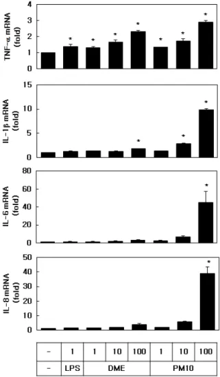
Figure 3. Effects of LPS, DME and PM10 on the expression of inflammatory cytokines in HEKs. The cells were exposed for 24 h to each material at the indicated concentrations (µg/mL). The mRNA levels of TNF-α, IL-1β, IL-6, and IL-8 were analyzed by qRT-PCR and normalized to that of GAPDH. Data are expressed as fold changes compared to control values. Means ± SEs, n = 3. *p < 0.05 vs. control.
있었고, 반응 혼합물 #3의 RTA는 그대로 남아 있었다.
반면 반응 혼합물 #2의 레스베라트롤은 RTA로 100%
전환되었음을 확인할 수 있었다.
레스베라트롤과 RTA의 용액 중 안정성을 비교하기 위하여 이들을 1% (w/w) 의 농도로 polyethylene glycol 400에 녹이고 5일간 햇빛에 노출시켰다. Figure 1의 좌 측 하단 그림에서 보듯이, 처음에 무색투명하였던 레 스베라트롤 용액이 햇빛 노출에 의해 짙은 주황색으로 변화하였다. 반면 RTA용액은 무색투명한 상태가 유지 되었다. 따라서 RTA가 레스베리트롤보다 태양광에 대 한 안정성이 우수한 것으로 확인되었다.
3.2. PM10의 염증 유발 효과
LPS, DME및 PM10이 HEK 세포의 생존율 및 염증 반응에 미치는 영향을 비교하였다. Figure 2에 도시된 바와 같이, LPS (1 µg/mL), DME (10-100 µg/mL) 및 PM10 (100 µg/mL)은 농도에 따라 세포 생존율을 감소 시켰다.
Figure 3에 보인 바와 같이 이들 물질들은 모두 1 µg/mL에서 TNF-α의 mRNA발현을 증가시켰다. PM10 은 같은 농도의 DME보다 더 강력하게 TNF-α, IL-1β, IL-6, IL-8과 같은 염증성 사이토카인의 mRNA 발현을 증가시켰다.
Figure 4. Effects of resveratrol, RTA, and NAC on the viability and the production of ROS of HEKs exposed to PM10. For viability test, cells were exposed to PM10 (100 µg/mL) for 24 h in the presence of resveratrol (RES), RTA or NAC at the indicated concentrations (µM), or vehicle only (control). NAC was used as a positive control. The cell viability was determined by the MTT assay. For ROS assay, cells were pretreated with RES, RTA or NAC at the indicated concentrations (µM). The cells were labeled with DCF-DA and exposed to PM10 (100 µg/mL) for 30 min. The dye was extracted from the cells and quantified by measuring fluorescence intensity. Data are expressed as percentages relative to control values. Means ± SEs, n = 3. *p < 0.05 vs. PM10 group.
Figure 5. Effects of resveratrol, RTA, and NAC on the mRNA expression of inflammatory cytokines of HEKs exposed to PM10. The cells were exposed to PM10 (100 µg/mL) for 24 h in the absence or presence of RES, RTA or NAC at the indicated concentrations (µM). The mRNA levels of TNF-α, IL-1β, IL-6, and IL-8 were analyzed by qRT-PCR and normalized to that of GAPDH. Data are expressed as percentages compared to the PM10 group. Means ± SEs, n = 3.
*p < 0.05 vs. PM10 group.
3.3. PM10에 노출된 세포의 생존율과 ROS 생성에 대한 레스베라트롤과 RTA의 영향
PM10 (100 µg/mL)에 노출된 HEK세포의 생존율과 ROS 생성에 대한 레스베라트롤과 RTA의 영향을 조사 했다. Figure 4에서 알 수 있듯이, 레스베라트롤(1-10 µM)과 RTA (10 µM)는 PM10 노출 조건 하에서 세포 생 존율을 회복시켰다. 레스베라트롤(10 µM)과 RTA (10 µM)는 또한 PM10이 유발한 ROS 생성을 감소시켰다.
NAC (10 µM)는 ROS 생성을 감소시켰지만, PM10 노출 조건 하에서는 세포 생존율에는 영향을 미치지 않았다.
3.4. PM10에 노출된 세포의 사이토카인 mRNA 발현에 대 한 레스베라트롤과 RTA의 영향
PM10 (100 µg/mL)에 노출된 HEK세포에서 TNF-α,
IL-1β, IL-6, IL-8 등의 염증성 사이토카인의 mRNA 발 현에 대한 레스베라트롤과 RTA의 효과를 조사했다.
Figure 5에서 볼 수 있듯이, PM10에 노출된 조건에서 레 스베라트롤(10 µM)과 RTA (10 µM) 모두 IL-6의 mRNA 발현을 감소시키는 반면, IL-1β의 발현을 증가시켰다.
TNF-α와 IL-8을 포함한 다른 사이토카인의 mRNA 발 현의 변화는 통계적으로 유의하지 않았다. 비교물질로 사용된 NAC (10 µM)는 PM10 노출 조건 하에서 사이 토카인의 발현에 큰 영향을 미치지 않았다.
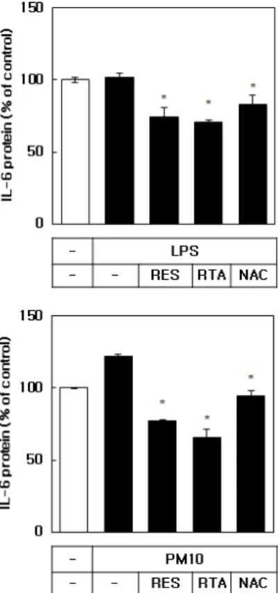
3.5. IL-6 단백질 발현에 대한 레스베라트롤과 RTA의 영향 LPS 및 PM10에 노출된 HaCaT 각질형성세포주에서 IL-6 단백질 발현에 대한 레스베라트롤과 RTA의 효과 를 조사했다. 세포 배양 배지의 IL-6 단백질 수준을 ELISA 방법으로 측정하였다. Figure 6에 보인 바와 같 이 IL-6의 단백질 발현이 LPS (1 µg/mL)에 의해서는 영향을 받지 않았고, PM10 (100 µg/mL)에 의해 증가하 였다. 레스베라트롤과 RTA는 PM10 처리 조건하에서 IL-6 단백질 발현을 PM10 처리하지 않은 대조군 수준 이하로 억제하였다. 비교물질로 사용된 NAC (10 µM) 도 IL-6 단백질의 발현을 유의하게 억제하였지만 레스 베라트롤과 RTA보다 효과가 미약하였다.
4. 고 찰
지속적인 대기 오염의 증가가 사람의 피부에 해로운 영향을 미친다는 직접적인 증거들이 제시되고 있다 [23,24]. 따라서, 1) 대기 오염 자체를 줄이고, 2) 대기 오염에 노출되는 빈도를 줄이며, 3) 대기 오염으로 인 해 야기된 피부 손상을 줄이기 위한 다양한 전략들이 필요하다.
본 연구의 결과는 레스베라트롤과 RTA가 PM10에 의한 세포 독성 및 ROS 생성을 완화시키는 데 효과적 이라는 것을 보여주었다. 레스베라트롤을 비롯한 다양 한 폴리페놀 화합물들은 ROS를 효과적으로 제거하고, 산화 스트레스와 관련된 여러 유전자의 발현을 조절한 다[25,26]. 레스베라트롤은 특히 대사 조절, 스트레스 반응, 노화 과정에 관여하는 sirtuins (NAD+ 의존성 단 백질 deacetylases /deacylases)의 활성화제로 주목받고 있다[27]. 따라서 레스베라트롤은 ROS 소거와 세포 신 Figure 6. Effects of resveratrol, RTA, and NAC on the IL-6
protein level expressed by HaCaT keratinocytes. The cells were exposed to LPS (1 µg/mL) or PM10 (100 µg/mL) for 48 h in the absence or presence of RES, RTA or NAC at 10 µM. The IL-6 protein level in the conditioned medium was analyzed by ELISA.
Data are expressed as percentages compared to the control values.
Means ± SEs, n = 3. *p < 0.05 vs. LPS or PM10 group.
호 전달 조절을 통해 PM10에 의한 세포 독성을 완화 시키는 것으로 사료된다. RTA는 세포 안의 에스터라 제 효소에 의해 레스베라트롤로 전환된 후 생물학적 활성을 보이는 것으로 사료된다[9].
본 연구팀의 선행연구에서 punicalagin, (-)-epi- gallocatechin gallate, dieckol 등의 폴리페놀 화합물이 PM10에 의해 유도된 TNF-α, IL-1β, IL-6, IL-8 등의 염증성 사이토카인의 발현을 모두 경감하였다[8-11].
그런데 본 연구에서 레스베라트롤과 RTA는 여러 사이 토카인 중 IL-6의 발현을 선택적으로 억제하였고, 다른 사이토카인의 발현에는 영향이 없거나 오히려 증가시 켰다. IL-6은 염증 및 자가 면역 과정을 자극하는 다양 한 생리 활성의 사이토카인이다[32]. 본 연구에서 레스 베라트롤과 RTA는 PM10에 의해 유도되는 IL-6의 mRNA 및 단백질 발현을 현저하게 억제시켰다. PM10 에 의한 세포 사멸을 억제하는 레스베라트롤과 RTA의 방어 작용이 IL-6의 발현 조절과 직접적인 관련성이 있는지에 대해서는 후속 연구가 필요하다.
여러 선행 연구들에서도 레스베라트롤은 세포의 종 류에 따라 특정 사이토카인의 발현을 감소시키거나 혹 은 반대로 증가시켰다[28]. 레스베라트롤은 세포 환경 에 따라 염증을 억제하거나 혹은 반대로 촉진하였으며 [29, 30], 세포 자살을 억제하거나 혹은 반대로 촉진하 였다[31]. 따라서 세포의 종류 및 세포 환경 등에 따라 서 레스베라트롤 및 RTA가 사이토카인의 발현을 조절 하는 방식이 다를 것이라 사료된다.
특정 사이토카인의 발현에 관여하는 특정 전사인자 의 활성은 다양한 수용체와 단백질 인산화 효소들이 관여하는 세포내 신호 전달 과정의 조절을 통해 증진 또는 억제가 가능하다. Aryl hydrocarbon receptor (AhR) 가 대기 미세 먼지의 독성 발현과 관련된 유전자 발현 을 조절한다고 알려져 있다[33]. 레스베라트롤은 AhR 길항제로서 AhR-의존적 유전자 발현에 영향을 미칠 수 있다[34,35]. 반면에 레스베라트롤이 간접적으로 AhR의 활성화를 야기할 수도 있다[36]. 현재의 정보로 는 레스베라트롤과 RTA가 어떤 구체적인 메커니즘을 통해 여러 사이토카인의 발현을 억제 또는 증진하는지 를 설명하기 어렵지만, 그 조절의 종합적인 결과가 세 포의 생존을 증진하는 방향으로 나타났다는 점이 중요 하다고 사료된다.
결론적으로, 본 연구에서 PM10이 DME 보다 더 강
하게 인간 표피 각질형성세포의 사멸을 유발하고 염증 반응을 일으킴을 보여 주었으며, 레스베라트롤과 RTA 가 PM10에 노출된 세포의 생존율을 회복시키고, ROS 생성을 경감시킬 뿐만 아니라 염증성 사이토카인의 발 현을 조절함을 보여 주었다. 따라서 레스베라트롤과 RTA가 대기 미립자에 의해 유도되는 피부 세포 손상 과 염증 반응을 조절하는 작용이 있다고 제안된다. 본 연구는 또한 유해한 유기 용매를 사용하지 않고 레스 베라트롤로부터 RTA를 합성하는 효율적인 방법을 제 시하였다. 그리고 RTA는 레스베라트롤에 비해 태양광 에 대한 우수한 안정성을 보여주었다. 따라서 레스베 라트롤과 RTA의 생물학적 활성은 동등하지만 장기간 보관하면서 사용하는 화장품에 적용하는 데는 안정성 이 우수한 RTA가 더 유리하다고 제안된다.
Acknowledgement
이 연구는 대한민국 중소벤처기업부의 창업성장기 술개발사업의 지원을 받았다(No S2486152).
Reference
1. J. D. Sacks, L. W. Stanek, T. J. Luben, D. O. Johns, B. J. Buckley, J. S. Brown, and M. Ross, Particulate matter-induced health effects: who is susceptible?, Environ. Health Perspect., 119(4), 446 (2012).
2. J. O. Anderson, J. G. Thundiyil, and A. Stolbach, Clearing the air: a review of the effects of particulate matter air pollution on human health, J. Med. Toxicol.,
8(2), 166 (2012).
3. K. E. Kim, D. Cho, and H. J. Park, Air pollution and skin diseases: Adverse effects of airborne particulate matter on various skin diseases, Life Sci., 152(1), 126 (2016).
4. A. Vierkotter, T. Schikowski, U. Ranft, D. Sugiri, M.
Matsui, U. Kramer, and J. Krutmann, Airborne particle exposure and extrinsic skin aging, J. Invest. Dermatol.,
130(12), 2719 (2010).
5. R. Bengalli, E. Molteni, E. Longhin, M. Refsnes, M.
Camatini, and M. Gualtieri, Release of IL-1 beta trig- gered by Milan summer PM10: molecular pathways in-
volved in the cytokine release, Biomed. Res. Int.,
2013(1), 158093 (2013).
6. D. Y. Cho, W. Le, D. T. Bravo, P. H. Hwang, B.
Illek, H. Fischer, and J. V. Nayak, Air pollutants cause release of hydrogen peroxide and interleukin-8 in a hu- man primary nasal tissue culture model, Int. Forum.
Allergy Rhinol., 4(12), 966 (2014).
7. R. Villarreal-Calderon, W. Reed, J. Palacios-Moreno, S. Keefe, L. Herritt, D. Brooks, R. Torres-Jardon, and L. Calderon-Garciduenas, Urban air pollution produces up-regulation of myocardial inflammatory genes and dark chocolate provides cardioprotection, Exp. Toxicol.
Pathol., 64(4), 297 (2012).
8. J. K. Seok, J. W. Lee, Y. M. Kim, and Y. C. Boo, Punicalagin and (-)-epigallocatechin-3-gallate rescue cell viability and attenuate inflammatory responses of human epidermal keratinocytes exposed to airborne particulate matter PM10, Skin Pharmacol. Physiol.,
31(3), 134 (2018).
9. J. W. Lee, J. K. Seok, and Y. C. Boo, Ecklonia cava extract and dieckol attenuate cellular lipid peroxidation in keratinocytes exposed to PM10, Evid.-Based Complementary Altern. Med., 2018(1), 8248323 (2018).
10. S. Park, J. K. Seok, J. Y. Kwak, H. J. Suh, Y. M.
Kim, and Y. C. Boo, Anti-inflammatory effects of pomegranate peel extract in THP-1 cells exposed to particulate matter PM10, Evid.-Based Complementary Altern. Med., 2016(1), 6836080 (2016).
11. A. Rauf, M. Imran, H. A. R. Suleria, B. Ahmad, D.
G. Peters, and M. S. Mubarak, A comprehensive re- view of the health perspectives of resveratrol, Food Funct., 8(12), 4284 (2017).
12. R. Yutani, R. Teraoka, and S. Kitagawa, Microemulsion using polyoxyethylene sorbitan tri- oleate and its usage for skin delivery of resveratrol to protect skin against UV-induced damage, Chem.
Pharm. Bull., 63(9), 741 (2015).
13. G. Fabbrocini, S. Staibano, G. De Rosa, V.
Battimiello, N. Fardella, G. Ilardi, M. I. La Rotonda, A. Longobardi, M. Mazzella, M. Siano, F. Pastore, V.
De Vita, M. L. Vecchione, and F. Ayala, Resveratrol-containing gel for the treatment of acne vulgaris: a single-blind, vehicle-controlled, pilot study, Am. J. Clin. Dermatol., 12(2), 133 (2011).
14. V. Karuppagounder, S. Arumugam, R. A.
Thandavarayan, V. Pitchaimani, R. Sreedhar, R.
Afrin, M. Harima, H. Suzuki, M. Nomoto, S.
Miyashita, K. Suzuki, and K. Watanabe, Resveratrol attenuates HMGB1 signaling and inflammation in house dust mite-induced atopic dermatitis in mice, Int. Immunopharmacol., 23(2), 617 (2014).
15. S. Caglayan Sozmen, M. Karaman, S. Cilaker Micili, S. Isik, Z. Arikan Ayyildiz, A. Bagriyanik, N.
Uzuner, and O. Karaman, Resveratrol ameliorates 2,4-dinitrofluorobenzene-induced atopic dermati- tis-like lesions through effects on the epithelium, PeerJ, 4(1), e1889 (2016).
16. R. A. Baxter, Anti-aging properties of resveratrol: re- view and report of a potent new antioxidant skin care formulation, J. Cosmet. Dermatol., 7(1), 2 (2008).
17. S. H. Kwon, H. R. Choi, Y. A. Kang, and K. C.
Park, Depigmenting effect of resveratrol is dependent on FOXO3a activation without SIRT1 activation, Int.
J. Mol. Sci., 18(6), 1213 (2017).
18. J. Park, J. H. Park, H. J. Suh, I. C. Lee, J. Koh, and Y. C. Boo, Effects of resveratrol, oxyresveratrol, and their acetylated derivatives on cellular melanogenesis, Arch. Dermatol. Res., 306(5), 475 (2014).
19. J. H. Ryu, J. K. Seok, S. M. An, J. H. Baek, J. S.
Koh, and Y. C. Boo, A study of the human skin-whitening effects of resveratryl triacetate, Arch.
Dermatol. Res., 307(3), 239 (2015).
20. Y. C. Boo, Clinical evaluation of skin whitening ef- fect of a cream containing resveratryl triacetate, Fragrance J. Korea, 2016(3), 72 (2016).
21. T. C. Hsieh, Y. C. Huang, and J. M. Wu, Control of prostate cell growth, DNA damage and repair and gene expression by resveratrol analogues, in vitro, Carcinogenesis., 32(1), 93 (2011).
22. M. M. Mojtahedi and S. Samadian, Efficient and rap- id solvent-free acetylation of alcohols, phenols, and
thiols using catalytic amounts of sodium acetate trihy- drate, J. Chem., 2013(1), 642479 (2013).
23. P. Puri, S. K. Nandar, S. Kathuria, and V. Ramesh, Effects of air pollution on the skin: A review, Indian J. Dermatol. Venereol. Leprol., 83(4), 415 (2017).
24. S. E. Mancebo and S. Q. Wang, Recognizing the im- pact of ambient air pollution on skin health, J. Eur.
Acad. Dermatol. Venereol., 29(12), 2326 (2015).
25. A. Carreras, M. L. Mateos-Martin, A.
Velazquez-Palenzuela, E. Brillas, S. Sanchez-Tena, M. Cascante, L. Julia, and J. L. Torres, Punicalagin and catechins contain polyphenolic substructures that influence cell viability and can be monitored by radi- cal chemosensors sensitive to electron transfer, J.
Agric. Food Chem., 60(7), 1659 (2012).
26. J. Joven, V. Micol, A. Segura-Carretero, C.
Alonso-Villaverde, J. A. Menendez, and P. Bioactive, Food components, polyphenols and the modulation of gene expression pathways: can we eat our way out of the danger of chronic disease?, Crit. Rev. Food Sci.
Nutr., 54(8), 985 (2014).
27. M. Gertz, T. T. N. Giang, F. Fischer, B. Suenkel, C.
Schlicker, B. Franzel, J. Tomaschewski, F. Aladini, C. Becker, D. Wolters, and C. Steegborn, A molec- ular mechanism for direct sirtuin activation by resver- atrol, PLoS One, 7(11), e49761 (2012).
28. J. Schwager, N. Richard, F. Widmer, and D.
Raederstorff, Resveratrol distinctively modulates the inflammatory profiles of immune and endothelial cells, BMC Complement. Altern. Med., 17(1), 309 (2017).
29. A. Cignarella, C. Minici, C. Bolego, C. Pinna, P.
Sanvito, R. M. Gaion, and L. Puglisi, Potential pro-inflammatory action of resveratrol in vascular smooth muscle cells from normal and diabetic rats, Nutr. Metab. Cardiovasc. Dis., 16(5), 322 (2006).
30. J. Walker, K. Schueller, L. M. Schaefer, M. Pignitter,
L. Esefelder, and V. Somoza, Resveratrol and its me- tabolites inhibit pro-inflammatory effects of lip- opolysaccharides in U-937 macrophages in plas- ma-representative concentrations, Food Funct., 5(1), 74 (2014).
31. M. Baarine, S. J. Thandapilly, X. L. Louis, F. Mazue, L. Yu, D. Delmas, T. Netticadan, G. Lizard, and N.
Latruffe, Pro-apoptotic versus anti-apoptotic proper- ties of dietary resveratrol on tumoral and normal car- diac cells, Genes Nutr., 6(2), 161 (2011).
32. J. Scheller, A. Chalaris, D. Schmidt-Arras, and S.
Rose-John, The pro- and anti-inflammatory properties of the cytokine interleukin-6, Biochim Biophys Acta.,
1813(5), 878 (2011).
33. T. Hidaka, E. Ogawa, E. H. Kobayashi, T. Suzuki, R.
Funayama, T. Nagashima, T. Fujimura, S. Aiba, K.
Nakayama, R. Okuyama, and M. Yamamoto, The aryl hydrocarbon receptor AhR links atopic dermatitis and air pollution via induction of the neurotrophic factor artemin, Nat. Immunol., 18(1), 64 (2017).
34. R. F. Casper, M. Quesne, I. M. Rogers, T. Shirota, A. Jolivet, E. Milgrom, and J. F. Savouret, Resveratrol has antagonist activity on the aryl hydro- carbon receptor: implications for prevention of dioxin toxicity, Mol. Pharmacol., 56(4), 784 (1999).
35. S. R. Beedanagari, I. Bebenek, P. Bui, and O.
Hankinson, Resveratrol inhibits dioxin-induced ex- pression of human CYP1A1 and CYP1B1 by inhibit- ing recruitment of the aryl hydrocarbon receptor com- plex and RNA polymerase II to the regulatory regions of the corresponding genes, Toxicol. Sci., 110(1), 61 (2009).
36. A. Mohammadi-Bardbori, J. Bengtsson, U. Rannug, A. Rannug, and E. Wincent, Quercetin, resveratrol, and curcumin are indirect activators of the aryl hy- drocarbon receptor (AHR), Chem. Res, Toxicol.,