http://dx.doi.org/10.4047/jkap.2012.50.1.21
ORIGINAL ARTICLE서론
치과용 골내 임플란트를 이용한 치료는 부분 또는 완전무치 악 환자의 구강기능 회복을 위한 예지성 높은 치료방법으로 인정받고 있다.1,2기능적이며 심미적으로 장기간 임플란트 치 료의 성공을 위해서는 여러 가지 요소가 관여하는데,3그 중에 서도 특히 임플란트 변연골을 보존하고 주변 연조직을 건강하 게 유지하는 것이 중요하다.4
초기 변연골 흡수의 원인으로 임플란트 수술 시 골막거상, 골삭제, 과도한 응력, 세균 침입 및 생물학적 폭경의 침범 등을 들 수 있다.5수술 시 시행하는 골막거상은 혈액공급에 영향을 줄 수 있지만, 망상골이 풍부한 경우에는 피질골 흡수에 미치 는 영향은 크지 않다.5임플란트 식립 드릴링으로 인한 외상이 초기 변연골 상실의 원인이라고 주장하기도 하지만,62차 수술 시 이전보다 골이 증가한 경우를 종종 확인할 수 있다.5임플란 트 주위골에 과하중이 전달될 경우 변연골 흡수가 야기되지 만,7,8비교적 약한 하중 또는 점진적인 하중은 오히려 골흡수를
막는다고 보고되고 있다.9-12세균은 자연치에서는 주변골상실 의 근본적인 원인이지만, 임플란트의 경우에서는 보조적인 인 자에 국한되며 초기 변연골 흡수의 원인으로 보기는 어렵다.13-15 임플란트에서도 골유착을 보호하기 위한 최소한의 연조직 즉, 생물학적 폭경이 형성될 때까지 변연골 흡수가 일어난다 고 밝혀진바 있으며,16임플란트에서 생물학적 폭경이 주변조 직에 미치는 영향에 대한 많은 연구가 있어 왔다.16-20임플란트 의 생물학적 폭경은 전체적으로는 안정적으로 존재하지만 시 간의 경과에 따라 접합상피 부착은 증가하고 결합조직 부착은 감소하는 특징을 보인다.19,20생물학적 폭경은 수직적 길이 외에 도 수평적 공간을 포함하여 인접한 임플란트 사이 거리가 3 mm 이하일 때는 생물학적 폭경을 확보하기 위해 골흡수가 발 생한다고 보고된 바 있다.21특히 변연골의 보존을 위해서는 생 물학적 폭경을 이루는 구성요소 중 접합상피보다는 결합조직 부착이 더욱 중요하다.22
연조직과 경조직의 경계에 위치하는 임플란트 경부의 디자 인은 연조직 반응과 변연골 보존에 영향을 미친다.17,18,22플랫폼
*교신저자: 전전영영찬찬
626-870 경상남도 양산시 물금읍 범어리 부산대학교 치의학전문대학원 치과보철학교실 055-360-5130: e-mail, [email protected] 원고접수일: 2011년 12월 23일 / 원고최종수정일: 2012년 1월 2일 / 원고채택일: 2012년 1월 5일
임플란트의 collar design이 변연골과 연조직에 미치는 영향
유현상∙강선녀∙정창모∙윤미정∙허중보∙전영찬*
부산대학교 치의학전문대학원 치과보철학교실
연구 목적: 임플란트 경부가 잘 설계된 경우 양호한 연조직 반응을 통해 변연골을 보존하는데 도움이 된다. 본 실험에서는 연, 경조직 경계부에 가장 가까이 위치하는 임플 란트의 collar design이 변연골 변화와 연조직 반응에 미치는 영향을 동물실험을 통해 알아보고자 하였다.
연구 재료 및 방법: 2마리의 건강한 Beagle dog에 임플란트 collar design만 다른 두 종류의 임플란트(Neobiotech Co. Seoul, Korea)를 식립하였다. Collar에 bevel 을 부여한 군(Bevel 군)과 "S"자 형태를 부여한 군(Bioseal 군)으로 나누어 마리 당 7개, 군당 7개, 총 14개의 임플란트를 무작위로 식립한 후 Healing abutment를 즉시 체결하였다. 디지털 표준구내 방사선사진을 이용해 4주 간격으로 총 12주간 근원심 변연골 변화를 관찰하였고, 12주에 희생하여 조직학적 분석을 통해 협설 변연골 흡수 및 임플란트 주변 연조직 반응 을 평가하였다. Mann-Whitney test를 통해 동일한 방사선 사진 촬영 시점에서 근원심 변연골 변화량 및 조직계측치를 군 간 비교하였고, Kruskal-Wallis test를 통해 방사선 사진 상 근원심 변연골 변화량이 시간에 따른 차이가 있는지 군 내 분석 한 후 Duncan test를 통해 사후 검증하였다(α=.05).
결과: 방사선학적 분석 결과 각 촬영 시점에서 두 군간 근원심 변연골 변화량의 차이를 보이지 않았다(P>.05). 군 내에서 시간에 따른 근원심 변연골의 흡수량을 분석한 결 과 Bevel 군에서는 시간에 따른 차이를 보이지 않았으나 (P>.05), Bioseal 군에서는 시간에 따른 차이를 보였으며, 4주 및 8주와 비교했을 때 12주에서 변연골의 증가를 보였다 (P<.05). 조직학적 분석 결과 협설측 변연골 흡수량에서 두 군간 차이를 보이지 않았으나 (P>.05), Bevel 군에 비해 Bioseal 군에서 더 견고한 결합조직부착을 관찰할 수 있었으 며, 생물학적 폭경의 값은 두 집단 간 차이를 보이지 않은 반면에 (P>.05), 접합상피부착은 Bevel 군에서 유의하게 길었고, 결합조직부착은 Bioseal 군에서 더 길게 나타났다 (P<.05).
결론:Bevel 군에 비해 Bioseal 군에서 결합조직부착은 길게 형성된 반면에 접합상피부착은 더 짧게 나타났으며, 생물학적 폭경과 초기 변연골 흡수에는 차이가 없음을 알 수 있었다. 연조직 반응의 차이가 실제 기능하중 하에서 변연골 변화에 미치는 영향에 대해서 향후 연구가 필요할 것으로 생각된다. (대한치과보철학회지 2012;50:21-8) 주요단어: Collar design; 변연골흡수; 생물학적 폭경; 결합조직부착; 접합상피부착; Bioseal
*이 논문은 부산대학교 자유과제 학술연구비 (2년)에 의하여 연구되었음.
스위칭은 식립된 임플란트의 플랫폼보다 작은 직경의 지대주 를 연결하는 술식으로서,23,24염증세포 침윤대를 내측으로 이동 시킬 뿐만 아니라 변연골에 가해지는 응력을 감소시켜 변연골 흡수를 감소시킬 수 있는 장점이 있다.25-27또한 임플란트 연조 직 관통부위의 오목한 디자인이 결합조직의 두께 증가와 견고 한 부착을 가능하게 하며,22임플란트 경부의 표면 roughness나 microthread 설계가 변연골 흡수를 감소시킨다는 보고가 있다.28,29 근자에는 이러한 보고들을 근거로 임플란트 경부가 디자인된 제품들이 임상에 많이 사용되고 있다.28-30
하지만 임플란트 경부 디자인에 관한 기존 연구들은 비교한 임플란트의 제조사가 다르거나, 크기뿐만 아니라 경부 이외에 다른 부분의 디자인에서도 어느 정도 차이가 존재하기 때문에 경부 디자인만의 영향을 파악하는 데는 다소의 제한점이 있
다.17,22,28-31또한 임플란트 경부 디자인 측면에 대한 연구에서도28,29
연, 경조직 경계부에 가장 가까운 임플란트 collar design에 관한 연구는 미흡한 편이다.
이에 본 연구에서는 임플란트 제조사, 크기 및 전체적인 디 자인이 동일하지만 collar 부위에 단순한 bevel 형태 또는 결합조 직 부착길이의 증가를 위한“S”자 형태를 갖는 임플란트를 제 작, 이용하여, 임플란트의 collar design이 변연골 변화와 연조직 반응에 미치는 영향을 동물실험을 통해 알아보고자 하였다.
연구 재료 및 방법 1. 연구 재료
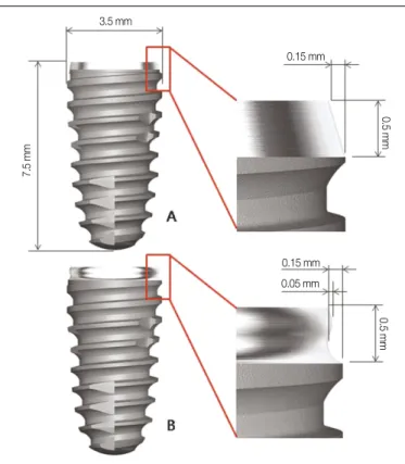
실험용 임플란트(Neobiotech Co., Seoul, Korea)는 두 가지의 collar 디자인을 가지고 있으며, collar 부위에 bevel만을 가지고 있 는 집단을 Bevel 군, “S”자 형태를 가지고 있는 집단을 Bioseal 군 으로 나누었다(Table 1). 나머지 부위는 internal cone 연결부를 가 지는 tapered-screw 형태로 동일하고 두 군 모두에서 collar 부위는 machined 표면이나 나머지 부위는 resorbable blasting media (RBM) 표면처리(Ra = 1.2 - 1.5) 되었으며 자세한 수치 및 형태는 Fig. 1과 같다.
2. 연구 방법
1) 실험 동물의 발치
본 연구에서는 2마리의 건강한 비글견(평균 2세, 평균 15 kg) 이 사용되었으며, 전남대학교 수의과대학 동물실험 윤리위원 회의 승인 하에 진행했다. 수술실은 소독상태를 유지하였으며 실험동물의 발치는 2% isoflurane (Isoflurane�, Choongwae Co.
Seoul, Korea)과 oxygen으로 흡입진정마취 하에 시행했으며 Lactated Ringer’s solution을 발치 종료 시까지 5 ml/kg/h 속도로 투여하였고 1:100,000 epinephrine이 포함된 2% lidocaine HCL (Yu- Han Co., Gunpo, Korea)을 이용하여 하악 편측에 1.8 ml씩 점막에 추가적인 국소침윤마취를 하였다. 양측의 하악 소구치 및 제 1
대구치를 발거한 후, 4-0 흡수성 봉합사(Vicryl�, Ethicon, Somerville, NJ, USA)로 봉합하였다.
2) 임플란트 식립(Fig. 2)
약 4주간 치유 후 발치와 동일한 방법으로 실험동물을 진정 마취 후 국소마취 하였다. 무치악 치조정 절개로 협설측 전층 판막을 박리하고 치조정을 평탄하게 하여 임플란트를 식립하 기 위한 골폭을 확보한 후, 3 mm 이상의 임플란트 사이 간격을 두어 마리 당 7개씩 무작위 배정 식립으로 총 14개의 임플란트 를 식립하였다. 임플란트 수술용 엔진(NSK Surgic XT, NSK, Tochigi-ken, Japan)의 출력토크를 30 Ncm로 설정하였으며, 엔진으 로 식립이 완료되지 않은 임플란트는 수동토크렌치(Neobiotech Co., Seoul, Korea)를 이용하여 collar-thread간의 경계부위가 치조정 과 일치되도록 육안으로 확인하면서 식립하였다. Healing abut- ment (ISH404, Neobiotech Co., Seoul, Korea)를 즉시 체결하고 4-0 흡 수성 봉합사(Vicryl�, Ethicon, Somerville, NJ, USA)로 봉합하였다.
Fig. 1. Design of implant fixtures used in this study: A, Bevel group; B, Bioseal group.
Table 1. Implant fixtures used in this study
Group N Description Length Diameter
(mm) (mm)
Bevel 7 bevel and machined surface 7.5 3.5 Bioseal 7 "S"-shaped and machined surface 7.5 3.5
3.5 mm
7.5 mm
0.15 mm
0.5 mm0.5 mm
0.15 mm 0.05 mm
3) 수술 후 관리
실험 동물의 발치 및 임플란트 식립 직후와 48시간 후 peni- cillin G procaine과 penicillin G benzathine (Deasung microbiological labs.
co., Seoul, Korea)을 근육 내 주사하였다(1 ml/5 kg). 술 후 1주간 2%
chlorhexidine 을 10 cc 시린지에 넣어 하루에 2회 구강 내 소독을 하고 4주 간격으로 스케일링을 하였으며 유동식 사료를 공급 하였다.
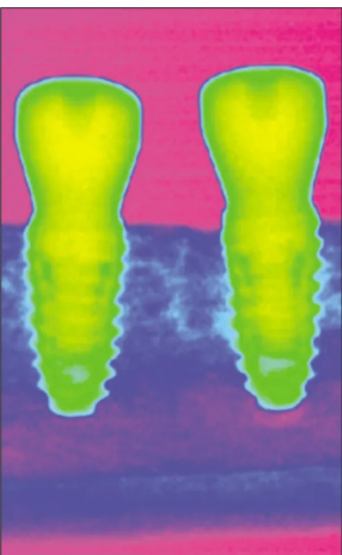
4) 방사선 계측
임플란트 식립 직후, 4주, 8주 그리고 12주에 진정마취 하에서 portable X-ray (Port II, Genoray Co., Sungnam, Korea)를 이용해 평행 촬영법으로 총 4회 방사선 사진을 촬영하였다. Digital X-ray soft- ware (CDX-View, PointNix, Seoul, Korea)를 이용하여 gray scale image를 color coding 한 후(Fig. 3), 임플란트 장경과 변연골 수준 (임플란트 플랫폼에서 변연골 최상방까지의 거리)을 근원심측 에서 각각 측정한 후, 실제 임플란트의 장경(7.5 mm)과 비교하 여 다음과 같이 변연골 흡수량을 계산하였다.
변연골 수준 (mm) =
방사선 사진 상 변연골 수준 (mm)
× 7.5 (mm) 방사선 사진 상 임플란트 장경 (mm)
변연골 흡수량 (mm) =
촬영 시 변연골 수준 (mm) - 수술 시 변연골 수준 (mm)
5) 조직시편 제작 및 계측
임플란트 식립 후 12주에 pentobarbital sodium (Entobar, Hanlim Pharm., Seoul, Korea)을 과량 주사하여 희생시키고 하악골을 절단 하여 블록을 채취하였다. 채취된 블록은 중성완충포르말린 (Sigma Aldrich, St. Louis, MO, USA)에 2주 동안 고정하고 알코올 의 농도를 순차적으로 높여 탈수한 후 Technovit 7200 레진 (Heraeus KULZER, South bend, IN, USA)에 포매하였다. 포매된 시 편은 임플란트의 중심부의 장축을 따라 EXAKT diamond cutter (KULZER EXAKT 300, EXAKT, Norderstedt, Germany)로 협설 방 Fig. 2. Surgical procedures. A: Flattening edentulous ridge, B: Drilling, C: After
fixation.
Fig. 3. Gray scale color coding for radiographic analysis.
A
C
B
향으로 약 400 μm 두께로 절단한 후 EXAKT grinding machine (KULZER EXAKT 400CS, EXAKT, Norderstedt, Germany)으로 연 마하여 30 μm 두께의 시편을 제작하였다. 시편은 hematoxylin &
eosin 염색 후 CCD camera (Polaroid DMC2, Polaroid Co., MA, USA) 가 부착된 광학현미경(Olympus BX, Olympus Optical Co., Tokyo, Japan)으로 ×12.5, ×50의 디지털영상을 얻어 저장한 후 SPOT Software V4.0 (Diagnostic Instrument Inc., Sterling Heights, MI, USA) 을이용하여협설측에다음과같은계측점을선정하였다(Fig. 4).
PM: marginal portion of mucosa aS: apical extension of sulcus
aJE: apical portion of junctional epithelium fBIC: first bone-implant contact
계측점을 이용하여 다음을 구하였다(Fig. 4).
JE: length of junctional epithelium (aS-aJE, mm) CT: length of connective tissue (aJE-fBIC, mm) BW: biologic width (PM-fBIC, mm)
임플란트 collar-thread간의 경계부위에서 fBIC (first bone-implant contact)까지의 길이를 협설측에서 측정하여 변연골 흡수량을 구하였다.
6) 통계 분석
본 연구는 SPSS (Ver. 17.0, SPSS, Chicago, IL, USA)를 사용하여 통계 처리하였다. 실험 결과에 대한 정규성 검정을 실시한 결 과 정규성을 만족하지 않아 비모수적 방법으로 분석하였다.
Mann-Whitney test를 통해 동일한 방사선 사진 촬영 시점에서 근원심 변연골 변화량 및 조직계측치를 군 간 비교하였고, Kruskal-Wallis test를 통해 방사선 사진 상 근원심 변연골 변화량
이 시간에 따른 차이가 있는지 군 내 분석 한 후 Duncan test를 통 해 사후 검증하였다. 유의 수준 5%에서 검정하였다.
결과
1. 임플란트 주위 변연골 변화
방사선 촬영을 통해 근원심 변연골 흡수량을 계산한 결과는 Table 2와 같으며, 각 촬영 시점에서 두 군간 차이를 보이지 않았 다(P>.05). 각군의대표적인방사선사진을Figs. 5, 6에나타내었다.
군 내에서 시간에 따른 근원심 변연골의 흡수량을 분석한 결 과 Bevel 군에서는 시간에 따른 차이를 보이지 않았으나(P>.05), Bioseal 군에서는 시간에 따른 차이를 보였으며, 4주 및 8주와 비 교했을 때 12주에서 변연골 증가를 보였다(P<.05) (Table 2).
Fig. 4. Histometric measurement. PM, marginal portion of mucosa; aS, apical exten- sion of sulcus; aJE, apical portion of junctional epithelium, fBIC, first bone-implant contact; JE, length of junctional epithelium (aS/aJE); CT, length of connective tis- sue (aJE/fBIC); BW, biologic width (PM/fBIC).
Table 2. Mesiodistal marginal bone resorption in radiographic analysis Group Marginal bone resorption (mm)
Week 4 Week 8 Week 12
Bevel 0.26 ± 0.29aA 0.34 ± 0.33aA 0.14 ± 0.31aA Bioseal 0.45 ± 0.30aB 0.36 ± 0.30aB 0.07 ± 0.26aA Different lowercase letters in the same column indicate significant differences between Bevel and Bioseal at the same time using Mann-Whitney test (P<.05).
Different uppercase letters in the same row indicate significant differences according to time using Kruskal-Wallis test and Duncan post-hoc test (P<.05).
Fig. 5. Radiographic analysis on Bevel group. The white arrows indicate mesiodis- tal marginal bone level.
Fig. 6. Radiographic analysis on Bioseal group. The white arrows indicate mesiodistal marginal bone level.
Week 4
Week 0 Week 8 Week 12
Week 4
Week 0 Week 8 Week 12
조직학적 분석을 통해 희생 시의 협설측 변연골 흡수량을 비 교한 결과를 Table 3에 나타내었으며, 두 군간 차이를 보이지 않 았다(P>.05).
2. 임플란트 주위 연조직 반응
조직시편을 관찰한 결과 Bevel 군에 비해 Bioseal 군에서 더 견 고한 결합조직부착을 관찰할 수 있었으며(Fig. 7), Bevel 군에서 는 2개의 시편에서 연조직이 임플란트에서 탈락되었으나, Bioseal 군에서는 연조직 부착이 탈락된 시편은 없었다(Fig. 8).
조직계측학적 분석 결과를 Table 4에 나타내었다. 접합상피부 착 (JE)과 결합조직부착(CT)에서 두 집단 간 통계적으로 차이를 보였으나(P<.05), 생물학적 폭경(BW)의 값은 두 집단 간 차이를 보이지 않았다(P>.05). 접합상피부착은 Bevel 군에서 유의하게 길었고, 결합조직부착은 Bioseal 군에서 더 길게 나타났다.
Table 4. Result of histometric analysis for soft tissue Group Histometric measurement (mm)
JE CT BW
Bevel 1.25 ± 0.19 1.09 ± 0.31 3.33 ± 0.34 Bioseal 0.84 ± 0.27 1.54 ± 0.20 2.83 ± 0.23
P 0.015 0.004 0.065
Mann-Whitney test was used to assess differences between Bevel and Bioseal. JE, length of junctional epithelium ; CT, length of connective tissue; BW, biologic width.
Table 3. Buccolingual marginal bone resorption in histometric analysis Group Marginal bone resorption (mm)
Buccal Lingual
Bevel 0.91 ± 0.17 0.25 ± 0.23
Bioseal 0.77 ± 0.20 0.23 ± 0.16
P 0.132 0.937
Mann-Whitney test was used to assess differences between Bevel and Bioseal.
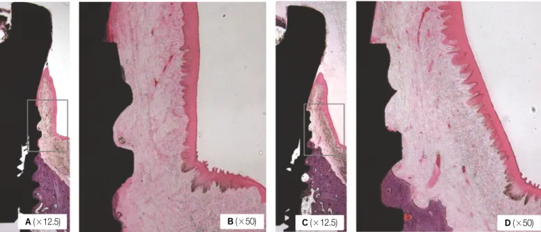
Fig. 7. Histological analysis of two groups. A: Bevel group. Hematoxylin & eosin staining, B: Detail of (A), C: Bioseal group. Hematoxilin & eosin staining, D: Detail of (C). The connective tissue of Bioseal group was more firm and dense. In addition, Bioseal group had more connective volume.
Fig. 8. Soft tissue detachment observed in Bevel group only. In Bioseal group, all specimens had tight soft tissue attachment.
A(×12.5) B(×50) C(×12.5) D(×50)
×50
고찰
본 연구에서는 임플란트 변연골의 변화 및 연조직 반응을 관 찰하기 위하여 방사선학적 분석 및 조직학적 분석을 시행하였 다. 본 연구에서 이용한 표준구내방사선사진의 경우 경조직 관찰은 용이한 반면 연조직은 관찰이 어려우며, 주기적인 촬 영을 통해 시간에 따른 변연골 변화를 관찰하는데 용이한 장 점을 가지지만 관찰 부위가 임플란트 근원심부위에 한정되어 실제 골흡수가 많은 협측피질골 부위를 평가할 수 없는 한계 를 지니고 있다.32이를 보완하기 위해 임플란트 식립 후 12주에 실험 동물을 희생시켜 협설방향으로 절단된 조직시편을 제작 한 후 광학현미경을 통해 협설측 변연골 및 주변 연조직을 관 찰하여 계측, 분석하였다.
임플란트의 식립 깊이는 임플란트 collar에서 연조직 부착을 유도하기 위해 collar-thread 경계부위가 치조정과 일치되도록 하였다. 그러나 collar-thread 경계부위가 치조정에 일치되도록 식립하더라도 육안으로 확인하기 어려운 오차가 존재할 수 있 으므로, 식립 직후 방사선사진을 촬영하여 변연골 수준을 구 하였고, 4주마다 측정한 변연골 수준과 비교하여 골변화량을 계산하였다.
방사선학적 분석을 통해 각 촬영시점에서 두 군의 근원심 변 연골 변화량을 비교한 결과 통계적으로 유의한 차이를 보이지 않았다. 조직계측학적 분석을 통해 협측과 설측의 변연골 흡 수량을 비교한 결과 Bevel 군에서 변연골 흡수량이 다소 많았지 만 두 군간 통계적으로 유의한 차이를 보이지 않았으며 이는 실험 동물을 희생시킨 시기에 해당하는 12주의 방사선학적 분 석 결과와 상응한다. 근원심 변연골 변화량을 군내 분석한 결 과 Bevel 군에서는 4주에서 변연골 흡수가 관찰된 이후 변연골 변화량이 시간에 따른 차이를 보이지 않은 반면에, Bioseal 군에 서는 4주에서 변연골 흡수가 관찰된 이후 12주에 변연골이 회 복되어 처음 식립 시와 비교하였을 때 단지 0.07 mm 의 변연골 흡수를 보였다. 따라서 임플란트 collar의“S”자 형태가 변연골 이 조기에 회복이 되는데 영향을 주는 것으로 보여진다. 다만 통계적으로 유의하지 않지만 4주의 변연골 흡수량이 Bevel 군 에 비해 Bioseal 군에서 다소 많았으며 향후 그 원인에 대한 추가 적인 연구가 필요하다고 생각된다.
각 군의 조직시편을 관찰하여 연조직을 비교한 결과 Bioseal 군에서 더욱 긴밀한 결합조직부착을 보였으며, 일부 Bevel 군 시편에서는 접합상피 및 결합조직이 임플란트 표면에서 탈락 되어 있었다. 조직시편 제작과정에서 연조직이 탈락할 수도 있지만, Bioseal 군에서는 탈락된 시편이 발견되지 않은 점을 고려하였을 때 Bioseal 군에서 좀 더 긴밀한 접합상피 및 결합조 직부착을 보였다고 생각된다.
연조직에 대한 조직계측학적 분석 결과 Bioseal 군에서 결합 조직부착은 길게 형성된 반면에 접합상피부착은 짧았다. 비글 견의 경우 임플란트에 부착하는 결합조직의 길이는 1 - 2 mm로
알려져 있는데,22Bioseal 군에서는 약 1.5 mm의 결합조직부착 을 보였고, 이는 1 mm의 값을 보인 Bevel 군에 비해 길었다. 따라 서 임플란트 collar에“S”자 형태를 부여하는 것이 더 긴 결합조 직부착을 유도한다고 생각되며, 곡면의 입체적인 외형을 고려 하였을 때 결합조직의 체적이 증가하는 효과도 함께 가진다고 여겨진다. 생물학적 폭경의 값은 Bevel 군에서 다소 길게 나타 났으나 통계적 유의성은 보이지 않았으며, 이는 임플란트 디 자인이 생물학적 폭경에 영향을 주지 않는다는 이전의 연구들 과 같은 경향을 보인다.22,33비록 통계학적으로 유의하지는 않지 만 Bioseal 군에서 생물학적 폭경의 값이 약 0.5 mm 짧은 것은 임 플란트 collar에“S”자 형태를 부여한 것이 플랫폼 스위칭 효과 를 보이기 때문이라고 생각된다. 임플란트에서의 생물학적 폭 경은 수직, 수평적인 공간을 모두 포함하는데,21“S”자 형태의 함요부가 0.15 mm 깊이로 환형으로 부여되어 있는 것을 고려한 다면 약 0.3 mm 정도의 플랫폼 스위칭 효과를 보인다고 볼 수 있어 이를 고려할 경우 두 군 사이의 생물학적 폭경의 값의 차 이는 더욱 줄어든다.
Bioseal 군에서 접합상피부착이 Bevel 군에 비해 짧은 것은 수 술 후 tissue remodeling 과정 동안 결합조직이 조기에 안정화 되어 상피조직의 하방이동을 막았기 때문이라고 생각된다. 상피조 직이 하방으로 이동할 경우 변연골 흡수를 일으킬 가능성이 커지므로 이를 막는 것은 변연골 보존에 중요하다.18,34방사선학 적 분석 결과 Bioseal 군에서 4주 까지 초기 변연골 흡수를 보인 후 지속적으로 변연골이 회복된다는 점을 비추어 보았을 때 결합조직의 조기 안정과 견고한 부착이 접합상피의 하방이동 과 변연골 흡수를 막는다고 미루어 볼 수 있다.
이번 연구를 통해 임플란트 collar에“S”자 형태를 부여하는 것이 bevel 만 부여한 것에 비해 양호한 연조직 반응을 보였음 을 알 수 있었다. 반면 실제 골흡수 양상에서는 차이를 보이지 않았으므로 연조직 반응의 차이가 실제 변연골 변화에 있어 차이를 보이는지 알아보기에는 다소 한계가 있었다. 임플란트 의 collar design에 따른 연조직 반응의 차이가 실제 기능하중 하 에서 변연골 변화에 미치는 영향에 대해서 향후 연구가 필요 할 것으로 생각된다.
결론
본 연구를 통하여 Bevel 군에 비해 Bioseal 군에서 결합조직부 착은 길게 형성된 반면에 접합상피부착은 더 짧게 나타났으 며, 생물학적 폭경과 초기 변연골 흡수에는 차이가 없음을 알 수 있었다.
참고문헌
1. Adell R, Lekholm U, Rockler B, Bra�nemark PI. A 15-year study of osseointegrated implants in the treatment of the eden- tulous jaw. Int J Oral Surg 1981;10:387-416.
2. Bra�nemark PI. Osseointegration and its experimental back- ground. J Prosthet Dent 1983;50:399-410.
3. Linkow LI, Rinaldi AW, Weiss WW Jr, Smith GH. Factors influencing long-term implant success. J Prosthet Dent 1990;63:64-73.
4. Quirynen M, Naert I, van Steenberghe D. Fixture design and over- load influence marginal bone loss and fixture success in the Bra�nemark system. Clin Oral Implants Res 1992;3:104-11.
5. Misch CE. Stress factors: influence on treatment. In: Misch CE. Dental Implant Prosthetics. St. Louis, Mosby; 2005. p.
71-90.
6. Haider R, Watzek G, Plenk H. Effects of drill cooling and bone structure on IMZ implant fixation. Int J Oral Maxillofac Implants 1993;8:83-91.
7. Isidor F. Loss of osseointegration caused by occlusal load of oral implants. A clinical and radiographic study in monkeys. Clin Oral Implants Res 1996;7:143-52.
8. Duyck J, R�nold HJ, Van Oosterwyck H, Naert I, Vander Sloten J, Ellingsen JE. The influence of static and dynamic loading on marginal bone reactions around osseointegrated im- plants: an animal experimental study. Clin Oral Implants Res 2001;12:207-18.
9. Piattelli A, Ruggeri A, Franchi M, Romasco N, Trisi P. An histologic and histomorphometric study of bone reactions to un- loaded and loaded non-submerged single implants in mon- keys: a pilot study. J Oral Implantol 1993;19:314-20.
10. Miyata T, Kobayashi Y, Araki H, Motomura Y, Shin K. The in- fluence of controlled occlusal overload on peri-implant tissue: a histologic study in monkeys. Int J Oral Maxillofac Implants 1998;13:677-83.
11. Miyata T, Kobayashi Y, Araki H, Ohto T, Shin K. The influence of controlled occlusal overload on peri-implant tissue. Part 3: A histologic study in monkeys. Int J Oral Maxillofac Implants 2000;15:425-31.
12. Miyata T, Kobayashi Y, Araki H, Ohto T, Shin K. The influence of controlled occlusal overload on peri-implant tissue. part 4: a histologic study in monkeys. Int J Oral Maxillofac Implants 2002;17:384-90.
13. Lekholm U, Ericsson I, Adell R, Slots J. The condition of the soft tissues at tooth and fixture abutments supporting fixed bridges.
A microbiological and histological study. J Clin Periodontol 1986;13:558-62.
14. Adell R, Lekholm U, Rockler B, Bra�nemark PI, Lindhe J, Eriksson B, Sbordone L. Marginal tissue reactions at osseointegrated titanium fixtures (I). A 3-year longitudinal prospective study. Int J Oral Maxillofac Surg 1986;15:39-52.
15. Rams TE, Roberts TW, Tatum H Jr, Keyes PH. The subgingival microbial flora associated with human dental implants. J Prosthet Dent 1984;51:529-34.
16. Berglundh T, Lindhe J. Dimension of the periimplant mucosa.
Biological width revisited. J Clin Periodontol 1996;23:971-3.
17. Bae EK, Chung MK, Cha IH, Han DH. Marginal tissue response to different implant neck design. J Korean Acad Prosthodont 2008;46:602-9.
18. Hermann F, Lerner H, Palti A. Factors influencing the preservation of the periimplant marginal bone. Implant Dent 2007;16:165-75.
19. Cochran DL, Hermann JS, Schenk RK, Higginbottom FL,
Buser D. Biologic width around titanium implants. A histometric analysis of the implanto-gingival junction around unloaded and loaded nonsubmerged implants in the canine mandible. J Periodontol 1997;68:186-98.
20. Hermann JS, Buser D, Schenk RK, Higginbottom FL, Cochran DL. Biologic width around titanium implants. A physiologically formed and stable dimension over time. Clin Oral Implants Res 2000;11:1-11.
21. Tarnow DP, Cho SC, Wallace SS. The effect of inter-implant dis- tance on the height of inter-implant bone crest. J Periodontol 2000;71:546-9.
22. Kim S, Oh KC, Han DH, Heo SJ, Ryu IC, Kwon JH, Han CH. Influence of transmucosal designs of three one-piece implant systems on early tissue responses: a histometric study in beagle dogs. Int J Oral Maxillofac Implants 2010;25:309-14.
23. Gardner DM. Platform switching as a means to achieving implant esthetics. N Y State Dent J 2005;71:34-7.
24. Lazzara RJ, Porter SS. Platform switching: a new concept in im- plant dentistry for controlling postrestorative crestal bone levels.
Int J Periodontics Restorative Dent 2006;26:9-17.
25. Maeda Y, Miura J, Taki I, Sogo M. Biomechanical analysis on platform switching: is there any biomechanical rationale? Clin Oral Implants Res 2007;18:581-4.
26. Quaresma SE, Cury PR, Sendyk WR, Sendyk C. A finite element analysis of two different dental implants: stress distribution in the prosthesis, abutment, implant, and supporting bone. J Oral Implantol 2008;34:1-6.
27. Kim DY, Kim TI, Seol YJ, Lee YM. Influence of platform switching on crestal bone resorption. J Korean Acad Periodontol 2008;38:135-42.
28. Lee SY, Piao CM, Koak JY, Kim SK, Kim YS, Ku Y, Rhyu IC, Han CH, Heo SJ. A 3-year prospective radiographic evaluation of marginal bone level around different implant systems. J Oral Rehabil 2010;37:538-44.
29. Yun HJ, Park JC, Yun JH, Jung UW, Kim CS, Choi SH, Cho KS.
A short-term clinical study of marginal bone level change around microthreaded and platform-switched implants. J Periodontal Implant Sci 2011;41:211-7.
30. de Almeida FD, Carvalho AC, Fontes M, Pedrosa A, Costa R, Noleto JW, Mourão CF. Radiographic evaluation of marginal bone level around internal-hex implants with switched platform: a clin- ical case report series. Int J Oral Maxillofac Implants 2011;26:
587-92.
31. Baumgarten H, Cocchetto R, Testori T, Meltzer A, Porter S. A new implant design for crestal bone preservation: initial observa- tions and case report. Pract Proced Aesthet Dent 2005;17:735-40.
32. Korean academy of oral and maxillofacial radiology. Intraoral ra- diography. In: Oral and Maxillofacial Radiology. 3rded. Seoul;
Narae publishing; 2001. p. 70-115.
33. Baffone GM, Botticelli D, Pantani F, Cardoso LC, Schweikert MT, Lang NP. Influence of various implant platform configurations on peri-implant tissue dimensions: an experimental study in dog. Clin Oral Implants Res 2011;22:438-44.
34. Abrahamsson I, Berglundh T, Lindhe J. The mucosal barrier fol- lowing abutment dis/reconnection. An experimental study in dogs.
J Clin Periodontol 1997;24:568-72.
Effects of implant collar design on marginal bone and soft tissue
Hyun-Sang Yoo, DDS, Sun-Nyo Kang, DDS, MSD, Chang-Mo Jeong, DDS, MSD, PhD, Mi-Jung Yun, DDS, MSD, Jung-Bo Huh, DDS, MSD, PhD, Young-Chan Jeon*, DDS, MSD, PhD
Department of Prosthodontics, School of Dentistry, Pusan National University, Yangsan, Korea
Purpose: The purpose of this study was to investigate the effects of implant collar design on marginal bone change and soft tissue response by an animal test. Materials and methods: Two types of Implant (Neobiotech Co. Seoul, Korea) that only differs in collar design were planted on two healthy Beagle dogs. The implants were divided into two groups, the first group with a beveled collar (Bevel Group) and the second group with "S" shaped collar (Bioseal group). Standardized intraoral radiographs were used to inves- tigate the mesio-distal change of the marginal bone. Histological analysis was done to evaluate the bucco-lingual marginal bone resorption and the soft tissue response adja- cent to the implant. Mann-Whitney test was done to compare the mesio-distal marginal bone change at equivalent time for taking the radiographs and the tissue measurements between the groups. Results: Radiographic and histological analysis showed that there was no difference in marginal bone change between the two groups (P>.05).
Histological analysis showed Bioseal group had more rigid connective tissue attachment than the Bevel group. There was no difference in biological width (P>.05). Bevel group showed significantly longer junctional epithelium attachment and Bioseal group showed longer connective tissue attachment (P<.05). Conclusion: For three months there were no differences in marginal bone change between the Bevel group and the Bioseal group. As for the soft tissue adjacent to the implant, Bioseal group showed longer connec- tive tissue attachment while showing shorter junctional epithelium attachment. There were no differences in biologic width. (J Korean Acad Prosthodont 2012;50:21-8) Key words: Collar design; Marginal bone resorption; Biologic width; Cnnective tissue attachment; Junctional epithelium attachment; Bioseal
*Corresponding Author: Young-Chan Jeon
Department of Prosthodontics, School of dentistry, Pusan National University, Beomeo-ri, Mulgeum-eup, Yangsan, 626-870, Korea +82 55 360 5130: e-mail, [email protected]
Article history
Received December 23, 2011 / Last Revision January 2, 2012 / Accepted January 5, 2012 ORIGINAL ARTICLE