VOLUME 12, NUMBER 3, September 2007
Trans-scaphoid Perilunate Fracture- Dislocations - Results of Open Reduction and Internal Fixation of the Scaphoid with a Volar Approach -
Yong Jin Kim, M.D., Sang Hyun Lee, M.D.
Handsurgery and Reconstructive Microsurgery Center Choonhae Hospital, Busan, Korea
Purpose: To review the clinical and radiological out- come of trans-scaphoid perilunate fracture dislocations treated with open reduction and internal fixation of the scaphoid with a volar approach.
Materials and Methods: Thirteen patients treated over 18 years for trans-scaphoid perilunate fracture-dis- locations were reviewed retrospectively at a mean of 53 months (1~17 years). The mean age of the patient was 27 years and 12 patients were men. All of the patients were dorsal dislocation and treated by open reduction with a volar approach. Fixation of the scaphoid was made by Herbert screw in 9 cases and K-wires in 4 cases.
Percutaneous K-wire fixation without ligament repair of the lunotriquetral joint was done in 9 patients. The func- tional outcome was determined by compairing the range of motion of the injured extremity with uninjured extremity, grip strength, ability to return pre-injury employment and overall patient satisfaction. Radiologic evaluation compromised time to scaphoid union, any changes in the carpal joint.
Results: Total range of motion is 84% of the uninjured
wrist and grip strength is 82% of the uninjured wrist. All of the scaphoid united primarily (average time to union:
14 weeks). None of the patients developed a DISI or VISI deformity over time.
Conclusion: A volar approach to the scaphoid pro- vides adequate exposure for reduction of the carpal bones, internal fixation of the scaphoid and possible carpal tunnel release. Although perilunate fracture-dislo- cations are challenging problems to treat, all of the patients had acceptable pain relief and achieved suffi- cient range of motion and strength to return to gainful employment.
Key Words: Perilunate, Fracture-dislocations, Volar approach
서 론
주상골 경유 월상골 골절 탈구는 완관절 골절 탈구 중 가장 흔한 형태이며 대부분 후방으로 탈구된다1. 주 상골 경유 월상골 골절 탈구를 성공적으로 치료하기 위해서는 만성적인 수근골의 불안정성 및 주상골 붕괴 를 막기 위해 수근골의 역학적인 정렬을 유지하면서 주상골 유합이 되어야 한다2-4. 치료 방법 중 도수 정복 술은 골절 부위의 정복과 유지, 수근골의 역학적인 정 렬 유지가 어려워5-7 현재는 대부분 관혈적 정복술 및 내 고정술로 치료하고 있다8,9. 관혈적인 정복술 시 골 절 부위나 손상 인대 부위의 충분한 노출을 위한 수술 적 접근법에 대하여서는 전방 접근법, 후방 접근법 또 는 전 후방 접근법을 동시에 사용하는 방법 등 저자에 따라 의견이 다양하다4,7.
저자들은 전방 접근법으로 주상골 경유 월상골 주위 골절 탈구를 관혈적 정복술 및 내 고정술로 치료한 방 사선 및 임상적인 결과를 보고하고자 한다.
주
주상 상골 골 경 경유 유 월 월상 상골 골 주 주위 위 골 골절 절 탈 탈구 구 -
- 전 전방 방 접 접근 근법 법을 을 이 이용 용한 한 관 관혈 혈적 적 정 정복 복술 술 및 및 내 내 고 고정 정술 술의 의 결 결과 과 - -
수부∙미세 재건 수술 센터, 춘해 병원 김용진∙이상현
통신저자: 김김 용용 진진
부산광역시 부산진구 범천1동 873 춘해병원 수부∙미세 재건 수술 센터
TEL: 051-638-8000, FAX: 051-645-8980 E-mail: [email protected]
연구 대상 및 방법
1989년부터 2006년까지 본원에서 치료한 주상골 경 유 월상골 주위 골절 탈구 환자 13명을 대상으로 하였 다. 술 후 추적 관찰 기간은 평균 53개월(1~17년)이 었다. 환자들의 평균 나이는 27세였으며 12명이 남자 였고 1명이 여자였다. 2례에서 요골 경상 돌기 골절이 있었으며 2례에서 수근관 증후군 증상이 있었다 (Table 1). 모든 환자는 후방 탈구였으며 전방 접근 법으로 개방성 정복술을 시행하였으며 2례에서는 수근
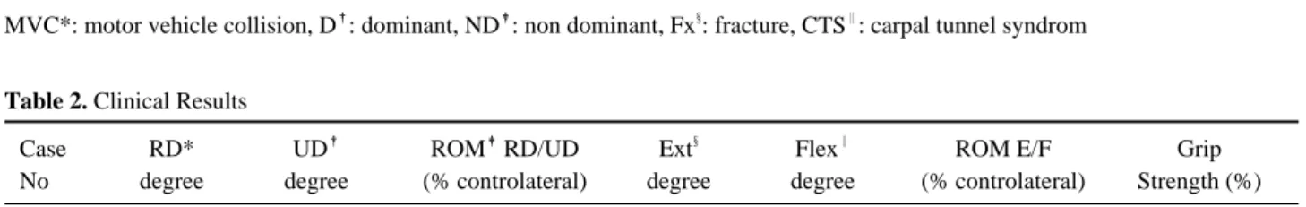
관 감압술을 같이 시행하였다. 주상골 골절의 내고정 방법으로는 9례에서는 Herbert screw를 이용하였고, 4례에서는 K-강선을 이용 하여 내고정 하였다. 전례 에서 월상 삼각골간 인대의 봉합은 시행하지 않았으며 9례에서는 경피적인 K-강선 고정을 시행하였다(Table 2). 기능적 평가는 손상받지 않은 수부를 기준으로 운 동 범위, 악력을 비교하였으며, 환자의 만족도, 다치 기 전의 직업 복귀 정도를 확인하였다. 방사선적으로 는 주상골 유합 시기 및 수근골 관절의 변화 정도를 비교하였다.
Table 1. Details of 13 Patients and Radiological Results
Gender Hand Scaphoid Lunotriquetal
Case No
/Age Cause
D�/ND� Associated fixation Fixation with
with K-wire
01 M/26 Fall D - Herbert screw +
02 F/30 Fall D Radial styloid Fx§ Herbert screw +
03 M/24 MVC* ND - Herbert screw +
04 M/21 MVC D - Herbert screw +
05 M/36 MVC D CTS‖ Herbert screw -
06 M/25 Fall ND - Herbert screw +
07 M/23 MVC ND - K-wire -
08 M/25 Fall ND Radial styloid Fx K-wire +
09 M/28 MVC D - Herbert screw -
10 M/29 MVC D - K-wire +
11 M/30 Fall D - Herbert screw +
12 M/28 Fall D CTS K-wire -
13 M/27 Sports ND - Herbert screw +
Average M/27
MVC*: motor vehicle collision, D�: dominant, ND�: non dominant, Fx§: fracture, CTS‖: carpal tunnel syndrom
Table 2. Clinical Results
Case RD* UD� ROM�RD/UD Ext§ Flex‖ ROM E/F Grip
No degree degree (% controlateral) degree degree (% controlateral) Strength (%)
01 10 15 80 60 55 80 80
02 12 20 85 60 50 80 80
03 20 25 90 70 60 90 90
04 20 15 80 65 60 80 90
05 18 25 80 60 60 85 80
06 13 20 85 55 50 75 80
07 18 25 88 65 60 90 80
08 12 18 85 60 60 80 80
09 12 18 84 60 50 75 80
10 12 16 80 50 50 70 80
11 14 18 80 55 50 75 80
12 16 20 85 60 55 80 80
13 18 25 90 60 55 80 90
Average 15 20 84 60 55 80 82
RD*: radial deviation, UD�: ulnar deviation, ROM�*: range of motion, EXT§: extension, FLEX‖: flexion
결 과
평균 운동 범위는 손상받지 않은 완관절의 운동 범 위 84%까지 회복하였으며, 평균 신전 각도는 60도였 으며 평균 굴곡 각도는 55도, 척측 변위는 20도, 요측 변위는 15도였다. 악력은 손상받지 않은 수부의 82%
까지 회복하였다(Table 2). 술 후 평균 골 유합 기간 은 14주였으며, 추시 관찰 기간 중 모든 환자에서 후 방 굴곡 중간 분절 불안정성과 전방 굴곡 중간 분절 불안정성은 보이지 않았다. 2례에서 핀 감염 소견이 있었으나 항생제 치료로 치유되었다. 모든 환자들은 수술 결과에 만족하였으며 완관절 운동 범위가 다소 제한을 보였으나 일상 생활 및 외상 전의 직업으로 복 귀가 가능하였다.
증례 보고
증례 1
21세 남자 환자로 교통 사고로 넘어지면서 우측 수
근부에 심한 동통과 부종으로 내원하였다. 내원 시 방 사선 검사상 후방 주상골 경유 월상골 주위 골절과 탈 구로 진단되어(Fig. 1) 전방 접근법으로 개방성 정복 술 후 Herbert 나사못으로 고정하였다(Fig. 2). 술 후 2년째 추시 관찰 상 골 유합은 이루어졌으며, 후방 굴곡 65도, 전방 굴곡 70도, 요측 굴곡 20도, 척측 굴곡 15도, 악력은 손상받지 않은 수부의 90%을 얻었 다(Fig. 3).
증례 2
26세 남자 환자로 작업 도중에 2 m 높이에서 떨어 지면서 우측 수근부에 심한 동통과 부종 및 운동 제한 으로 내원하였다. 방사선 검사상 후방 주상골 경유 월 상골 주위 골절과 탈구로 진단되었다(Fig 4). 전방 접 근법으로 개방성 정복술 후 K-강선으로 고정하였으며 월상골 삼각골은 경피적으로 K-강선 고정술을 시행 하였다(Fig. 5). 술 후 3년째 추시 관찰 상 골 유합 소견은 이루어졌으며(Fig. 6) 후방 굴곡 60도, 전방 굴곡 55도, 요측 굴곡 10도, 척측 굴곡 15도, 악력은
Fig. 1. Preoperative posteroanterior (PA) and lateral radiographs showing a dorsal transscaphoid perilunate fracture-dislocation in a 21-year- old man.
Fig. 2. Postoperative posteroanterior (PA) and lateral radiographs showing anatomic reduction and the internal fixation using a canu- lated Herbert screw with a volar approach.
Fig. 3. Range of motion at the 2 years follow-up visit. Wrist motion: dorsiflexion 65�, volar flexion 70�radial deviation. 20�, ulnar deviation. 15�(total 170�), Grip strength: 90% of uninjured wrist.
Fig. 4. Preoperative posteroanterior (PA) and lateral radiographs showing a dorsal transscaphoid perilunate fracture-dislocation in a 26-year-old man.
Fig. 5. Postoperative posteroanterior (PA) and later- al radiographs showing anatomic reduction and the internal fixation using a K-wire with a volar approach and percutaneous K-wire fixation in lunotriquetral joint.
손상받지 않은 수부의 80%을 얻었다(Fig. 7).
고 찰
치료가 늦어지거나 제대로 되지 않은 월상골 탈구나 주상골 경유 월상골 주위 골절 탈구는 관절 운동 장 애, 만성적인 수근관 증후군, 수근관절의 불안정성 및 외상성 관절염, 주상골 월상골 진행성 붕괴 등으로 인 해 불량한 예후를 나타내게 된다4,9. 이러한 문제점을 막기 위한 성공적인 치료를 위해서는 수근골의 역학적 인 정렬을 유지하면서 주상골 유합이 되는 것이 가장 중요하다2-4. 치료 방법 중 고식적인 방법인 도수 정복 술 후 석고 고정술은 역학적인 수근골의 정렬을 유지 하기가 힘들어 만족할만한 결과를 얻기가 힘들며7 현재 는 더 적극적인 치료 방법으로 관혈적 정복술 및 내
고정술이 보다 나은 결과를 나타낸다고 한다8,9. 주상골 경유 월상골 주위 골절 탈구에서 수술적 접근 시 탈구와 주상골 골절의 충분한 정복 및 내고정과 인 대 손상에 대해 인대 봉합을 할 수 있는 시야 확보가 중요하며 각각의 방법에 대해서 의견이 많다4,7. 전방 접근법의 장점은 원위 주상골 골절에서 충분한 시야 확 보가 가능하며 전방 인대 및 관절막 손상 시 인대 봉합 에 유리하며 수근관 증후군이 동반되는 경우 수근관 감 압술이 가능하다고 한다10,11. 후방 접근법은 후방 관절 막 및 월상 삼각골 인대 봉합에 유리하며 근위 수근골 과 중간 수근골 관절 부위에 좋은 시야가 확보된다고
한다12,13. 전후방 접근법은 각각의 장점을 다 취할 수
있다고 한다4. 1978년에 Gree과 O’Brien14은 주상골 과 월상골 해리가 있을 경우 개방성 정복술을 하여야 하며 주상골 경유 월상골 탈구 시 전방 접근법을, 월상 Fig. 7. Range of motion at the 3 years follow-up visit. Wrist motion: dorsiflexion 60�, volar flexion 55�, radial deviation 10�, ulnar
deviation 15�(total 140�), Grip strength: 80% of uninjured wrist.
Fig. 6. Postoperative 3 years, posteroanterior (PA) and lateral radiographs show healing of the scaphoid fracture and anatomical carpal alignment.
골 전방 탈구 시 후방 접근법 또는 전후방 접근법을 하 는 것을 권유하였다. 1987년에는 Adkison과 Chap- man5은 월상골과 수근골의 관계에 이상이 있을 경우 후방 접근법을 이용한 개방성 정복술을 권유하였고, Moneim 등15은 후방 접근법만으로도 주상골 골절의 정복이 충분하였으며 전방 접근법을 이용한 인대 봉합 을 하지 않아도 결과에 영향을 미치지 않았다고 하였 다. Cooney 등8은 필요에 따라 주상골을 고정할 수 있 는 전후방 접근법이 좋다고 하였지만 Viegas 등11은 전방 접근법만으로도 주상골 고정이 가능하였으며 만 족할만한 결과를 얻었다고 하였으며 Inoue 등16도 전방 접근만으로도 충분히 만족할만한 결과를 얻었다고 하 였다. 저자들은 모든 예에서 전방 접근법으로 탈구 정 복을 하면서 주상골 골절 부위를 고정하였으며 2례에 서는 수근관 감압술을 동시에 실시하였다.
탈구 정복 후 정복 유지 및 수근골 정렬을 위한 내 고정 방법으로 주로 K-강선을 사용하지만 일시적인 나사못 고정도 술 후 완관절 운동 결과에 큰 차이가 없다는 보고도 있다17. 주상골 골절의 내고정물로는 Herbert 나사못10, K-강선, Acutrack 나사못18,19, AO 3.5 mm cannulated 나사못20, Herbert- Whipple 나사못10 등이 있으며 주상골 내에 완전히 들어가는 Herbert 나사못, Acutrack 나사못이 주로 사용되고 있다. Herbert 나사못 고정 방법은 경험적, 기술적 어려움은 있으나 술 후 석고 고정 기간을 줄일 수 있고16 불유합의 합병증도 예방할 수 있는데 비해3 K-강선 고정 방법은 수술 시 간단히 고정할 수 있으 나 강한 고정력을 줄 수 없어 골간 인대 치료에 도움 이 되지 않으며21 술 후 석고 고정 기간이 길다는 단점 이 있다16. 월상골 주위 탈구 시 가장 손상이 많이 오 는 골간 인대는 주상골 월상골 골간 인대이나21,22, 주 상골 경유 월상골 주위 골절 탈구는 주상골 골절이 동 반 되기에 주상 월상골 인대 손상이 발생하는 경우는 흔지 않으며 월상 삼각골 인대 손상은 발생할 수 있다
1,3. 이러한 골간 인대 손상 유무는 수술 시 눈으로 확 인하며 가능한 인대 봉합은 인대 손상 직후에 하는 것 이 좋다고 하나 인대 봉합은 기술적으로 어려운 경우 가 많으며 어느 시기 이내에 골간 인대 재건을 하는게 좋은지는 발표된 게 없다3. 월상 삼각골간 인대 손상으 로 인해 월상 삼각골간 관절 간격이 현저하게 벌어진 9례에서 인대 재건술 없이 경피적인 K-강선 고정으로 일시적인 내고정을 실시하였다. 주상골 경유 월상골 주위 골절을 동반한 탈구에 대하여 많은 보고가 있지 만 아직까지 수술 결과에 대한 환자의 만족도에 대하 여 후향적으로 비교할 수 있는 객관적인 자료가 없으 며 수술 방법이나 수술적 접근법에 대하여서도 술자의 경험에 의존하는 경우가 많아 여기에 대한 객관적인
연구가 더 필요할 것으로 사료된다.
결 론
저자들은 주상골 경유 월상골 주위 골절 탈구 환자 에 대해 전방 접근법을 이용하여 탈구의 정복 및 주상 골의 내 고정술을 시행하여 비교적 만족스러운 결과를 얻을 수 있었다.
참고문헌
01) Herzberg G, Comtet JJ, Linscheid RL, Amadio PC, Cooney WP, Stalder J. Perilunate dislocations and frac- ture-dislocations: a multicenter study. J Hand Surg. 1993;
18:768-79.
02) White RE Jr., Omer GE Jr. Transient vascular compromise of the lunate after fracture-dislocation or dislocation of the carpus. J Hand Surg. 1984;9:181-4.
03) Knoll VD, Allan C, Trumble TE. Trans-scaphoid perilu- nate fracture dislocations: results of screw fixation of the scaphoid and lunotriquetral repair with a dorsal approach.
J Hand Surg. 2005;30:1145-52.
04) Sotereanos DG, Mitsionis GJ, Giannakopoulos PN, Tomaino MM, Herndon JH. Perilunate dislocation and fracture dislocation: a critical analysis of the volar-dorsal approach. J Hand Surg. 1997;22:49-56.
05) Adkison JW, Chapman MW. Treatment of acute lunate and perilunate dislocations. Clin Orthop Relat Res. 1982;
199-207.
06) Apergis E, Maris J, Theodoratos G, Pavlakis D, Antoniou N. Perilunate dislocations and fracture-dislocations.
Closed and early open reduction compared in 28 cases.
Acta Orthop Scand Suppl. 1997;275:55-9.
07) Melone CP Jr., Murphy MS, Raskin KB. Perilunate injuries. Repair by dual dorsal and volar approaches. Hand Clin. 2000;16:439-48.
08) Cooney WP, Bussey R, Dobyns JH, Linscheid RL.
Difficult wrist fractures. Perilunate fracture-dislocations of the wrist. Clin Orthop Relat Res. 1987;136-47.
09) Herzberg G, Forissier D. Acute dorsal trans-scaphoid per- ilunate fracture-dislocations: medium-term results. J Hand Surg. 2002;27:498-502.
10) Herbert TJ, Fisher WE. Management of the fractured scaphoid using a new bone screw. J Bone Joint Surg Br.
1984;66:114-23.
11) Viegas SF, Bean JW, Schram RA. Transscaphoid frac- ture/dislocations treated with open reduction and Herbert
screw internal fixation. J Hand Surg. 1987;12:992-9.
12) Mayfield JK. Mechanism of carpal injuries. Clin Orthop Relat Res. 1980;45-54.
13) Moneim MS. Management of greater arc carpal fractures.
Hand Clin. 1988;4:457-67.
14) Green DP, O’Brien ET. Open reduction of carpal disloca- tions: indications and operative techniques. J Hand Surg.
1978;3:250-65.
15) Moneim MS, Hofammann KE 3rd, Omer GE. Trans- scaphoid perilunate fracture-dislocation. Result of open reduction and pin fixation. Clin Orthop Relat Res.
1984;227-35.
16) Inoue G, Tanaka Y, Nakamura R. Treatment of trans- scaphoid perilunate dislocations by internal fixation with the Herbert screw. J Hand Surg. 1990;15:449-54.
17) Souer JS, Rutgers M, Andermahr J, Jupiter JB, Ring D.
Perilunate fracture-dislocations of the wrist: comparison of temporary screw versus K-wire fixation. J Hand Surg.
2007;32:318-25.
18) Bond CD, Shin AY, McBride MT, Dao KD. Percutaneous screw fixation or cast immobilization for nondisplaced scaphoid fractures. J Bone Joint Surg Am. 2001;83:483-8.
19) Slade JF 3rd, Jaskwhich D. Percutaneous fixation of scaphoid fractures. Hand Clin. 2001;17:553-74.
20) Wozasek GE, Moser KD. Percutaneous screw fixation for fractures of the scaphoid. J Bone Joint Surg Br. 1991;
73:138-42.
21) Trumble T, Verheyden J. Treatment of isolated perilunate and lunate dislocations with combined dorsal and volar approach and intraosseous cerclage wire. J Hand Surg.
2004;29:412-7.
22) Minami A, Ogino T, Ohshio I, Minami M. Correlation between clinical results and carpal instabilities in patients after reduction of lunate and perilunar dislocations. J Hand Surg. 1986;11:213-20.