http://www.ecevr.org/ 159
CLINICAL
EXPERIMENTAL VACCINE
RESEARCH
Introduction
Rabies is an important zoonosis that results in more than 60,000 human deaths around the world every year. Once its clinical symptoms are evident, death is almost inevita- ble. Mass vaccination of dogs, including stray dogs in high-risk areas, can reduce the incidence of rabies in both animals and humans [1]. In South Korea, the rabies virus (RABV) isolates are clustered into an Arctic-like lineage based on its molecular and bi- ological characteristics and has been circulated by animals bitten by raccoon dogs (Nyctereutes procynoide korensis) and badgers (Meles meles) since the 1990s [2]. The distribution of oral bait vaccines with either live attenuated RABV (SAG2 strain), re- combinant adenovirus (ONRAB), or vaccinia-rabies glycoprotein (V-RG) is the most effective method of eliminating rabies in wild animals [3-5]. The World Health Organi- zation (WHO) has recommended that live rabies vaccines should not cause any ad- verse symptoms in either target or nontarget species including rodents, wild animals,
© Korean Vaccine Society.
This is an Open Access article distributed under the terms of the Creative Commons Attribution Non-Com- mercial License (http://creativecommons.org/licenses/
by-nc/4.0) which permits unrestricted non-commercial use, distribution, and reproduction in any medium, pro- vided the original work is properly cited.
K O R E A N V A C C I N E S O C I E T Y
K O R E A N V A C C I N E S O C I E T Y K O R E A N
A C C I N E O C I E T Y V
S
Clin Exp Vaccine Res 2016;5:159-168 http://dx.doi.org/10.7774/cevr.2016.5.2.159 pISSN 2287-3651 • eISSN 2287-366X
Dong-Kun Yang1, Ha-Hyun Kim1, Sung-Suk Choi1, Jong-Tack Kim2, Kang-Bok Lee3, Seong Heon Lee1, In-Soo Cho1
1Viral Disease Division, Animal and Plant Quarantine Agency, MAFRA, Gimcheon; 2College of Veterinary Medicine, Kangwon National University, Chuncheon; 3Jeonnam Wildlife Management Center, Suncheon, Korea Received: May 11, 2016
Revised: June 15, 2016 Accepted: June 30, 2016
Corresponding author: Dong-Kun Yang, PhD, DVM Viral Disease Division, Animal and Plant Quaran- tine Agency, 177 Hyeoksin 8-ro, Gimcheon 39660, Korea
Tel: +82-54-912-0785, Fax: +82-54-912-0812 E-mail: yangdk@korea.kr
No potential conflict of interest relevant to this article was reported.
This work was supported financially by a grant (B1543083-2016-18-01) from Animal, and Plant Quarantine Agency, Ministry of Agriculture, Food and Rural Affairs (MAFRA), Republic of Korea.
Purpose: The development of a genetically modified live rabies vaccine applicable to wild raccoon dogs is necessary for the eradication of rabies in Korea. Thus, we constructed a recom- binant rabies virus (RABV) called the ERAGS strain, using a reverse genetic system and evalu- ated its safety and efficacy in mice and its safety and immunogenicity in raccoon dogs.
Materials and Methods: ERAGS, which has Asn194Ser and Arg333Glu substitutions in the glycoprotein, was constructed using site-directed mutagenesis. Mice were inoculated with the ERAGS strain (either 105.0 or 107.0 FAID50/mL) via intramuscular (IM) or intracranial injections and then challenged with a virulent RABV. Raccoon dogs were administered the ERAGS strain (108.0 FAID50/mL) either orally or via the IM route and the immunogenicity of the strain was evaluated using fluorescent antibody virus neutralization tests.
Results: The ERAGS strain inoculated into murine neuroblastoma cells reached 107.8 FAID50/mL at 96-hour post-inoculation. The virus was not pathogenic and induced complete protection from virulent RABV in immunized 4- and 6-week-old mice. Korean raccoon dogs immunized with the ERAGS strain via IM or oral route were also safe from the virus and developed high titer levels (26.4-32.8 IU/mL) of virus-neutralizing antibody (VNA) at 4 weeks post-inoculation.
Conclusion: The ERAGS RABV strain was effectively protective against rabies in mice and produced a high VNA titer in raccoon dogs.
Keywords: Rabies virus, Mouth, Vaccines, Raccoon dogs
Safety and immunogenicity of
recombinant rabies virus (ERAGS)
in mice and raccoon dogs
Dong-Kun Yang et al • Recombinant rabies virus (ERAGS) induces complete protection
160 http://www.ecevr.org/ http://dx.doi.org/10.7774/cevr.2016.5.2.159
or other domestic species [6,7].
Currently, an inactivated vaccine is used to prevent rabies in animals, and the use of this vaccine will soon be expanded in an attempt to completely eliminate the occurrence of dog- mediated human rabies. However, its high cost is threatening the achievement of 70% vaccination coverage in developing countries [6]. On the other hand, live attenuated rabies vac- cines are more cost-effective and simpler to manufacture. For example, the Evelyn-Rokitnicki–Abelseth (ERA) strain propa- gated in primary porcine kidney cell cultures or Vero cells has been used to immunize dogs and cattle, and shows protec- tive immunogenicity [8]. Although live rabies vaccine strains can also be given to wild animals via oral administration, there use is no longer recommended by world organizations in- volved in animal health. Furthermore, the ERA strain used in live vaccines cannot be administered to cats or wild carni- vores in South Korea due to safety concerns [7,9].
RABV is a single-stranded negative sense RNA virus belong- ing to Lyssavirus of the family Rhabdoviridae. The genome consists of five genes that encode a nucleoprotein (N), phos- phoprotein (P), matrix protein (M), glycoprotein (G), and large protein (L). Three of these proteins (N, P, and L) form a ribonucleoprotein complex that serves as a template for virus transcription and replication. When associated with the N and L proteins, the P protein acts as a non-catalytic cofactor of the viral RNA polymerase [10]. The G protein, which repre- sents the major surface protein of the virion, is associated with an interaction with the receptor binding site, fusion to facili- tate virus entry into host cells, and the induction of a neutral- izing antibody [11].
The G protein is also responsible for virulence. Either the arginine (Arg) or lysine (Lys) residue at position 333 of the G protein determines RABV virulence [12]. When the Arg333 residue in glycoprotein RABVs is substituted with glutamic acid (Glu), isoleucine (Ile), glycine (Gly), methionine (Met), or serine (Ser), the variants are less pathogenic or avirulent in adult mice following intracranial (IC) inoculation [13,14]. How- ever, after more than three back passages of these recombi- nant RABVs in suckling mice, a single mutation (asparagine:
Asn to Lys) at position 194 of the G protein occurs and incre- ases virulence [15]. Recombinant RABVs rescued with a sin- gle mutation at position 333 have the risk of reverting to a vir- ulent virus. However, this risk can be circumvented by con- structing an RABV with multiple mutations that involve Glu at position 333 and Asn at position 194, which would be more appropriate as a live vaccine because it cannot revert into a
pathogenic virus and thus will be useful for animals [15].
Therefore, we constructed a novel recombinant RABV termed the ERAGS strain that contains two mutations at positions 194 and 333 of the G protein. Its safety and efficacy was evalu- ated in mice, and its safety and immunogenicity was assessed in raccoon dogs.
Materials and Methods
Cells and viruses
BHK/T7-9 cells were grown in Dulbecco’s modified Eagle medium (DMEM) with 600 ng/mL hygromycin, 10% heat-in- activated fetal bovine serum (FBS), and several antibiotics (100 IU/mL penicillin, 10 μg/mL streptomycin, and 0.25 μg/
mL amphotericin B) and then maintained in 3% FBS [16].
Murine neuroblastoma (NG108-15) cells were maintained in DMEM supplemented with 5% FBS and then placed in a 5%
CO2 incubator at 37°C. The ERA strain of RABV that was in- troduced from Canada in 1974 was used as a safety compari- son for the ERAGS strain. A challenge virus standard (CVS) strain, CVSN2c, which is a virulent RABV, was used to assess the efficacy of ERAGS in mice while another CVS strain, CVS11, which is a fixed RABV, was used for the fluorescent assay vi- rus neutralizing (FAVN) tests in raccoon dogs.
Construction of the recombinant RABV
Full-length cDNA modified with Ser and Glu amino acid mu- tations at positions 194 and 333 of the G gene of the ERA strain was cloned into the pTM1 vector. Briefly, restriction enzyme sites of Pst I and Dra III were located at positions 5,407 and 7,422 in the full-length cDNA. After the digestion of cDNA with two restriction enzymes, the Asn (AAT) to Ser (TCC; Asn194S- er) and Arg (AGA) to Glu (GAA; Arg333Glu) mutations were conducted at positions 194 and 333 of the G gene in the DNA fragment via site-directed mutagenesis (Fig. 1). After the mod- ified DNA fragment was inserted into the original plasmid, the modified full-length cDNA was constructed and desig- nated the pUC19 ERAGS plasmid.
The N, P, and L genes obtained from the ERA strain were also cloned into the same vector and named EN, EP, and EL, respectively. To recover the recombinant RABV (the ERAGS strain) from cloned full-length cDNA and helper plasmids, the full-length modified genome plasmid (2 μg), EN (0.25 μg), EP (0.02 μg), and EL (0.25 μg) were mixed with 9 μL X-treme GENE HP DNA transfection reagent (Roche Diagnostic, Mann- heim, Germany) and transfected into BHK/T7-9 cells grown
http://www.ecevr.org/ 161
http://dx.doi.org/10.7774/cevr.2016.5.2.159
in a 24-well tissue culture plate. Following transfection, the cells were screened for cytopathic effects for 5 days (Fig. 2).
Identification of the recombinant RABV
The second-passaged recombinant ERAGS strain was propa- gated in BHK/T7-9 cells and infected cells were fixed using 80% chilled acetone for 15 minutes. To stain the cells, they were reacted with a specific monoclonal antibody (Median Diagnostics, Chuncheon, Korea) against the N protein of the
RABV for 45 minutes and then stained with fluorescence iso- thiocyanate (FITC)-conjugated goat anti-mouse IgG+IgM (KPL, Gaithersburg, MD, USA). After being washed in phos- phate buffered saline (PBS, pH 7.2), the cells were examined via fluorescent microscopy (Nikon, Tokyo, Japan) and a com- mercial rapid immunodiagnostic assay (RIDA) kit was used according to the manufacturer’s instruction as a supplemen- tary identification measure (Bionote, Hwaseong, Korea). Brief- ly, a mixture of 50 µL supernatant of the ERAGS strain and 50 µL proprietary buffer was applied to the sample well of the RIDA kit. The appearance of two lines was considered a posi- tive result.
Propagation of the ERAGS strain in NG108-15 cells
A monolayer of NG108-15 cells in a 25 cm2 tissue culture flask was infected with either the ERA or ERAGS strain at a multi- plicity of infection of 0.01. The flasks were incubated at 37°C and frozen at 12-, 24-, 36-, 48-, 72-, 96-, and 120-hour post- infection. After being thawed and frozen three times, the ERA and ERAGS strains were titered in a 96-well microplate (ten- fold dilution) and the viral titer was determined by an indi- rect fluorescent assay calculated using the method of Reed and Muench.
Safety and potency of RABVs in mice
Mice (4- and 6-week-old BALB/c type) were divided into seven groups consisting of either four or eight animals each. Groups 1 and 2 received intramuscular (IM) inoculations with the ERAGS strain (105.0 and 107.0 FAID50/mL, respectively), group 3 received an IM inoculation with the ERA strain (107.0 FAID50/
- 21 - 1
2
Fig. 1. Full-length plasmid map for the construction of the ERAGS strain. The full- 3
length cDNA plasmid was modified using site-directed mutagenesis at positions 194 4
and 333 of the glycoprotein in the ERA strain.
5
Fig. 1. Full-length plasmid map for the construction of the ERAGS strain. The full-length cDNA plasmid was modified using site-directed mutagenesis at positions 194 and 333 of the glycoprotein in the ERA strain.
Fig. 2. Cytopathic effects (×200) of BHKT7-9 cells (A) infected with the ERAGS strain and normal cells (B). BHKT7-9 cells infected with the virus were detached from the plate.
6
7
A B
8
Fig. 2. Cytopathic effects (200×) of BHKT7-9 cells (A) infected with the ERAGS strain
9
and normal cells (B). BHKT7-9 cells infected with the virus were detached from the
10
plate.
11 6
7
A B
8
Fig. 2. Cytopathic effects (200×) of BHKT7-9 cells (A) infected with the ERAGS strain
9
and normal cells (B). BHKT7-9 cells infected with the virus were detached from the
10
plate.
11
B A
Dong-Kun Yang et al • Recombinant rabies virus (ERAGS) induces complete protection
162 http://www.ecevr.org/ http://dx.doi.org/10.7774/cevr.2016.5.2.159
mL), groups 4 and 5 received IC inoculations with the ERAGS strain (105.0 and 107.0 FAID50/mL, respectively), group 6 recei- ved an IC inoculation with the ERA strain (107.0 FAID50/mL), and group 7 was the control group and did not receive an in- oculation. Each mouse was inoculated with either 30 μL (IC) or 100 μL (IM) of the virus and mice showing central nervous system symptoms were humanely euthanized. The body wei- ghts of the mice were measured after inoculation and their survival rates were observed for 16 days after the inoculation.
To determine the efficacy of the ERAGS strain, mice that sur- vived after immunization were challenged with a lethal 100 μL IM dose of 25 LD50/0.1 mL CVSN2c strain in the right leg.
Changes in the body weights of the mice were measured and the survival rates of the mice were observed for 14 days after the challenge.
Safety and immunogenicity of the ERAGS strain in raccoon dogs
Raccoon dogs (4 months old) that were sero-negative against RABV were divided into three groups. Group 1 included six raccoon dogs that received oral administration of the ERAGS strain (1 mL, 108.0 FAID50/mL), group 2 included four animals that received IM inoculations with the same strain and dose, and group 3 included three animals that did not receive any treatment as a control group. All raccoon dogs were monitor- ed daily for clinical signs of rabies such as abnormal behavior, nervous prostration, anxiety, agitation, aggression, and pa- ralysis. Two and four weeks after inoculation, the body weight of each raccoon dog was measured and blood was collected from each subject, including controls. The titers of the serum samples are expressed in international units per milliliter (IU/
mL) and were compared to the titers of a rabies-positive stan- dard serum (WHO). The minimum protective titer has been determined to be 0.5 IU/mL FAVN [6,17].
Serological assay
A virus neutralizing antibody (VNA) test was determined us- ing the FAVN test [17] with a positive reference serum sample obtained from the WHO (adjusted to 0.5 IU/mL) used as a positive control. Briefly, 50 µL volumes of serum and positive and negative controls were distributed in four consecutive wells and then serially diluted threefold. Then the CVS11 RA- BV strain (approximately 100 FAID50/50 μL) was added to each well and incubated for 1 hour at 37°C. Next, 50 μL BHK-21 cell suspension (4×10⁵ cells/mL) was added to each well and the microplates were incubated for 72 hours in a humidified
incubator with 5% CO2 at 37°C. The cells within the micro- plates were fixed with 100 µL cold acetone (-20°C) for 20 min- utes, successively washed three times with PBS, reacted with a specific monoclonal antibody against the N protein of RABV for 45 minutes at 37°C, and then stained with FITC-conjugat- ed goat/anti-mouse IgG+IgM. Following a wash in PBS, the microplates were air-dried and examined at 200× using a flu- orescent microscope (Nikon). The titers of the serum samples were expressed in IU/mL by comparing the results to those of the positive standard.
Statistical analysis
All data are expressed as means±standard deviations and all statistical analyses of the VNA titers and body weights were conducted with IBM SPSS version 19.0 for Windows (IBM Corp., Armonk, NY, USA). Differences between the groups were analyzed with a one-way analysis of variance (ANOVA) followed by Tukey’s post hoc tests, and for more detailed in- vestigations, unpaired Student’s t-tests were performed for each individual time point. p-values of <0.05 were consid- ered to indicate statistical significance.
Results
Construction of recombinant RABV
After transfection with a mixture of the full-length genome mutated at positions 194 and 333 in the G gene of the ERA strain and three helper plasmids (EN, EP, and EL), the recom- binant RABV (ERAGS strain) was successfully rescued in BHK/
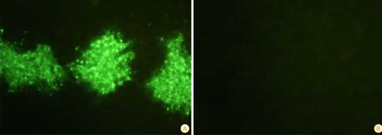
T7-9 cells. The BHK/T7-9 cells inoculated with the ERAGS strain were fixed with cold acetone and strained using a spe- cific antibody against the N protein of RABV. RABV-specific fluorescence appeared in the cytoplasm of the cells infected with the rescued virus indicating that the specific fluorescence in cytoplasm was RABV (Fig. 3). Additional tests, including the use of the RIDA kit, confirmed that the rescued virus was RABV (Fig. 4).
Growth properties and genetic stability
After three successive passages with the rescued ERAGS strain, the kinetics of the growth properties of the ERA and ERAGS strains were analyzed to determine whether the two muta- tions affected the replication ability of the ERAGS strain in NG108-15 cells. The growth curve of the ERAGS strain was similar to that of ERA strain but it had a slightly higher titer (107.8 FAID50/mL vs. 107.3 FAID50/mL) (Fig. 5). Both strains had
Dong-Kun Yang et al • Recombinant rabies virus (ERAGS) induces complete protection
http://www.ecevr.org/ 163
http://dx.doi.org/10.7774/cevr.2016.5.2.159
the highest titer at 96-hour post-inoculation, which indicated that the mutations at positions 194 and 333 in the G protein of the ERA strain did not influence the growth properties.
Safety and efficacy of the ERAGS strain in mice
The body weights of mice inoculated with either the ERA or ERAGS strain via either the IM or IC route were measured on designated days after the inoculation. The average body wei- ght of mice that received ERA IM slowly decreased until 10 days after inoculation (Fig. 6) and the average weight of those that received ERA IC showed a similar trend until 15 days. In contrast, the weights of the IM and IC groups inoculated with different titers of the ERAGS strain increased without any drastic weight loss.
Regarding survival, all mice inoculated with the ERA strain, whether IM or IC, succumbed to rabies and died 10 or 15 days post-inoculation, respectively. In contrast, all mice inoculat- ed with ERAGS (both IM and IC) and all control mice surviv- ed (Figs. 6, 7). All mice in the ERAGS group were subsequent-
ly challenged with a highly virulent RABV strain, CVSN2c, on day 15 post-immunization. The average body weights of the mice slightly increased and their survival rates did not change for 14 days after the challenge (Fig. 8) but the average weight of control mice inoculated with virulent RABV began to de- crease rapidly 7-8 days post-challenge and all mice died 9-10 days post-challenge. Thus, the ERA strain was pathogenic whereas the ERAGS strain was non-pathogenic and safe for adult mice.
Safety and immunogenicity of the ERAGS strain in raccoon dogs
All raccoon dogs were sero-negative for rabies VNA prior to inoculation, and sero-converted at 2-4 weeks after its admin- Fig. 3. Immunofluorescence (×200) of the ERAGS strain using an indirect fluorescent assay test with monoclonal antibodies against the nucleo- protein of the rabies virus (A) and normal cells (B). Recombinant rabies virus-specific fluorescent sites appeared in the cytoplasm of the infected BHK/T7-9 cells.
- 23 - 13
A B
14
Fig. 3. Immunofluorescence (200×) of the ERAGS strain using an indirect fluorescent
15
assay test with monoclonal antibodies against the nucleoprotein of the rabies virus (A)
16
and normal cells (B). Recombinant rabies virus-specific fluorescent sites appeared in the
17
cytoplasm of the infected BHK/T7-9 cells.
18
- 23 - 12
13
A B
14
Fig. 3. Immunofluorescence (200×) of the ERAGS strain using an indirect fluorescent
15
assay test with monoclonal antibodies against the nucleoprotein of the rabies virus (A)
16
and normal cells (B). Recombinant rabies virus-specific fluorescent sites appeared in the
17
cytoplasm of the infected BHK/T7-9 cells.
18
B A
- 24 - 19
20
21
Fig. 4. Results of the rapid immunodiagnostic assay (RIDA) kit following the
22
application of the ERAGS strain supernatant. The “C” and “T” in the RIDA kit stand
23
refer to the “control” and “test” lines. The appearance of two lines was considered to be
24
a positive result.
25 26
Fig. 4. Results of the rapid immunodiagnostic assay (RIDA) kit fol- lowing the application of the ERAGS strain supernatant. The “C” and
“T” in the RIDA kit stand refer to the “control” and “test” lines. The appearance of two lines was considered to be a positive result.
9 8 7 6 5 4 3 2 1
0 12 24 36 48 72 96 120
Hours postinoculation Viral titer (log10 FAID50/mL)
ERA ERAGS
Fig. 5. Multistep growth curves of the ERA and ERAGS strains in NG108-15 cells. Both strains revealed the highest viral titer at 96- hour post-inoculation.
Dong-Kun Yang et al • Recombinant rabies virus (ERAGS) induces complete protection
164 http://www.ecevr.org/ http://dx.doi.org/10.7774/cevr.2016.5.2.159
istration. Neither vaccinated nor non-vaccinated raccoon dogs exhibited any clinical signs of rabies during the test period. At 2 weeks post-inoculation, animals that received IM inocula- tions of the ERAGS strain developed a higher VNA titer (32.8 IU/mL) against rabies than those that were orally inoculated (3.27 IU/mL). However, all raccoon dogs that were immu- nized with the ERAGS strain (either orally or IM) exhibited similar mean VNA titers that ranged from 26.4 to 32.8 IU/mL at 4 weeks after inoculation (Fig. 9). Three animals in group 3 remained sero-negative against RABV throughout the test, which confirmed that no contact transmission occurred be- tween the vaccinated groups and the control group (Fig. 9A).
The body weights of those that received oral inoculations with the ERAGS were slightly heavier than those that received IM inoculations. The average body weight of group 3 increased during the same period (Fig. 9B).
Discussion
The use of oral immunizations with a rabies bait vaccine has greatly contributed to the elimination of rabies in wild ani- mals. For example, a live attenuated rabies vaccine (the SAG2 strain), which was isolated by selecting a monoclonal anti- body, has been shown to be safe and immunogenic based on Fig. 6. Changes in body weights in 4-week-old (A) and 6-week-old (B) mice inoculated with either the ERA or ERAGS strains via either the in- tramuscular (IM) or intracranial (IC) route. The weights of mice inoculated with the ERAGS strain continued to increase whereas those of mice inoculated with ERA progressively decreased, and these mice eventually died.
- 26 - 33
4-week-old mice 6-week-old mice
34
35
A B
36
Fig. 6. Changes in body weights in 4-week-old (A) and 6-week-old (B) mice inoculated
37
with either the ERA or ERAGS strains via either the intramuscular (IM) or intracranial
38
(IC) route. The weights of mice inoculated with the ERAGS strain continued to increase
39
whereas those of mice inoculated with ERA progressively decreased, and these mice
40
eventually died.
41
30 25 20 15 10 5
0 1 2 4 6 7 8 10 13 14 15 16
Days after inculation
Body weight (g)
- 26 - 33
4-week-old mice 6-week-old mice
34
35
A B
36
Fig. 6. Changes in body weights in 4-week-old (A) and 6-week-old (B) mice inoculated
37
with either the ERA or ERAGS strains via either the intramuscular (IM) or intracranial
38
(IC) route. The weights of mice inoculated with the ERAGS strain continued to increase
39
whereas those of mice inoculated with ERA progressively decreased, and these mice
40
eventually died.
41
ERAGS IM7.0 ERAGS IM5.0 ERA IM7.0 ERA IC7.0 ERAGS IC5.0
ERAGS IC7.0 Control
- 26 - 33
4-week-old mice 6-week-old mice
34
35
A B
36
Fig. 6. Changes in body weights in 4-week-old (A) and 6-week-old (B) mice inoculated
37
with either the ERA or ERAGS strains via either the intramuscular (IM) or intracranial
38
(IC) route. The weights of mice inoculated with the ERAGS strain continued to increase
39
whereas those of mice inoculated with ERA progressively decreased, and these mice
40
eventually died.
41
35 30 25 20 15 10 5
0 1 2 4 6 7 8 10 13 14 15 16
Days after inoculation
Body weight (g)
- 26 - 33
4-week-old mice 6-week-old mice
34
35
A B
36
Fig. 6. Changes in body weights in 4-week-old (A) and 6-week-old (B) mice inoculated
37
with either the ERA or ERAGS strains via either the intramuscular (IM) or intracranial
38
(IC) route. The weights of mice inoculated with the ERAGS strain continued to increase
39
whereas those of mice inoculated with ERA progressively decreased, and these mice
40
eventually died.
41
- 26 - 33
4-week-old mice 6-week-old mice
34
35
A B
36
Fig. 6. Changes in body weights in 4-week-old (A) and 6-week-old (B) mice inoculated
37
with either the ERA or ERAGS strains via either the intramuscular (IM) or intracranial
38
(IC) route. The weights of mice inoculated with the ERAGS strain continued to increase
39
whereas those of mice inoculated with ERA progressively decreased, and these mice
40
eventually died.
41
- 26 - 33
4-week-old mice 6-week-old mice
34
35
A B
36
Fig. 6. Changes in body weights in 4-week-old (A) and 6-week-old (B) mice inoculated
37
with either the ERA or ERAGS strains via either the intramuscular (IM) or intracranial
38
(IC) route. The weights of mice inoculated with the ERAGS strain continued to increase
39
whereas those of mice inoculated with ERA progressively decreased, and these mice
40
eventually died.
41
ERAGS IM7.0 ERAGS IM5.0 ERA IM7.0 ERA IC7.0 ERAGS IC5.0
ERAGS IC7.0 Control
B A
Fig. 7. Survival in 4-week-old (A) and 6-week-old (B) mice inoculated with either the ERA or ERAGS strain via either the intramuscular (IM) or intracranial (IC) route. Mice inoculated with ERA (both IM and IC) died 10 and 16 days post-inoculation, respectively. In contrast, 100% of mice inoculated with ERAGS (both IM and IC) survived, as did controls.
- 27 - 42
4-week-old mice 6-week-old mice
43
44
A B
45
Fig. 7. Survival in 4-week-old (A) and 6-week-old (B) mice inoculated with either the
46
ERA or ERAGS strain via either the intramuscular (IM) or intracranial (IC) route. Mice
47
inoculated with ERA (both IM and IC) died 10 and 16 days post-inoculation,
48
respectively. In contrast, 100% of mice inoculated with ERAGS (both IM and IC)
49
survived, as did controls.
50
120 100 80 60 40 20
0 1 2 4 6 7 8 10 13 14 15 16
Days after inoculation
Survival rate (%)
- 26 - 33
4-week-old mice 6-week-old mice
34
35
A B
36
Fig. 6. Changes in body weights in 4-week-old (A) and 6-week-old (B) mice inoculated
37
with either the ERA or ERAGS strains via either the intramuscular (IM) or intracranial
38
(IC) route. The weights of mice inoculated with the ERAGS strain continued to increase
39
whereas those of mice inoculated with ERA progressively decreased, and these mice
40
eventually died.
41
ERAGS IM7.0 ERAGS IM5.0 ERA IM7.0 ERA IC7.0 ERAGS IC5.0
ERAGS IC7.0 Control
120 100 80 60 40 20
0 1 2 4 6 7 8 10 13 14 15 16 Days after inoculation
Survival rate (%)
- 26 - 33
4-week-old mice 6-week-old mice
34
35
A B
36
Fig. 6. Changes in body weights in 4-week-old (A) and 6-week-old (B) mice inoculated
37
with either the ERA or ERAGS strains via either the intramuscular (IM) or intracranial
38
(IC) route. The weights of mice inoculated with the ERAGS strain continued to increase
39
whereas those of mice inoculated with ERA progressively decreased, and these mice
40
eventually died.
41
- 26 - 33
4-week-old mice 6-week-old mice
34
35
A B
36
Fig. 6. Changes in body weights in 4-week-old (A) and 6-week-old (B) mice inoculated
37
with either the ERA or ERAGS strains via either the intramuscular (IM) or intracranial
38
(IC) route. The weights of mice inoculated with the ERAGS strain continued to increase
39
whereas those of mice inoculated with ERA progressively decreased, and these mice
40
eventually died.
41
- 26 - 33
4-week-old mice 6-week-old mice
34
35
A B
36
Fig. 6. Changes in body weights in 4-week-old (A) and 6-week-old (B) mice inoculated
37
with either the ERA or ERAGS strains via either the intramuscular (IM) or intracranial
38
(IC) route. The weights of mice inoculated with the ERAGS strain continued to increase
39
whereas those of mice inoculated with ERA progressively decreased, and these mice
40
eventually died.
41
ERAGS IM7.0 ERAGS IM5.0 ERA IM7.0 ERA IC7.0 ERAGS IC5.0
ERAGS IC7.0 Control
B A
Dong-Kun Yang et al • Recombinant rabies virus (ERAGS) induces complete protection
http://www.ecevr.org/ 165
http://dx.doi.org/10.7774/cevr.2016.5.2.159
Fig. 8. Changes in body weights (A, B) and survival rates (C, D) in 4- and 6-week-old mice that were immunized with the ERAGS strains via either the intramuscular (IM) or intracranial (IC) route and then challenged with a highly pathogenic rabies virus strain (CVSN2c). The average body weight increased for 14 days after the challenge and the survival rate did not change for 14 days after the challenge.
- 28 - 51
52
A B
53 54
55
C D
56
Fig. 8. Changes in body weights (A, B) and survival rates (C, D) in 4- and 6-week-old
57
mice that were immunized with the ERAGS strains via either the intramuscular (IM) or
58
intracranial (IC) route and then challenged with a highly pathogenic rabies virus strain
59
(CVSN2c). The average body weight increased for 14 days after the challenge and the
60
survival rate did not change for 14 days after the challenge.
61 62
B
D A
C
- 28 - 51
52
A B
53 54
55
C D
56
Fig. 8. Changes in body weights (A, B) and survival rates (C, D) in 4- and 6-week-old
57
mice that were immunized with the ERAGS strains via either the intramuscular (IM) or
58
intracranial (IC) route and then challenged with a highly pathogenic rabies virus strain
59
(CVSN2c). The average body weight increased for 14 days after the challenge and the
60
survival rate did not change for 14 days after the challenge.
61 62
30 28 26 24 22 20 18
1 2 5 6 7 8 9 10 11 14 Days after challenge
Body weight (g)
- 28 - 51
52
A B
53 54
55
C D
56
Fig. 8. Changes in body weights (A, B) and survival rates (C, D) in 4- and 6-week-old
57
mice that were immunized with the ERAGS strains via either the intramuscular (IM) or
58
intracranial (IC) route and then challenged with a highly pathogenic rabies virus strain
59
(CVSN2c). The average body weight increased for 14 days after the challenge and the
60
survival rate did not change for 14 days after the challenge.
61 62
ERAGS IM7.0 ERAGS IM5.0 ERAGS IC5.0 ERAGS IC7.0
Control (challenge) Control
34 32 30 28 26 24 22 20 18
1 2 5 6 7 8 9 10 11 14 Days after challenge
Body weight (g)
- 28 - 51
52
A B
53 54
55
C D
56
Fig. 8. Changes in body weights (A, B) and survival rates (C, D) in 4- and 6-week-old
57
mice that were immunized with the ERAGS strains via either the intramuscular (IM) or
58
intracranial (IC) route and then challenged with a highly pathogenic rabies virus strain
59
(CVSN2c). The average body weight increased for 14 days after the challenge and the
60
survival rate did not change for 14 days after the challenge.
61 62
- 28 - 51
52
A B
53 54
55
C D
56
Fig. 8. Changes in body weights (A, B) and survival rates (C, D) in 4- and 6-week-old
57
mice that were immunized with the ERAGS strains via either the intramuscular (IM) or
58
intracranial (IC) route and then challenged with a highly pathogenic rabies virus strain
59
(CVSN2c). The average body weight increased for 14 days after the challenge and the
60
survival rate did not change for 14 days after the challenge.
61 62
ERAGS IM7.0 ERAGS IM5.0 ERAGS IC5.0 ERAGS IC7.0
Control (challenge) Control
- 28 - 51
52
A B
53 54
55
C D
56
Fig. 8. Changes in body weights (A, B) and survival rates (C, D) in 4- and 6-week-old
57
mice that were immunized with the ERAGS strains via either the intramuscular (IM) or
58
intracranial (IC) route and then challenged with a highly pathogenic rabies virus strain
59
(CVSN2c). The average body weight increased for 14 days after the challenge and the
60
survival rate did not change for 14 days after the challenge.
61 62
120 100 80 60 40 20 0
1 2 5 6 7 8 9 10 11 12 14 Days after challenge
Survival rate (%)
- 28 - 51
52
A B
53 54
55
C D
56
Fig. 8. Changes in body weights (A, B) and survival rates (C, D) in 4- and 6-week-old
57
mice that were immunized with the ERAGS strains via either the intramuscular (IM) or
58
intracranial (IC) route and then challenged with a highly pathogenic rabies virus strain
59
(CVSN2c). The average body weight increased for 14 days after the challenge and the
60
survival rate did not change for 14 days after the challenge.
61 62
ERAGS IM7.0 ERAGS IM5.0 ERAGS IC5.0 ERAGS IC7.0
Control (challenge) Control
- 28 - 52
A B
53 54
55
C D
56
Fig. 8. Changes in body weights (A, B) and survival rates (C, D) in 4- and 6-week-old
57
mice that were immunized with the ERAGS strains via either the intramuscular (IM) or
58
intracranial (IC) route and then challenged with a highly pathogenic rabies virus strain
59
(CVSN2c). The average body weight increased for 14 days after the challenge and the
60
survival rate did not change for 14 days after the challenge.
61 62
120 100 80 60 40 20 0
1 2 5 6 7 8 9 10 11 12 14 Days after challenge
Survival rate (%)
- 28 - 51
52
A B
53 54
55
C D
56
Fig. 8. Changes in body weights (A, B) and survival rates (C, D) in 4- and 6-week-old
57
mice that were immunized with the ERAGS strains via either the intramuscular (IM) or
58
intracranial (IC) route and then challenged with a highly pathogenic rabies virus strain
59
(CVSN2c). The average body weight increased for 14 days after the challenge and the
60
survival rate did not change for 14 days after the challenge.
61 62
- 28 - 51
52
A B
53 54
55
C D
56
Fig. 8. Changes in body weights (A, B) and survival rates (C, D) in 4- and 6-week-old
57
mice that were immunized with the ERAGS strains via either the intramuscular (IM) or
58
intracranial (IC) route and then challenged with a highly pathogenic rabies virus strain
59
(CVSN2c). The average body weight increased for 14 days after the challenge and the
60
survival rate did not change for 14 days after the challenge.
61 62
ERAGS IM7.0 ERAGS IM5.0 ERAGS IC5.0 ERAGS IC7.0
Control (challenge) Control
Fig. 9. The immune responses and body weights of raccoon dogs that received the ERAGS strain via either the oral or intramuscular (IM) route (26.4 and 32.8 IU/mL, respectively). At 4 weeks post-inoculation, high virus-neutralizing antibody titers were induced in all immunized raccoon dogs (A). The body weights of those that received oral administrations significantly increased whereas those that received IM administrations did not (B). Each bar represents the mean±standard error of the mean of three independent samples. Different lowercase letters indicate signifi- cant differences (p < 0.05, Tukey’s post hoc test). Asterisks (*p < 0.05) and double asterisks (**p < 0.01) in each panel indicate significant differ- ences from corresponding control groups without any treatment (unpaired t-test). FAVN, fluorescent antibody virus neutralization.
50
40
30
20
10
0 0 2 4
Week after administration
Mean FAVN titer (IU/mL)
Oral IM Control
a a a a a
a* c*
cb
* *c 5.0
4.5
4.0
3.5
3.0
2.5
0 2 4
Week after administration
Mean body weight (kg)
**
*
*
Oral IM Control
B A
Dong-Kun Yang et al • Recombinant rabies virus (ERAGS) induces complete protection
166 http://www.ecevr.org/ http://dx.doi.org/10.7774/cevr.2016.5.2.159
the absence of adverse clinical signs and salivary excretion in animals [18-20]. Thus, this vaccine has been registered as an oral rabies vaccine for wild animals in Europe and dogs in In- dia. However, highly attenuated RABVs, such as the HEP-Flury and SPBNGA strains, revert to virulence following a single amino acid substitution to Arg at position 333 in the G pro- tein after multiple passages in suckling mice [21,22]. This can lead to potential safety issues. The V-RG oral rabies vaccine meets the WHO safety requirements for animals and has play- ed a key role in controlling rabies in wild animals but it has also raised safety concerns after two cases of human vaccinia infection [23].
Thus, because safer candidates are necessary, several re- combinant RABVs constructed using reverse genetic systems have been developed under names such as the rERAG333E, rE- RAG3G, SPBNGA, Ni-CE (G333Glu), and ERAG333 strains [15,24-27]. These recombinant RABVs are based on the con- cept that the Arg at position 333 of the G protein of RABV is responsible for virulence. If the Arg is substituted with other amino acids, including Glu, Leu, and Gly, then its viral viru- lence is greatly decreased in adult mice [14,27]. It has also been reported that single mutated recombinant RABV strains con- taining a Glu at position 333 do not exhibit pathogenicity in mice older than 7 days [27]. A previous study from our research group found that the ERAG3G strain, which was developed based on the above mentioned concepts, is safe for several types of animals, including 4-week-old mice, and induces a high VNA titer against RABV in cats, dogs, and raccoon dogs [25]. However, because it is possible that the single mutated RABV strain can revert to pathogenic RABV strains, safer RABV vaccine strains are required to implement rabies eradication programs for wild animals.
Thus, we generated a novel recombinant RABV strain (called ERAGS), which is mutated at positions 194 and 333 of the G protein of the ERA strain. The ERA and ERAGS strains grew at similar rates in NG108-15 cells and reached over 107.5 FAID50/ mL at 96-hour post-inoculation. The growth property of the ERAGS strain seems to be similar to those of other recombi- nant RABVs, such as the rERAG333E and Ni-CE (G333Glu) strains [15,24,26].
The pathogenicity of RABV has been measured in mice in- oculated via a variety of routes. The important factors involved in RABV virulence include the age of the mice, the inoculation route, and the virus strain [28]. To determine whether the ERAGS strain was safe for mice, 4- and 6-week-old mice were inoculated with different titers of either the ERA or ERAGS
strains via either the IM or IC routes. Mice that received the ERA strain with a titer of 107.0 FAID50/mL via either route ex- hibited virulence 6 days after inoculation. On the other hand, mice inoculated with the ERAGS strain containing either 105.0 or 107.0 FAID50/mL via either route were observed for 16 days and did not exhibit any clinical signs. Thus, the safety of the ERAGS strain is similar to that of recombinant RABVs [24-26].
In addition, the mean body weight of the mice did not de- crease after the inoculation indicating that the ERAGS strain is non-pathogenic in 4- and 6-week-old mice.
The efficacy of the ERAGS strain was also evaluated in im- munized mice using a virulent RABV challenge. The mice in- oculated with the ERAGS strain were challenged with a 25 LD50/0.03 mL dose of rabies IC and all mice survived for 14 days. Furthermore, there were no clinical signs or decreases in body weight in these mice, indicating that a single immu- nization with different titers of the ERAGS strain may confer complete protection against an RABV challenge.
Raccoon dogs are primarily responsible for rabies trans- mission in South Korea and the safety and immunogenicity of any oral vaccine candidate should be evaluated in the main target animals. VNA is a critical protective component against lethal RABV infections [29] and the WHO considers a titer of 0.5 IU/mL rabies VNA to be the minimum protective antibody titer in carnivores [6]. Mucosal-related lymphoid tissues, in- cluding the tonsils, are a primary site of infection and this is replicated after the administration of an oral vaccine strain in dogs [30,31]. The present study showed that all raccoon dogs that received a 108.0 FAID50/mL dose via the oral route did not exhibit any adverse behavioral effects and developed high VNA titers that ranged from 13.7 to 41.6 IU/mL at 4 weeks post- administration. These findings suggest that raccoon dogs treat- ed with the ERAGS strain can be protected from a challenge inoculation with virulent RABV, which provides a basis for the use of the rabies bait vaccine strain. Future studies from our research group will extend these findings to dogs, cats, and cattle to assess whether the ERA strain that has been used for prevention of rabies in South Korea since the 1980s can be replaced. In addition, back passages of the ERAGS strain in suckling mice will be conducted to elucidate the likelihood that this strain will revert to pathogenic RABV.
In conclusion, the ERAGS strain, which is mutated at posi- tions 194 and 333 of the G protein, was safe and efficacious for use in 4- and 6-week-old mice. In addition, a single im- munization with the ERAGS strain via both the IM and oral routes induced a high VNA titer against RABV in raccoon dogs.
http://www.ecevr.org/ 167
http://dx.doi.org/10.7774/cevr.2016.5.2.159
Therefore, the ERAGS strain may be sufficiently safe and ef- fective for use as a live vaccine or oral bait vaccine in raccoon dogs.
ORCID
Dong-Kun Yang http://orcid.org/0000-0001-5765-3043 Ha-Hyun Kim http://orcid.org/0000-0001-6473-0035 Sung-Suk Choi http://orcid.org/0000-0001-5124-9736 Jong-Tack Kim http://orcid.org/0000-0002-6388-550X Kang-Bok Lee http://orcid.org/0000-0002-6215-326X Seong Heon Lee http://orcid.org/0000-0002-6024-3798 In-Soo Cho http://orcid.org/0000-0002-2692-3191
References
1. Seneschall C, Luna-Farro M. Controlling rabies through a multidisciplinary, public health system in Trujillo, La Lib- ertad, Peru. Pathog Glob Health 2013;107:361-6.
2. Hyun BH, Lee KK, Kim IJ, et al. Molecular epidemiology of rabies virus isolates from South Korea. Virus Res 2005;
114:113-25.
3. Mahl P, Cliquet F, Guiot AL, et al. Twenty year experience of the oral rabies vaccine SAG2 in wildlife: a global review.
Vet Res 2014;45:77.
4. Brown LJ, Rosatte RC, Fehlner-Gardiner C, et al. Oral vac- cination and protection of red foxes (Vulpes vulpes) against rabies using ONRAB, an adenovirus-rabies recombinant vaccine. Vaccine 2014;32:984-9.
5. Fehlner-Gardiner C, Rudd R, Donovan D, Slate D, Kempf L, Badcock J. Comparing ONRAB(R) AND RABORAL V- RG(R) oral rabies vaccine field performance in raccoons and striped skunks, New Brunswick, Canada, and Maine, USA. J Wildl Dis 2012;48:157-67.
6. World Health Organization. WHO Expert Consultation on Rabies. World Health Organ Tech Rep Ser 2005;931:1-88.
7. Faber M, Dietzschold B, Li J. Immunogenicity and safety of recombinant rabies viruses used for oral vaccination of stray dogs and wildlife. Zoonoses Public Health 2009;56:
262-9.
8. Lawson KF, Chiu H, Crosgrey SJ, Matson M, Casey GA, Campbell JB. Duration of immunity in foxes vaccinated orally with ERA vaccine in a bait. Can J Vet Res 1997;61:
39-42.
9. Yang DK, Go TO, Nam YH, et al. Antibody response in Ko- rean raccoon dogs inoculated with inactivated rabies vac-
cines. J Bacteriol Virol 2012;42:242-6.
10. Albertini AA, Schoehn G, Weissenhorn W, Ruigrok RW.
Structural aspects of rabies virus replication. Cell Mol Life Sci 2008;65:282-94.
11. Raux H, Flamand A, Blondel D. Interaction of the rabies virus P protein with the LC8 dynein light chain. J Virol 2000;
74:10212-6.
12. Dietzschold B, Wunner WH, Wiktor TJ, et al. Characteriza- tion of an antigenic determinant of the glycoprotein that correlates with pathogenicity of rabies virus. Proc Natl Acad Sci U S A 1983;80:70-4.
13. Seif I, Coulon P, Rollin PE, Flamand A. Rabies virulence:
effect on pathogenicity and sequence characterization of rabies virus mutations affecting antigenic site III of the glycoprotein. J Virol 1985;53:926-34.
14. Tuffereau C, Leblois H, Benejean J, Coulon P, Lafay F, Fla- mand A. Arginine or lysine in position 333 of ERA and CVS glycoprotein is necessary for rabies virulence in adult mice.
Virology 1989;172:206-12.
15. Faber M, Faber ML, Papaneri A, et al. A single amino acid change in rabies virus glycoprotein increases virus spread and enhances virus pathogenicity. J Virol 2005;79:14141-8.
16. Ito N, Takayama-Ito M, Yamada K, Hosokawa J, Sugiyama M, Minamoto N. Improved recovery of rabies virus from cloned cDNA using a vaccinia virus-free reverse genetics system. Microbiol Immunol 2003;47:613-7.
17. Cliquet F, Aubert M, Sagne L. Development of a fluores- cent antibody virus neutralisation test (FAVN test) for the quantitation of rabies-neutralising antibody. J Immunol Methods 1998;212:79-87.
18. Cliquet F, Gurbuxani JP, Pradhan HK, et al. The safety and efficacy of the oral rabies vaccine SAG2 in Indian stray dogs.
Vaccine 2007;25:3409-18.
19. Cliquet F, Robardet E, Must K, et al. Eliminating rabies in Estonia. PLoS Negl Trop Dis 2012;6:e1535.
20. Lafay F, Benejean J, Tuffereau C, Flamand A, Coulon P.
Vaccination against rabies: construction and character- ization of SAG2, a double avirulent derivative of SADBern.
Vaccine 1994;12:317-20.
21. Takayama-Ito M, Inoue K, Shoji Y, et al. A highly attenuat- ed rabies virus HEP-Flury strain reverts to virulent by sin- gle amino acid substitution to arginine at position 333 in glycoprotein. Virus Res 2006;119:208-15.
22. Dietzschold ML, Faber M, Mattis JA, Pak KY, Schnell MJ, Dietzschold B. In vitro growth and stability of recombi- nant rabies viruses designed for vaccination of wildlife.
Dong-Kun Yang et al • Recombinant rabies virus (ERAGS) induces complete protection
168 http://www.ecevr.org/ http://dx.doi.org/10.7774/cevr.2016.5.2.159
Vaccine 2004;23:518-24.
23. Rupprecht CE, Blass L, Smith K, et al. Human infection due to recombinant vaccinia-rabies glycoprotein virus. N Engl J Med 2001;345:582-6.
24. Shuai L, Feng N, Wang X, et al. Genetically modified ra- bies virus ERA strain is safe and induces long-lasting pro- tective immune response in dogs after oral vaccination.
Antiviral Res 2015;121:9-15.
25. Yang DK, Nakagawa K, Ito N, et al. A single immunization with recombinant rabies virus (ERAG3G) confers complete protection against rabies in mice. Clin Exp Vaccine Res 2014;3:176-84.
26. Nakagawa K, Ito N, Masatani T, et al. Generation of a live rabies vaccine strain attenuated by multiple mutations and evaluation of its safety and efficacy. Vaccine 2012;30:
3610-7.
27. Bankovskiy D, Safonov G, Kurilchuk Y. Immunogenicity of the ERA G 333 rabies virus strain in foxes and raccoon dogs.
Dev Biol (Basel) 2008;131:461-6.
28. Mebatsion T. Extensive attenuation of rabies virus by si- multaneously modifying the dynein light chain binding site in the P protein and replacing Arg333 in the G protein.
J Virol 2001;75:11496-502.
29. Franka R, Wu X, Jackson FR, et al. Rabies virus pathogen- esis in relationship to intervention with inactivated and attenuated rabies vaccines. Vaccine 2009;27:7149-55.
30. Orciari LA, Niezgoda M, Hanlon CA, et al. Rapid clearance of SAG-2 rabies virus from dogs after oral vaccination. Vac- cine 2001;19:4511-8.
31. Ogra PL, Faden H, Welliver RC. Vaccination strategies for mucosal immune responses. Clin Microbiol Rev 2001;14:
430-45.