Etiology of Hypokalemic Paralysis in Korea:
Data from a Single Center
Jung-Kook Wi, M.D., Hong Joo Lee, M.D., Eun Young Kim, M.D., Joo Hee Cho, M.D., Sang Ouk Chin, M.D., Sang Youl Rhee, M.D., Ju-Young Moon, M.D., Sang-Ho Lee, M.D., Kyung-Hwan Jeong, M.D.,
Chun-Gyoo Ihm, M.D., Tae-Won Lee, M.D.
Recognizing the underlying causes of hypokalemic paralysis seems to be essen- tial for the appropriate management of affected patients and their prevention of recurrent attacks. There is, however, a paucity of documented reports on the etio- logy of hypokalemic paralysis in Korea. We retrospectively analyzed 34 patients with acute flaccid weakness due to hypokalaemia who were admitted during the 5-year study period in order to determine the spectrum of hypokalemic paralysis in Korea and to identify the differences in clinical parameters all across the causes of hypokalemic paralysis. We divided those 34 patients into 3 groups; the 1st group, idiopathic hypokalemic periodic paralysis (HPP), the 2nd, thyrotoxic periodic paraly- sis (TPP), and the 3rd group, secondary hypokalemic paralysis (HP) without TPP.
Seven of the patients (20.6%) were diagnosed as idiopathic HPP considered the sporadic form, and 27 patients (79.4%) as secondary HP. Among the patients diagnosed as secondary HP, 16 patients (47.1%) had TPP. Patients of secondary hypokalemic paralysis without TPP required a longer recovery time compared with those who had either idiopathic HPP or TPP. This is due to the fact that patients of secondary HP had a significantly negative total body potassium balance, whereas idiopathic HPP and TPP were only associated with intracellular shift of potassium.
Most of the TPP patients included in our study had overt thyrotoxicosis while 3 patients had subclinical thyrotoxicosis. This study shows that TPP is the most common cause of hypokalemic paralysis in Korea. And we suggest that doctors should consider the presence of TPP in patients of hypokalemic paralysis even if they clinically appear to be euthyroid state.
Key Words: Hypokalemic periodic paralysis; Thyrotoxic periodic paralysis; Hypo- kalemic paralysis
Department of Internal Medicine, College of Medicine, Kyung Hee University, Seoul, Korea
Received: June 11, 2012 Accepted: August 21, 2012
Corresponding Author: Tae-Won Lee, M.D.
Department of Internal Medicine, College of Medicine, Kyung Hee University, 1 Hoegi-dong, Dongdaemun- gu, Seoul 130-701, Korea
Tel: +822-958-8200, Fax: +82-2-958-8187 E-mail: [email protected]
This is an Open Access article distributed under the terms of the Creative Commons Attribution Non-Commercial License(http://creativecommons.org/licenses/by-nc/3.0/) which permits unrestricted non-commercial use, distribution, and reproduction in any medium, provided the original work is properly cited.
Introduction
Hypokalemic paralysis is an important cause of acute flaccid paralysis that ranges from mild muscle weakness to severe paralysis with life-threatening cardiac arrhyth- mia and respiratory paralysis1,2). A number of underlying etiologies have been described, such as thyrotoxicosis, renal tubular acidosis (RTA), Gitelman-syndrome, barium poiso- ning, diuretics and diarrhea; however, in some cases, it is difficult to identify the exact etiology of hypokalemic paralysis. Misdiagnosis may lead to mismanagement, in- terference in appropriate treatments, and to failure for the
prevention of recurrent attacks.
Hypokalemic periodic paralysis (HPP) is the most com- mon cause of hypokalaemic paralysis in Caucasians3). HPP includes familial and sporadic form. Familial HPP is an autosomal dominant hereditary disorder while the cause of sporadic HPP remains unknown. Most cases of HPP in Western countries are the familial forms while most cases of HPP identified in Asians are sporadic4). Most Asian patients of sporadic HPP have been diagnosed as idiopathic HPP, also called primary HPP. In Asian popula- tion, however, the most common cause of hypokalemic paralysis is the thyrotoxic periodic paralysis (TPP)5).
We are aware that there are only a few reported documents
regarding the etiology of hypokalemic paralysis in Korea.
In this study, we report a spectrum of underlying etiol- ogies, clinical features, and consequences for the patients of hypokalemic paralysis admitted at a single center in Korea. We also identify the differences in clinical parame- ters in relation to certain causes of the hypokalemic paraly- sis to guide a selection of appropriate treatment strategies and to take preventive measures against recurrent attacks.
Materials and Methods
1. Patients and methodsWe retrospectively identified 34 patients with acute flaccid weakness due to hypokalaemia who were admit- ted to the Kyung-Hee Medical Center from Jan. 1, 2006 to the end of Dec. 2011. A definitive diagnosis of hypo- kalemic paralysis was confirmed based on the following criteria: (1) a transient and severe paralytic attack of the limb observed at the hospital by a physician, (2) hypoka- lemia during the paralytic attack(s) as defined by a serum potassium level: <3.5 mmol/L. Patients with Guillaine Barré syndrome, acute transverse myelitis, cerebrovascu- lar attack, chronic kidney disease or end stage renal disease on dialysis were excluded. Patients whose chief complaint was not the paralysis of the limb were also excluded.
A detailed medical history was obtained and neurolo- gical examination was performed on all patients. The family history of similar disease was recorded such as reports of weakness, thyroid disease, drug intake, diarrhea, vom- iting, hypertension, and kidney disease. Muscle strength was assessed on a scale of 0 to 5 with using the Medical Research Council (MRC) scale. On admission, complete blood counts, blood urea nitrogen (BUN), serum creati- nine, serum electrolytes (sodium, potassium, bicarbonate, chloride, calcium, inorganic phosphate), and thyroid func- tion tests (TSH, Free T4 and T3 level) were obtained in all patients. If hyperthyroidism was suspected, auto-anti- bodies such as anti-thyroglobluin (anti-TG) antibody, an- ti-thyroid microsomal antibody (TMS-Ab) and thyrotro-
pin binding inhibitory immunoglobulin (TBII) were mea- sured. Patients of hyperchloremic metabolic acidosis with normal anion gap in the absence of gastrointestinal loss and a fasting urine pH >5.5 were regarded as having RTA.
The presence of metabolic alkalosis (serum bicarbonate
>29 mmol/L) with hypokalemia (serum potassium <3.0 mmol/L), hypomagnesemia (serum magnesium <2.5 mg/
dL), and hypocalciuria (urinary calcium <0.05 mmol/kg/
day) were regarded as indicative of Gitelman syndrome.
The patients were initially divided into 2 groups; the first were patients of idiopathic hypokalemic periodic pa- ralysis and the second were those diagnosed as secondary hypokalemic paralysis, including TPP, diuretics or lico- rice induced hypokalemic paralysis, Gitelman syndrome, primary hyperaldosteronism, and distal RTA (dRTA). Be- cause TPP has been recognized as the most common cause of hypokalemic paralysis in Asia and the clinical charac- teristics of TPP are more similar to idiopathic HPP than other causes of secondary hypokalemic paralysis, so we further divided the second group into TPP and non-TPP secondary HP.
2. Statistical analysis
Continuous variables were expressed as mean ± stand- ard deviation (SD) and were compared to using Student’s T-test or Kruskal-Wallis test. Categorical variables were compared to using the Chi-squared test or two-by-K test.
All statistical analyses were conducted by using the SPSS 17.0 statistical software (Chicago, IL, USA). Differences were considered significant if the p-value less than 0.05.
Results
Those 34 patients with hypokalemic paralysis had a mean age of 38.59±16.92 years. Six were female. Seven of these patients (20.6%) were diagnosed as idiopathic hypokalemic periodic paralysis considered to be the spo- radic form and 27 patients (79.4%) were categorized as
Fig. 1. Etiology of hypokalemic paralysis from a single center in Korea for 5 years. HPP, hypoka- lemic periodic paralysis; TPP, thyrotoxic periodic paralysis; dRTA, distal renal tubular acidosis.
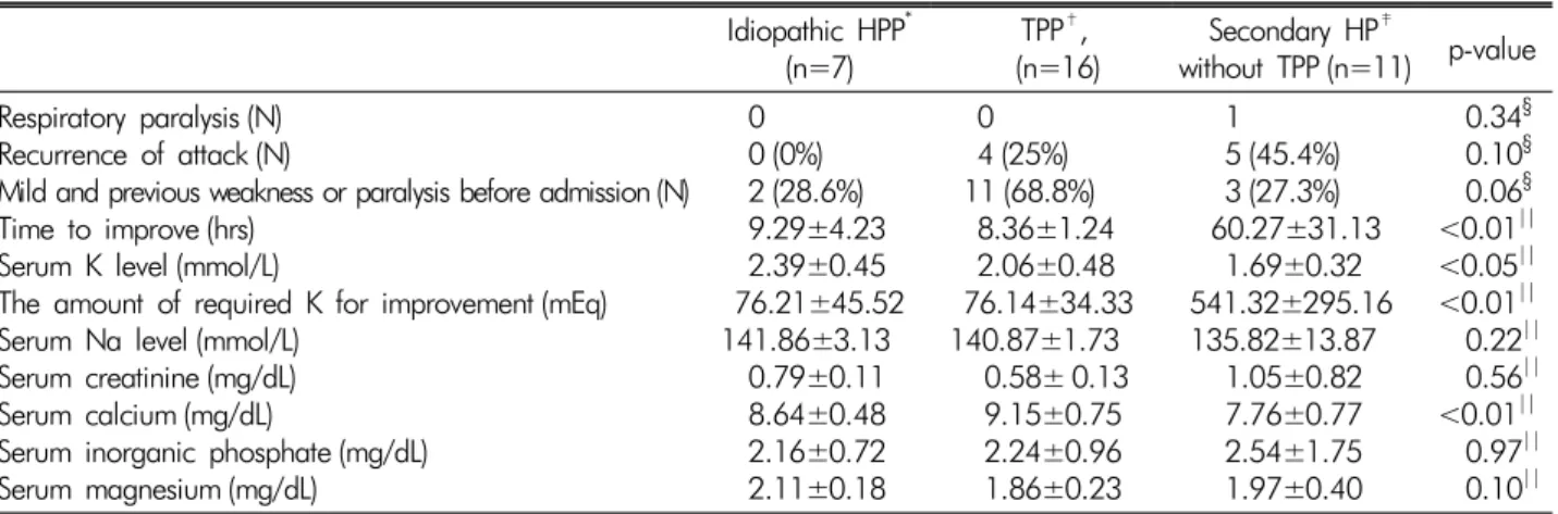
Table 1. Comparison of the clinical and biochemical parameters in idiopathic HPP, TPP and secondary HP without TPP Idiopathic HPP*
(n=7) TPP†,
(n=16) Secondary HP‡
without TPP (n=11) p-value Respiratory paralysis (N)
Recurrence of attack (N)
Mild and previous weakness or paralysis before admission (N) Time to improve (hrs)
Serum K level (mmol/L)
The amount of required K for improvement (mEq) Serum Na level (mmol/L)
Serum creatinine (mg/dL) Serum calcium (mg/dL)
Serum inorganic phosphate (mg/dL) Serum magnesium (mg/dL)
0 0 (0%) 2 (28.6%) 9.29±4.23 2.39±0.45 76.21±45.52
141.86±3.13 0.79±0.11 8.64±0.48 2.16±0.72 2.11±0.18
0 4 (25%)
11 (68.8%) 8.36±1.24 2.06±0.48 76.14±34.33 140.87±1.73 0.58± 0.13 9.15±0.75 2.24±0.96 1.86±0.23
1 5 (45.4%) 3 (27.3%) 60.27±31.13 1.69±0.32 541.32±295.16 135.82±13.87 1.05±0.82 7.76±0.77 2.54±1.75 1.97±0.40
0.34§ 0.10§ 0.06§
<0.01||
<0.05||
<0.01||
0.22||
0.56||
<0.01||
0.97||
0.10||
*HPP: hypokalemic periodic paralysis,†TPP: Thyrotoxic periodic paralysis,‡HP: hypokalemic paralysis, §: Two-by-K test, ||: Kruskal-Wallis test having secondary hypokalemic paralysis. Of the secon-
dary HP group, 16 patients (47.1%) were confirmed as having TPP. The secondary HP seen in the other patients was due to diuretics (n=4, 11.8%), licorice (n=2, 5.9%), Gitelman syndrome (n=2, 5.9%) or dRTA (n=2, 5.9%).
Fig. 1 shows the spectrum of hypokalemic paralysis in our hospital. Table 1 shows the clinical characteristics of the affected patients in each of the three groups. The paralytic attack in all of those in the idiopathic HPP group was caused by the first event in their life time.
In the TPP group, 11 patients (68.8%) had previously expe- rienced mild weakness or paralysis of the limb, and 8 patients (50%) were showing repetitive paralysis before admission. In secondary hypokalemic paralysis without
TPP, almost all of the patients (81.9%) had recurrences of the paralytic attack. In the idiopathic HPP and TPP groups, the mean recovery time (defined as resolution time of the paralysis or hypokalemia) was 9.29 hrs (SD±
4.23) and 8.36 hrs (SD±1.24), respectively. On the other hand, in the secondary HPP group, the mean recovery time was 60.27 hrs (SD±31.13).
Table 2 presents the clinical features of patients in the thyrotoxic and idiopathic HPP group. The mean age was 31.38 years and 30.29 years, respectively. In TPP, 75%
of the patients were in their 20-30’S (Fig. 2) and 50%
of the patients had identifiable precipitating causes such as exercise, alcohol intake, carbohydrate-rich meal or up- per respiratory infection (URI). Patients in the idiopathic
Fig. 2. Age distribution of thyrotoxic periodic paralysis.
Table 2. Clinical features of patients in the thyrotoxic and idiopathic hypokalemic periodic paralysis groups
TPP* Idiopathic HPP† p-value
Number (Male : female) 16 (16 : 0) 7 (7 : 0) -
Age (years) 31.38±8.87 30.29±7.16 0.40‡
Positive family history (N) 1 0 -
Respiratory paralysis (N) 0 0 -
Recurrence of attacks (N) 3 0 0.22§
Age at first attack N (%) N (%) 0.42§
11-20 1 (6.3%) 1 (14.3%)
21-40 12 (75%) 6 (85.7%)
>41 3 (18.6%) 0 (0%)
Precipitating cause of first attack N (%) N (%) -
Exercise 3 (18.6%) 1 (14.3%)
High carbohydrate intake 1 (6.3%) 0 (0%)
Alcohol intake 3 (18.6%) 3 (42.9%)
URI 1 (6.3%) 1 (14.3%)
Unknown 8 (50%) 2 (28.6%)
Time of first attack N (%) N (%) 0.28§
Day (6 a.m.– 2 p.m.) 2 (12.5%) 2 (28.6%)
Night (2 p.m.– 10 p.m.) 10 (62.5%) 5 (71.4%)
During sleep (10 p.m. – 6 a.m.) 4 (25%) 0 (0%)
Recovery time after treatment (hrs) 8.36±4.63 9.29±4.23 0.63‡
<12 hrs 14 (87.5%) 4 (57.1%) 0.14§
12 hrs <t <24 hrs 2 3 (42.9%)
>24 hrs 0 0
*TPP: thyrotoxic periodic paralysis,†HPP: hypokalemic periodic paralysis,‡: Student’s T-test,§: Two-by-K test
HPP and TPP groups had similar clinical features.
Almost all patients (87.5%) with TPP recovered within 24 hours, but only 57.1% of the patients with idiopathic HPP recovered within this time period. Among the pa- tients of the idiopathic HPP group, the paralytic attack
usually happened in the morning or afternoon, but among the TPP group, the paralytic attack mainly occurred in the afternoon or while in sleep.
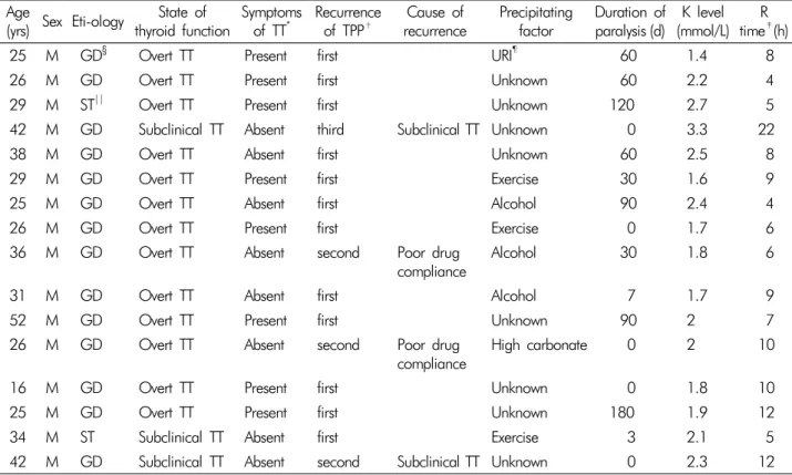
Table 3 shows the clinical features of TPP in this study.
From among 50%(8/16) of the patients, clinical signs and
Table 3. Clinical features of thyrotoxic periodic paralysis Age
(yrs) Sex Eti-ology State of
thyroid function Symptoms
of TT* Recurrence
of TPP† Cause of
recurrence Precipitating
factor Duration of
paralysis (d) K level (mmol/L) R
time‡(h)
25 M GD§ Overt TT Present first URI¶ 60 1.4 8
26 M GD Overt TT Present first Unknown 60 2.2 4
29 M ST|| Overt TT Present first Unknown 120 2.7 5
42 M GD Subclinical TT Absent third Subclinical TT Unknown 0 3.3 22
38 M GD Overt TT Absent first Unknown 60 2.5 8
29 M GD Overt TT Present first Exercise 30 1.6 9
25 M GD Overt TT Absent first Alcohol 90 2.4 4
26 M GD Overt TT Present first Exercise 0 1.7 6
36 M GD Overt TT Absent second Poor drug
compliance Alcohol 30 1.8 6
31 M GD Overt TT Absent first Alcohol 7 1.7 9
52 M GD Overt TT Present first Unknown 90 2 7
26 M GD Overt TT Absent second Poor drug
compliance High carbonate 0 2 10
16 M GD Overt TT Present first Unknown 0 1.8 10
25 M GD Overt TT Present first Unknown 180 1.9 12
34 M ST Subclinical TT Absent first Exercise 3 2.1 5
42 M GD Subclinical TT Absent second Subclinical TT Unknown 0 2.3 12
*TT: thyrotoxicosis, †TPP: thyrotoxic periodic paralysis, ‡R time: recovery time, §GD: Grave’s disease, ||ST: subacute thyroiditis,
¶URI: upper respiratory infection
symptoms of hyperthyroidism were absent at the time of the TPP diagnosis. The mean serum TSH level was 0.11±0.56 μU/mL (reference range: 0.30-4.00 μU/mL) in 16 patients. The serum T3 and FT4 levels were 247.75±
83.38 ng/dL (reference range: 80-200 ng/dL) and 3.12±
1.23 ng/dL (reference range: 0.77-1.94 ng/dL), respectively.
Among the TPP group, only 3 out of the 16 patients (18.6
%) had a previous history of hyperthyroidism. Two patients were poorly compliant with their medications and 1 patient was in a euthyroid state despite having a TSH level lower than the reference range. Of the 16 patients with TPP, 13(81.3%) had clinically overt hyperthyroidism and Graves’
disease at presentation while the other 3 (29.7%) had sub-clinical hyperthyroidism. Either positive TBII or anti- TG antibodies, or both were detected in all patients diag- nosed with Graves’ disease. After discharge, 2 out of 3 patients with sub-clinical hyperthyroidism were diagno- sed as subacute thyroiditis.
Discussion
The episodes of hypokalemic paralysis of 27 (79.4%) from among the patients in our study were secondary to the pre-existing condition, including dRTA, Gitelman syndrome, primary hyperaldosteronism, diuretic or licorice induced HPP, and thyrotoxicosis. Idiopathic (primary) hypokalemic periodic paralysis was occurred from only 20.6% of the patients. Secondary hypokalemic paralysis was due to thyrotoxicosis from 47.1% of the patients; diu- retics from 11.8%, and dRTA, Gitelman syndrome and licorice each represented 5.9%, and primary hyperaldos- teronism from 2.9%. The etiology of hypokalemic paraly- sis has been shown varied across from different ethnic and geographical areas6-7). In a study from Taiwan, the majority of hypokalemic paralysis cases were secondary to pre-existing conditions (68%), although some sporadic
and familial cases were identified. The underlying etiolo- gies in that study included TPP (40.2%), sporadic (29.8%), familial (2.1%), primary aldosteronism (6.2%), renal tub- ular alkalosis, Bartter and Gitelman syndrome (6.2%), di- uretic use (3.1%), and ingestion of toluene blue (3.1%)4), which is consistent with previous studies demonstrating TPP as the most common cause in Chinese and Japanese patients. Contrastingly, in Caucasian and Indian, familial HPP is the most common cause of hypokalemic paralysis, whereas in the Asian population, TPP is the most com- mon cause5-6,14). There had been a paucity of reports docu- mented on the etiology of hypokalemic paralysis in Korea, and therefore this might be the first to report that TPP is the most common cause of hypokalemic paralysis in Korea.
Earlier work has shown that mutations in the CACNA1S gene, which encodes the alpha 1 sub-unit of the skeletal muscle L-type voltage-dependent calcium channel, are responsible for the majority (70%) of the cases of familial HPP8,9). Missense mutations in the SCN4A gene, which encodes the skeletal muscle voltage-gated sodium channel alpha sub-unit, account for approximately 10% of the cases. Each mutation has different penetrance of pheno- type, which also depends on gender10-11). Several Korean cases of familial HPP have been reported, but cases of previously identified gene mutations are rare. Even with- out genetic analysis associated with calcium channel or sodium channel in this study, the idiopathic HPP in our study could be regarded as a sporadic form in view of the absence of the familial history or previous symptoms.
This is consistent with previous reports that most of the HPP patients in Western countries were of familial forms, whereas those reported from Asia were predominantly sporadic4). This raises the possibility that HPP in different ethnic settings may be linked to different types of muta- tions or to different environmental factors12). We were not able to investigate for the presence of mutation in patients with idiopathic HPP, and it seems clear that more genetic studies should be necessary.
We divided the patients with secondary hypokalemic
paralysis into two groups, because TPP was clearly the most common cause of hypokalemic paralysis in Korea and it had a distinct clinical character from the other causes of secondary HP. The recurrence of paralytic attacks, presence of respiratory paralysis as a clinical feature, and most serum electrolyte (sodium, creatinine, inorganic pho- sphate or magnesium) levels were similar in all groups.
There were significant differences in the serum potassium and serum calcium levels, and in the required potassium for the improvement among the three groups. In accord- ance with earlier reports, patients of secondary hypoka- lemic paralysis required a larger amount of potassium for the clinical improvement and had a longer recovery time compared with those who have idiopathic HPP or TPP13). Patients of secondary hypokalemic paralysis had a signi- ficantly negative total body potassium balance, whereas idiopathic HPP and TPP were associated with only intra- cellular shift of potassium, thus requiring a less amount of supplemental potassium14).
The precipitating causes of paralytic attacks were sim- ilar across all groups, including exercise, URI and alcohol intake. The patient’s ages at first attack were also similar.
Premoni- tory symptoms such as mild weakness and para- lysis of both lower limbs, were more common in the TPP group than the idiopathic HPP group, which may repre- sent one of the most important clinical differences be- tween two groups.
The incidence of TPP among Asians, the most frequen- tly affected population, is approximately 2%. While pre- viously TPP was largely unknown in the West, the num- ber of reported cases in Western countries has increased recently15). All reported TPP patients were sporadic cases without a family history of TPP, with the exception of one patient. This feature is important for differentiating TPP from familial hypokalemic periodic paralysis, which is more common among Caucasians16). Since most cases of idiopathic HPP in Korea were the sporadic forms, it seemed difficult to distinguish between TTP and the idio- pathic HPP only by means of the presence or absence of
familial history and the previous symptoms.
Despite the higher female incidence of thyrotoxicosis, TPP predominantly appears to affect males, and the ratio of male to female is 20:1.16 All of our cases were male.
The youngest case was 16 and the oldest was 52 years old;
most of the cases appeared between in the third or forth decades of life. No relationship between the age-onset of the disease and the complete recovery time was found in our study. The reasons for the male preponderance and the tendency of TPP onset between 20-40 years old remains unclear17-18). In hyperthyroid males, significantly increased plasma testosterone levels have been reported19). In Graves’disease, the plasma concentration of 17-β-estra- diol is increased in males but remains unchanged in fema- les20). The potassium channels in smooth muscle cells are acti- vated by testosterone and estrogen in animal studies21,22). Furthermore, the age distribution of TPP patients coin- cides with the commonly observed age distribution of not only hyperthyroidism but the rapid increase in testoster- one production. However, currently, we do not have any concrete evidence to prove the effects of sex hormones and potassium channels on TPP; this might be an impor- tant direction for future research.
The leading predictor of TPP in large case series was Graves’ disease23-24).
We should take note of that Graves' disease was also the most common cause of TPP in our study. Subacute- thyroiditis was also an etiological factor in our study. A thyrotoxic state is generally considered necessary for the induction of TPP, however, some studies have reported that acute paralysis had occurred in even in a euthyroid state after a treatment of the thyrotoxicosis25-26). Most of the patients included in our study had overt thyrotox- icosis while 3 patients had only subclinical thyrotoxicosis.
We suggest that doctors should consider TPP even if a patient is under taking medicine for thyrotoxicosis or is having the possibility of subclinical hyperthyroidism, i.e., an apparent euthyroid state. Accordingly, those with hy- perthyroidism and TPP should not be presumed to have
Graves’ disease, for some of them may experience hypo- thyroidism after subacute thyroiditis.
The patients with secondary HPP without TPP in our study required a longer recovery time compared with those who have idiopathic HPP or TPP. Those patients with secondary HP had a significant negative total body potas- sium balance, and required larger amounts of potassium compared with idiopathic HPP and TPP groups.
Because of the limitations on the retrospective data, the relatively small number of patients and restriction to a single center in this study, further prospective large- scale studies are required for delineating the spectrum of etiologies, clinical findings, treatment outcomes, and predictors of hypokalemic periodic paralysis.
In conclusion, our study revealed that, in Korea, idio- pathic (primary) hypokalemic periodic paralysis occurred in 20.6% of the patients. The most common cause of hypo- kalemic paralysis in Korea was thyrotoxicosis (47.1%).
References
1. Ahlawat SK, Sachdev A: Hypokalemic paralysis. Postgrad Med J 75:193-197, 1999
2. Kalita J, Nair PP, Kumar G, et al.: Renal tubular acidosis presenting as respiratory paralysis: Report of a case and review of literature. Neurol India 58:106-108, 2010 3. Stedwell RE, Allen KM, Binder LS: Hypokalemic paraly-
ses: a review of the etiologies, pathophysiology, presen- tation, and therapy. Am J Emerg Med 10:143-148, 1992 4. Lin SH, Lin YF, Halperin ML: Hypokalemia and paralysis.
Q J Med 94:133-139, 2001
5. Ko GTC, Chow CC, Yeung VTF, Chan HHL, Li JKY, Cockram CS: Thyrotoxic periodic paralysis in a Chinese population. Q J Med 89:463-468, 1996
6. Ober KP: Thyrotoxic periodic paralysis in the United States. Report of 7 cases and review of the literature.
Medicine (Baltimore) 71:109-120, 1992
7. Sinharay R: Hypokalaemic thyrotoxic periodic paralysis in an Asian man in the United Kingdom. Emerg Med J 21:120-121, 2004
8. Sternberg D, Maisonobe T, Jurkat-Rott K, et al.: Hypo- kalemic periodic paralysis type 2 caused by mutations
at codon 672 in the muscle sodium channel gene SCN4A.
Brain 124:1091-1099, 2001
9. Fontain B, Vele-Santos JM, Jurkat-Rott K, et al.: Mapping of hypokalemic periodic paralysis locus to chromosome 1q31-q32 in three European families. Nat Genet 6:267- 272, 1994
10. Grosson CL, Esteban J, Mckenna-Yasek D, Gusella JF, et al.: Hypokalemic periodic paralysis mutations: confir- mation of mutation and analysis of founder effect. Neuro- muscul Disord 6:27-31, 1966
11. Fouad G, Dalakas M, Servidei S, et al.: Genotype-pheno- type correlations of DHP receptor alpha 1-subunit gene mutations causing hypokalemic periodic paralysis. Neu- romusc Disord 7:33-38, 1997
12. Jurkat-Rott K, Mitrovic N, Hang C, et al.: Voltage-sensor sodium channel mutations cause hypokalemic periodic paralysis type 2 by enhanced inactivation and reduced current. Proc Natl Acad Sci USA 97:9549-9554, 2000 13. Lin SH, Chiu JS, Hsu CW, et al.: A simple and rapid
approach to hypokalemic paralysis. Am J Emerg Med 21:
487-491, 2003
14. Pradeep Kumar Maurya, Jayantee Kalita: Usha Kant Misra Postgrad. Med J 86:692-695, 2010
15. Miyashita Y, Monden T, Yamamoto K, et al.: Ventricular fibrillation due to severe hypokalemia induced by steroid treatment in a patient with thyrotoxic periodic paralysis.
Internal Medicine 45:11-13, 2006
16. Lin SH, et al.: Thyrotoxic periodic paralysis. Mayo Clinic Proceedings 80:99-105, 200
17. Kung AW: Clinical review: thyrotoxic periodic paralysis:
a diagnostic challenge. The Journal of Clinical Endocri- nology and Metabolism 91:2490-2495, 2006
18. McFadzean AJ, Yeung R: Periodic paralysis complicating thyrotoxicosis in Chinese. British Medical Journal 1:451- 455, 1967
19. Gordon GG, Southren AL: Thyroid-hormone effects on steroid-hormone metabolism. Bulletin of the New York Academy of Medicine 53:241-259, 1977
20. Olivo J, Gordon GG, Rafii F, et al.: Estrogen metabolism in hyperthyroidism and in cirrhosis of the liver. Steroids 26:47-56, 1975
21. Deenadayalu VP, White RE, Stallone JN, et al.: Testos- terone relaxes coronary arteries by opening the largecon- ductance, calciumactivated potassium channel. American Journal of Physiology. Heart and Circulatory Physiology 281:1720-1727, 2001
22. White RE, Han G, Maunz M, et al: Endotheliuminde- pendent effect of estrogen on Ca(2+)-activated K(+) channels in human coronary artery smooth muscle cells.
Cardiovascular Research 53:650-661, 2002
23. Manoukian MA, Foote JA, Crapo LM: Clinical and met- abolic features of thyrotoxic periodic paralysis in 24 epi- sodes. Archives of Internal Medicine 159:601-606, 1991 24. Silva MR, Chiamolera MI, Kasamatsu TS, Cerutti JM,
Maciel RM: Thyrotoxic hypokalemic periodic paralysis, an endocrine emergency: clinical and genetic features in 25 patients. Arquivos Brasileiros de Endocrinologiae Meta- bologia 48:196-215, 2004
25. Rone JK, Brietzke SA: Euthyroid thyrotoxic periodic paralysis. Military Medicine 156:434-436, 1991 26. Coates JT, Mirick MJ, Rubino FJ: Thyrotoxic periodic
paralysis with relapse during the euthyroid state. Wiscon- sin Medical Journal 86:20-22, 1987