INTRODUCTION
Inflammation is nonspecific response to tissue injury or microbial in- vasion, and its pathologic processes are complex. Inflammatory re- sponse is regulated by immune cells releasing different types of cyto- kines and inflammatory enzymes [1]. Macrophages are known to be critical immune cells in various inflammation responses [2]. Macro- phages, which are activated by lipopolysaccharides (LPS), secrete various inflammatory cytokines and enzyme such as tumor necrosis factor (TNF)-α, Nitric Oxide (NO), inducible nitric oxide synthase (iNOS), cy- clooxygenase (COX)-2, interleukin (IL)-1β and prostaglandin E2 (PGE2) [3]. RAW 264.7 macrophages are widely used in inflammation models to assess anti-inflammatory effect and usually treated with LPS to in- duce inflammatory response.
LPS are the components of the outer membrane of Gram-negative
bacteria. They act as endotoxins, leading to tissue injury. Activated LPS increase the production of pro-inflammatory cytokines, as previously mentioned, from macrophages or monocytes [4]. NO, an endogenous free radical, plays an important role in host defense, immunity and modulation of inflammatory responses, and is produced by a family of NOS such as endothelial NOS (eNOS), neuronal NOS (nNOS) and iNOS. In particular, iNOS, which is induced by LPS, produces large amount of nitric oxide [5].
The overproduction of nitric oxide can promote tissue injury and contribute to the progression of diseases such as sepsis, ulcerative colitis, diabetes, rheumatoid arthritis, osteoarthritis, and other diseases [6,7].
Several studies have reported that the inhibition of nitric oxide produc- tion through the down-regulation of iNOS expression, has anti-inflam- matory effects [8,9]. In addition, experimental animal models studies have also have shown that the reduced production of iNOS attenuates
Anti-Inflammatory Effects of Chrysanthemum indicum Water Extract in RAW 264.7 Cell as a Whole Plant
Kyoungah Kang
Department of Nursing, Kunsan National University, Gunsan, Korea
Purpose: Chrysanthemum indicum (CHI) has been used for edible and medical purposes for a long time in Korea. The purpose of this study was to evaluate the anti-inflammatory effects of CHI water extract in lipopolysaccharides (LPS)-induced RAW 264.7 macro- phage cells. Methods: To investigate the anti-inflammatory effects on LPS-induced RAW 264.7 macrophage cells, CHI extract as a whole plant was used in this study. RAW 264.7 cells were treated with various concentrations of CHI extract (1, 10, and100 μg/mL). Af- ter that Nitric Oxide (NO), inducible nitric oxide synthase (iNOS), interleukin (IL)-1β, cyclooxygenase (COX)-2 and prostaglandin E2
(PGE2) expression level were measured. Results: CHI extract significantly suppressed the LPS-induced NO production and decreased the level of iNOS, IL-1β, COX-2 messenger ribonucleic acid (mRNA) expression and also the down regulation of PGE2 expression in a dose-dependent manner. Conclusion: The present study suggested that CHI extract can be substituted for anti-inflammatory drugs and provide a safe and effective non pharmacological therapeutic approach.
Key Words: Anti-inflammation; Chrysanthemum indicum; RAW 264.7 cell
Corresponding author: Kyoungah Kang
Department of Nursing, Kunsan National University, 558 Daehak-ro, Gunsan 54150, Korea Tel: +82-63-469-1999 Fax: +82-63- 469-7429 E-mail: [email protected]
* This paper was supported by research funds of Kunsan National University.
Received: October 30, 2015 Revised: November 5, 2015 Accepted: November 9, 2015
This is an Open Access article distributed under the terms of the Creative Commons Attribution Non-Commercial License (http://creativecommons.org/licenses/by-nc/3.0) which permits unrestricted non-commercial use, distribution, and reproduction in any medium, provided the original work is properly cited.
systemic lupus erythematosus (SLE), Crohn’s disease and osteoarthritis [10-12]. COX is inflammatory enzyme to convert arachidonic acid to prostaglandins which are divided into two subtypes such as COX-1 and COX-2. Among them, COX-2 is related to inflammatory response.
PGE2, which is produced by COX-2, is the cause of pain and fever, and also well-known to promote angiogenesis in cancer conditions [13].
Currently nonsteroidal anti-inflammatory drugs (NSAIDs) are the most commonly used for the treatment of inflammatory diseases through the suppression of COX activity, but it is well known that they have gastrointestinal side effects such as indigestion, stomach upset or gastritis and other common side effects of NSAIDs include headache, raised liver enzymes, dizziness, hypertension [14]. In order to reduce those side effects, recent studies are being focused on searching new anti- inflammatory agents, which are based on biosynthesis using a herb, such as green tea, mushroom and compositae plant [2,15,16]. Among them, compositae plant is a flowering plant which spreads all over the world and has been used for edible and medical purposes for a long time in Korea. And there are several types of compositae plants such as Den- dranthema zawadskii var. latilobum, Dandelion (Taraxacum officinale) and Chrysanthemum indicum (CHI) [17]. Recent studies have demon- strated that CHI has high anti-inflammatory and antioxidant effect through the down-regulation of IL, TNF-α expression. But most of the previous studies were used a specific part of a compositae plant such as stem, leaf and flower, which were extracted using an ethanol [8,16-19].
Few of existing studies, except one that analyzed the antioxidative effects of Dandelion [20] that is one kind of CHI, have attempted to examine the anti-inflammatory effect of CHI using water. Instead, most of them used ethanol to examine the anti-inflammatory effect of CHI focusing on a specific part of the plant such as root, stem, or leaves. Therefore, the present study was aimed to verify whether all components of CHI have anti-inflammatory effect, when they were extracted under the same conditions and the result of this study will provide an evidenced-base of safe and effective non-pharmacological therapeutic approach.
MATERIALS AND METHODS
1. Reagents
LPS and Dulbecco’s modified Eagle medium (DMEM) were pur- chased from Sigma-Aldrich Corporation (St. Louis, USA). Primary anti- bodies for iNOS, COX-2, actin, and the peroxidase-conjugated second-
ary antibodies were purchased from Santa Cruz Biotechnology (Santa cruz, CA, USA).
2. Sample preparation
The dried flowers of CHI were extracted with distilled water (dH2O) (100g, dry weight, whole plant), and the extract was then concentrated under reduced pressure. The decoction was filtered through a 0.45 μm filter, lyophilized and stored at -20°C. The lyophilized powder was dis- solved in 0.1% dimethyl sulfoxide (DMSO) and then diluted in phos- phate buffered saline (PBS).
3. Cell culture
RAW 264.7 cells were obtained from Korean Cell Line Bank (KCLB, Seoul, Korea). The cells were cultured in a 5% CO2 incubator at 37°C with DMEM supplemented with 10% fetal bovine serum (FBS) and 1% ampi- cillin and streptomycin. The entire reagents for cell culture were pur- chased from Life Technologies (Grand Island, NY, USA). Treatments with LPS and/or CHI extract were carried out under serum-free conditions.
4. NO assay and cell viability assay
In order to determine the NO production, RAW 264.7 cells were add- ed to a 96-well plate at a density of 3×10⁴ cells/well and cultured for 16 hours in DMEM medium. Cells were treated with various concentra- tions (1, 10, and100 μg/mL) of CHI extract and then incubated with LPS (100 ng/ml) for 24 hours. The supernatant (50 µL) was harvested and mixed with an equal volume of Griess reagent (Sigma, MO, USA) in a 96 well plate for 10 minutes at a room temperature. Finally, the absorbance values were measured at 570 nm using a microplate reader (Molecular Devices, CA, USA). At this time, sodium nitrite was used as a standard to calculate NO concentration. The 3-(4, 5-Dimethylthiazol-2-yl)-2, 5-diphenyltetrazolium bromide (MTT) assay was used for the effect of CHI on cell viability. Cells were cultured in a 96-well plate at a density of 3×10⁴ cells/well and treated with various concentrations (1, 10, and 100 μg/mL) of CHI extract for 24 hours. And then the supernatant was dis- carded and MTT solution 100 µL (5 μg /mL) was added to each well and incubated for 1 hour. at 37°C then the absorbance values were measured at 570 nm using a microplate reader.
5. Real time polymerase chain reaction (RT-PCR)
iNOS, COX-2, and IL-1β messenger ribonucleic acid (mRNA) expres-
sion levels were measured by real time PCR. RAW 264.7 cells were cul- tured in a 6-well plate for 24 hours then they were treated with both CHI extract (1, 10, and 100 μg/mL) and LPS (100 ng/mL) and cultured for an- other 24 hours. The harvested cells were washed with PBS, total RNA was isolated with Tri- reagent (Trizol) according to the manufacturer’s instructions, and 1 μg of complementary deoxyribonucleic acid (cDNA) from the RNA samples were synthesized using Superscript II Reverse Transcriptase and oligo-dT (Invitrogen, CA, USA). The synthesized cDNA and SYBR green mixture reagent (Bio-rad, CA USA) were mixed and they were incubated at 94°C for 45 seconds, at 60°C for 30 seconds, and at 72°C for 30 seconds, respectively. This process was repeated 26 cycles to amplify them and glyceraldehyde 3-phosphate dehydrogenase (GAPDH) was used as an reference gene. Forward and reverse primers were synthesized on known gene sequences RT-PCR was performed us- ing a CFX 96 Real -Time PCR Detection System (Bio-Rad, CA, USA), according to the manufacturer’s instructions. The primers used in this
experiment were shown in Table 1.
6. Measurement of PGE2
The sandwich enzyme-linked immunosorbent assay (ELISA) kits used for PGE2 were obtained from R&D Systems (Cayman Chemical, MI, USA). RAW 264.7 cells were added at 3×10⁴ cells/well and incubat- ed in an incubator in the same manner as NO measurement. And then the culture medium was discarded and serum free media was treated with both CHI extract and LPS (100 ng/ml), at the same time, under the condition of 5 % CO2, 37°C and 24 hrs. Media from culture medium was harvested and centrifuged at 12,000 rpm for 10 minutes to discard cell debris. PGE2 level in the culture supernatants was measured by ELI- SA kit, according to manufacturer’s direction.
7. Statistical analysis
Experiments were performed at least 3 times. All data were presented as mean±SD and statistical significance was determined by Student’s t- test. p<.05 was considered as statistically significant.
RESULT
1. Effects of CHI extract on LPS-induced NO production & cell viability
The effects of CHI extract on LPS-induced nitrite production in RAW 264.7 cells were measured for the amount of nitrite released into the culture medium. CHI extract significantly reduced the LPS-stimu- lated nitrite production, in a dose-dependent manner. Particularly, ni- trite production was suppressed more than 70 % at 100 μg/mL CHI ex- Table 1. The Sequence of Primers used in the Real Time PCR
Gene Primer sequences (5´"3´)
iNOS CCG TCC ACA GTA TGT GAG GA (sense) GAA CTC CAA GGT GGC AGC A (anti-sense) IL-1β CCG TGG ACC TTC CAG GAT GAG (sense)
ACC AGT TGG GGA ACT CTG CAG (anti-sense) COX-2 GAA GGG ACA CCC TTT CAC AT (sense)
ACA CTC TAT CAC TGGC ATC C (anti-sense) GAPDH TGA AGG TCG GAG TCA ACG GAT TTG GT (sense)
CAT GTG GGC CAT GAG GTC CAC CAC (anti-sense) PCR= Polymerase chain reaction; iNOS= Inducible nitric oxide synthase;
IL-1β = Interleukin-1β; COX-2 = Cyclooxygenase-2; GAPDH = Glyceraldehyde 3-phosphate dehydrogenase.
30 25 20 15 10 5 0
* *
**
- + + + +
- - 1 10 100
LPS (100 ng/mL) CHI (μg/mL)
Nitricoxide (μM)
1.5 1.2 0.9 0.6 0.3 0
- + + + +
- - 1 10 100
LPS (100 ng/mL) CHI (μg/mL)
Cell viability (ratio)
Figure 1. Effect of CHI extract on LPS-induced nitric oxide (A) and cell viability (B) in RAW 264.7 cell line. Cells were treated LPS and indicated con- centration of CHI extract for 24 hours. CHI extract significantly reduced the LPS-stimulated nitrite production compared with LPS alone treated group in a dose-dependent manner. There was no cytotoxicity on RAW 264.7 cells with CHI extract up to 100 μg/mL.
*p< .05, **p< .01 compared to LPS stimulation. CHI= Chrysanthemum indicum; LPS= Lipopolysaccharides.
(A) (B)
tract, compared with only LPS treated group (Figure 1A). The cytotoxic effect of CHI extract on LPS-induced RAW 264.7 cells was assessed us-
ing the MTT assay. There was no cytotoxicity on RAW 264.7 cells with CHI extract up to 100 μg/mL and cell viability was not significantly al- tered by doses of CHI extract (Figure 1B).
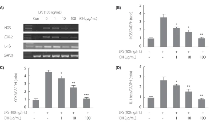
2. Effects of CHI extract on mRNA expression levels of iNOS, COX-2 and IL-1β
To verify the inhibiting mechanism of CHI on NO production, this study used real time PCR to measure mRNA expression levels of iNOS.
In addition, it also confirmed mRNA expression levels of COX-2 and IL-1β, which are believed to cause inflammation. As a result, it was found that CHI inhibits iNOS mRNA expression levels, depending on concentration. In particular, the experimental group treated with 100 μg/mL of CHI showed statistically significant difference in the inhibitive effect on inflammatory response from the control group. As for its effect on COX-2, CHI also reduced the mRNA expression of COX-2 1.5 times at 100 μg/mL than by LPS, which increased the expression 4.5 times. In indicates that CHI extract also have anti-inflammatory effect on COX-2 mRNA expression, and the decrease of IL-1β by CHI extract was also observed (Figure 2).
3,000 2,500 2,000 1,500 1,000 500 0
*
*
**
- + + + +
- - 1 10 100
LPS (100 ng/mL) CHI (μg/mL)
PGE2 (μM)
Figure 3. Effects of CHI extract on protein expression of PGE2. Cells were treated with 1, 10, and 100 μg/mL of CHI extract and 100 ng/mL of LPS for 24 hours. Then 100 μL of the cell suspension were collected and PGE2 protein were quantified by ELISA. PGE2 protein expression on LPS-induced RAW 264.7 cells was dramatically decreased in CHI extract treated groups in a concentration dependent manner compared with LPS treated group.
*p< .01, **p< .001 compared to LPS stimulation. CHI= Chrysanthemum indicum;
PGE2= Prostaglandin E2; LPS= Lipopolysaccharides; ELISA= Enzyme-linked immunosorbent assay.
LPS (100 ng/mL)
iNOS COX-2 IL-1β GAPDH
(CHI, μg/mL)
Con 0 1 10 100
5 4 3 2 1 0
* *
**
- + + + +
- - 1 10 100
LPS (100 ng/mL) CHI (μg/mL)
iNOS/GADPH (ratio)
5 4 3 2 1 0
*
**
***
- + + + +
- - 1 10 100
LPS (100 ng/mL) CHI (μg/mL)
COX2/GAPDH (ratio)
4 3 2 1 0
*
**
**
- + + + +
- - 1 10 100
LPS (100 ng/mL) CHI (μg/mL)
IL-1 beta/GADPH (ratio)
Figure 2. Effect of CHI extract on LPS-induced inflmmatory cytokien expression in RAW 264.7 cell line. The iNOS, COX-2 and IL-1β mRNA expression were analysed by real time PCR (A). All gene expressions were normalized using GAPDH as a reference gene. The iNOS (B), COX-2 (C) and IL-1β (D) mRNA expression were significantly decreased in CHI extract treated group compared with LPS treated group.
*p< .05, **p< .01, ***p< .001 compared to LPS stimulation. CHI=Chrysanthemum indicum; LPS= Lipopolysaccharides; iNOS= Inducible nitric oxide synthase;
COX-2= Cyclooxygenase-2; IL-1β = Interleukin -1β; PCR= Polymerase chain reaction; GAPDH= Glyceraldehyde 3-phosphate dehydrogenase.
(A)
(C)
(B)
(D)
3. Effects of CHI extract on protein expression of PGE2
PGE2 is produced through COX-2 activation. To verify whether CHI extract has anti-inflammatory effect on PGE2 as it does on COX-2 ex- pression in Figure 2. Sandwich ELISA was used to measure the expres- sion levels of PGE2 protein. As expected, PGE2 protein expression on LPS-induced RAW 264.7 cells was dramatically decreased in CHI ex- tract treated groups in a concentration dependent manner. In particular, the effect was significant even at 1 μg/mL, which reconfirms the anti-in- flammatory effect of CHI (Figure 3).
DISCUSSION
This study, we tried to find out the effects of non-pharmacological agent on the expression level of inflammatory mediators and to provide an evidence-based of non-pharmacological therapeutic approach. In or- der to investigate the anti-inflammatory effects on LPS-induced RAW 264.7 macrophage cells, CHI extract as a whole plant was used in this study. Recent studies reported that CHI extract significantly inhibited the expression of pro-inflammatory cytokines but most of the previous studies were used a specific part of CHI such as roots, stem, leaf and flower, which was extracted using an ethanol [18,19,21]. There was no study using whole plant extracts of chrysanthemum with water. For this reasons, present study would find out anti-inflammatory effects of CHI, which was extracted using water and used a whole plant, including roots, stems, leaves, and flowers.
Inflammation is a complex response regulated by a variety of im- mune cells through the releasing of different types of cytokines and in- flammatory enzymes such as of TNF-α, NO, iNOS, COX-2, IL-1β, and PGE2 [1]. In inflammatory response macrophages play a key role by reg- ulating inflammatory mediators such as NO, PGE2 and pro-inflamma- tory cytokines and also as host-defense mechanism [22]. RAW 264.7 macrophage is widely used in inflammation models to assess anti-in- flammatory effect. In this study, therefore RAW 264.7 macrophage cells were treated with LPS to induce inflammatory response. It is well known that the overproduction of inflammatory mediators is the cause of cancer or inflammatory diseases. Therefore, such anti-inflammatory medicine as steroids or NSAIDs has been used in order to inhibit the ac- tivity of anti-inflammatory mediators. Furthermore, NSAIDs drugs are used a long term in general and high doses are required to treat chronic inflammation disease so there are many side effects such as indigestion,
stomach upset, gastritis, headache and dizziness. Therefore, many stud- ies have made efforts to develop natural plant components that can sub- stitute these drugs to reduce side effects [2,15]. NO is produced by L-ar- ginin and plays an important role in modulating inflammatory re- sponses, also is involved in the activation of vasodilation, neurotrans- mitter system and immunity. The excessive amounts of NO are the cause of serious tissue injury. Particularly, iNOS, which is induced by LPS, produces large amount of NO [5].
In this study, CHI extract inhibited mRNA expression of iNOS and IL-1β as well as NO production. These results demonstrates that CHI extract suppresses iNOS expression and down regulation of iNOS and IL-1β enzymatic activity as a result of reduced NO production.
Several previous studies reported that compositae plant such as Stevia rebaudiana and dandelion has anti-inflammatory activity which acts via the suppression of NO and iNOS production [20,23]. A previous study which analyzed the anti-inflammatory effects of the isolated com- pounds from the flowers of CHI. The extract of CHI was rich in pheno- lic compounds and this compounds decreased iNOS and COX-2 pro- tein expression in a dose dependent manner [18]. COX-2 is related to biosynthesis of the PGE2 in inflammatory responses. Therefore, medi- cines that are currently used for the treatment of inflammation through the down regulation of PGE2 take advantage of the inhibition of COX-2 production and activity [13]. The results of the present study demon- strated that CHI extract suppressed PGE2 protein expression on LPS-in- duced RAW 264.7 cells significantly. This result is associated with the down-regulation of COX-2 mRNA. In several studies using natural plants such as green tea, Mori Folium and phloretin from apple wood, it was reported that they have anti-inflammatory effects through inhibit- ing of COX-2 and PGE2 expression [9,15,24]. In addition, the study that used ethanol to extract CHI by 25, 50,100, and 200 μg/mL, respectively, to analyze its anti-inflammatory effect reported that the CHI extract re- duced the activation of such anti-inflammatory cytokines as NO, COX- 2, TNF-α, and PGE2 depending on concentration [19]. However the present study used a lower concentration (1, 10, and 100 μg/mL) than in the previous studies and also confirmed the concentration-dependent effect of CHI extract on anti-inflammation response.
As a result of this study, it is demonstrated that CHI water extract has anti-inflammatory effect as the ethanol extract, also this study observed that the whole pant of CHI has the same effect as specific parts of the plant. Therefore, the present study proposed the scientific ground that
CHI water extract is a safer and more effective non-pharmacologic ther- apeutic approach to the treatment of inflammation. Therefore, it is be- lieved that this result can be used as the basic data for nursing education for patients with inflammation disease. Furthermore, the mechanism of anti-inflammatory effects of CHI still remains to be defined through more in vivo study. In conclusion, CHI water extract acts as an anti-in- flammatory agent in inflammatory conditions through the inhibition of NO production and down regulation of inflammatory mediators, iNOS, IL-1β, COX-2, and PGE2. This present study suggested that CHI water extract can be substituted for anti-inflammatory drugs and provide safer and more effective non pharmacological therapeutic approach.
CONCLUSION
This study was aimed to evaluate whether all components extract of CHI have anti-inflammatory effect, when they were extracted under the same conditions. RAW 264.7 cells were treated with various concentra- tions of CHI extract (1, 10, and100 μg/mL), and mRNA expression and protein levels of inflammatory mediators were confirmed. CHI extract significantly inhibited the levels of NO, iNOS, IL-1β, COX-2, and PGE2
in a dose-dependent manner. According to these result, CHI extract can substitute anti-inflammatory drugs and provide safer and more effective non pharmacological therapeutic approach. However, this study ana- lyzed only the effects of several pro-inflammatory cytokines. Therefore, further studies are necessary to be conducted to confirm down signaling pathways associated with these cytokines.
REFERENCE
1. Braun CA, Anderson CM. Pathophysiology functional alterations in human health. Philadelphia: Lippincott; 2007. p. 38-52.
2. Chen JN, de Mejia EG, Wu JSB. Inhibitory effect of a glycoprotein isolated from golden oyster mushroom (pleurotus citrinopileatus) on the lipopolysaccharide- induced inflammatory reaction in RAW 264.7 macrophage. Journal of Agricul- tural and Food Chemistry. 2011;59(13):7092-7097. http://dx.doi.org/10.1021/
jf201335g
3. Olefsky JM, Glass CK. Macrophages, inflammation, and insulin resistance. An- nual Review of Physiology. 2010;72:219-246. http://dx.doi.org/10.1146/an- nurev-physiol-021909-135846
4. Su GL. Lipopolysaccharides in liver injury: Molecular mechanisms of kupffer cell activation. American Journal of Physiology. 2002;283(2):256-265. http://
dx.doi.org/10.1152/ajpgi.00550.2001
5. Kubes P. Inducible nitric oxide synthase: A little bit of good in all of us. Gut.
2000;47:6-9. http://dx.doi.org/10.1136/gut.47.1.6
6. Tracey KJ. The inflammatory reflex. Nature. 2002;420(6917):853-859. http://
dx.doi.org/10.1038/nature01321
7. Nathan. C. Points of control in inflammation. Nature. 2002;420(6917):846-852.
http://dx.doi.org/10.1038/nature01320
8. Byun MW. Anti-inflammatory activity of austroinulin from stevia rebaudiana in LPS-induced RAW 264.7 cells. Journal of the Korean Society of Food Science and Nutrition. 2012;41(4):456-461. http://dx.doi.org/10.3746/jkfn.2012.41.4.456 9. Park SM, Byun SH, Kim YW, Cho IJ, Kim SC. Inhibitory effect of Mori Folium
ethanol extract on pro-inflammatory mediator in lipopolysaccharide - activat- ed RAW 264.7 cells. The Korea Journal of Herbology. 2012; 27(3):31-38. http://
dx.doi.org/10.6116/kjh.2012.27.3.31
10. Oates JC, Ruiz P, Alexander A, Pippen A MM, Gilkeson GS. Effect of late mod- ulation of nitric oxide production on murine lupus. Clinical Immunology and Immunopathology. 1997;83(1):86-92.
11. Zingarelli B, Szabo C, Salzman AL. Reduced oxidative and nitrosative damage in murine experimental colitis in the absence of inducible nitric oxide synthase.
Gut. 1999;45(2):199-209. http://dx.doi.org/10.1136/gut.45.2.199
12. Boileau C, Martel-Pelletier J, Brunet J, Tardif G, Schrier D, Flory C, et al. Oral treatment with PD-0200347, an alpha~2delta ligand, reduces the development of experimental osteoarthritis by inhibiting metalloproteinases and inducible nitric oxide synthase gene expression and synthesis in cartilage chondrocytes.
Arthritis and Rheumatology. 2005;52(2):488-500. http://dx.doi.org/10.1002/
art.20809
13. Kim JY, Jung KS, Jeong HG. Suppressive effects of the kahweol and cafestol on cyclooxygenase-2 expression in macrophages. FEBS Letters. 2004;569(1- 3):321-326. http://dx.doi.org/10.1016/j.febslet.2004.05.070
14. Crofford LJ. Use of NSAIDs in treating patients with arthritis. Arthritis Re- search and Therapy. 2013;15(Suppl 3):S2.
15. Kim MY, Son IP, Kim SY, Song YS, Chang HK, Yoo SJ, et al. Anti-inflammatory effect of green tea cell water in activated RAW 264.7 cells with lipopolysaccha- ride. Journal of Asthma, Allergy and Clinical Immunology. 2012;32(2):115-121.
16. Lee E. Anti-inflammatory activity of chrysanthemum indicum L. extract in lipo- polysaccharide-treated rats. Natural Product Sciences. 2009;15(2):55-59.
17. Woo JH, Shin SL, Lee CH. Antioxidant effects of ethanol extracts from flower species of compositae plant. Journal of the Korean Society of Food Science and Nutrition. 2010;39(2):159-164. http://dx.doi.org/10.3746/jkfn.2010.39.2.159 18. Luyen BT, Tai BH, Thao NP, Cha JY, Lee HY, Lee YM, et al. Anti-inflammatory
components of chrysanthemum indicum flowers. Bioorganic and Medicinal Chemistry Letters. 2015;25(2):266-269. http://dx.doi.org/10.1016/j.bmcl.2014.
11.054
19. Cheon MS, Yoon TS, Lee DY, Choi GY, Moon BC, Lee AY, et al. Chrysanthe- mum indicum Linné extract inhibits the inflammatory response by suppressing NF-κB and MAPKs activation in lipopolysaccharide-induced RAW 264.7 macrophages. Journal of Ethnopharmacology. 2009;122(3):473–477. http://
dx.doi.org/10.1016/j.jep.2009.01.034
20. Kang MJ, Shin SR, Kim KS. Antioxidative and free radical scavenging activity of water extract from dandelion (Taraxacum officinale). Korean Journal of Food Preservation. 2002;9(2):253-259.
21. Cheng W, Li J, You T, Hu C. Anti-inflammatory and immunomodulatory activi- ties of the extracts from the inflorescence of chrysanthemum indicum linne. Jour- nal of Ethnopharmacology. 2005;101(1-3):334-337. http://dx.doi.org/10.1016/
j.jep.2005.04.035
22. Kumar V. Innate lymphoid cells: New paradigm in immunology of inflamma- tion. Immunology Letters. 2014;157(1-2):23-37. http://dx.doi.org/10.1016/
j.imlet.2013.11.003
23. Lee MY, Lee JA, Seo CS, Ha H, Lee H, Son JK, et al. Anti-inflammatory activity of angelica dahurica ethanolic extract on RAW 264.7 cells via upregulation of heme oxygenase-1. Food and Chemical Toxicology. 2011;49(5):1047-1055.
http://dx.doi.org/10.1016/j.fct.2011.01.010
24. Ahmed S, Rahman A, Hasnain A, Lalonde M, Goldberg VM, Haqqi TM.
Green tea polyphenol epigallocatechin-3-gallate inhibits the IL-1b-induced ac- tivity and expression of cyclooxygenase-2 and nitric oxide synthase-2 in human chondrocytes. Free Radical Biology and Medicine. 2002;33(8):1097-1105.