www.jkfs.or.kr
101
비스포스포네이트 복용과 관련된 비전형 척골 골절에 대한 치료 - 증례 보고 -
김동수ㆍ박지강 ㆍ최의성ㆍ정호승ㆍ홍석현ㆍ안병현
충북대학교 의과대학 충북대학교병원 정형외과학교실
Treatment of Atypical Ulnar Fracture Associated with Bisphosphonate Therapy
- A Case Report -
Dong-Soo Kim, M.D., Ji-Kang Park, M.D., Ph.D. , Eui-Sung Choi, M.D., Ph.D., Ho-Seung Jeong, M.D., Seok-Hyun Hong, M.D., Byung-Hyun Ahn, M.D.
Department of Orthopaedic Surgery, Chungbuk National University Hospital, Chungbuk National University College of Medicine, Cheongju, Korea
Received December 4, 2019 Revised December 18, 2019 Accepted March 29, 2020 Correspondence to:
Ji-Kang Park, M.D., Ph.D.
Department of Orthopaedic Surgery, Chungbuk National University Hospital, Chungbuk National University College of Medicine, 776, 1Sunhwan-ro, Seowon-gu, Cheongju 28644, Korea Tel: +82-43-269-6077
Fax: +82-43-274-8719 E-mail: [email protected] Financial support: None.
Conflict of interests: None.
Bisphosphonates can cause atypical fractures when taken for a long time. Atypical fractures appear mainly as femoral subtrochanteric or shaft fractures. On the other hand, reports of atypical fractures in the proximal ulna are relatively rare, with a high proportion of nonunion cases. This paper reports a case of nonunion after fixation for atypical fractures of the proximal ulna.
Key Words: Atypical ulnar fracture, Nonunion, Bisphosphonate, Bone graft
Copyright © 2020 The Korean Fracture Society. All rights reserved.
This is an Open Access article distributed under the terms of the Creative Commons Attribution Non-Commercial License (http://creativecommons.org/licenses/by-nc/4.0) which permits unrestricted non-commercial use, distribution, and reproduction in any medium, provided the original work is properly cited.
CASE REPORT
J Korean Fract Soc 2020;33(2):101-104ISSN 1225-1682 (Print)ㆍISSN 2287-9293 (Online) https://doi.org/10.12671/jkfs.2020.33.2.101
비스포스포네이트는 골다공증 치료 및 골다공증성 골절 예방에 주로 사용되며 장기간 복용 시 비전형적 골절의 위험 이 있다.1) 비전형적 골절은 주로 대퇴골 전자하부 혹은 간부 에 발생하며 많은 환자 보고를 통해 치료에 대한 가이드라인 이 제시되고 있다.2) 이에 비해 척골의 비전형 골절은 드물게 보고되고 있으며 치료에 대한 합의는 없는 상태이다. 또한 척
골의 비전형 골절치료 후 발생한 불유합에 대한 치료 및 결과 에 대한 보고도 없는 상태이다. 5년간 비스포스포네이트 복 용을 하였던 84세 여자 환자로 우측 척골에 발생한 비전형 골절에 대해 금속 내고정 후 발생한 불유합에 대해 치료한 경 험을 문헌고찰과 함께 보고하고자 한다.
Journal of the Korean Fracture Society Vol. 33, No. 2, April 2020
102
증례 보고
84세 여자 환자가 우측 전완부 통증으로 지역 2차 의료기 관에 내원하였다. 과거력상 내원 5년 전부터 지역 1차 의료기 관에서 골다공증으로 5년간 매주 Risedronate 35 mg을 복용 하였다. 또한 뇌기능 개선제(choline alfoscerate)와 고혈압으 로 칼슘채널 차단제를 복용 중이었다. 단순 방사선 소견상 척 골 근위 1/3 부위 분쇄가 없는 횡적 골절이 관찰되었으며, 가 장자리 경화 및 주변 부 피질골 비대가 있었다(Fig. 1). 비스포 스포네이트 복용으로 인한 비전형 골절로 진단받았다. 골 소 파술 및 정복 후 잠김 금속판(locking plate)을 이용하여 금속 내고정술을 시행받았다. 이후 X-ray상 특이 소견이 없었으
나, 수술 3개월째 식사를 하던 중 식탁에 좌측 팔을 살짝 부 딪힌 후 금속 고정물 파손이 발생하였다(Fig. 2). 재입원하여 다시 골 소파술 후 rush pin 및 locking plate로 재고정을 하 였으며, 골절 사이 공간에 DBM (Genex DS; Biocomposites, Wales, England) 적용하였다(Fig. 3). 이후 단순 방사선 추시 시 불유합이 지속되었다.
이후 본원에 의뢰되었으며, 내원 당시 시행한 방사선 소견 상 우측 척골 근위 1/3 부위에 불유합 및 금속 해리소견이 관 찰되었다(Fig. 4). 혈청 검사상 비타민 D 저하(6.9 ng/ml), 부
A B
R R
Fig. 1. Initial anteroposterior (A) and lateral (B) radiographs show a transverse fracture of the right proximal ulna with cortical thickening and marginal sclerosis.
R
Fig. 2. After 3 months, there was no evidence of union, but metal breakage of the plate had occurred.
A B
R
Fig. 3. Anteroposterior (A) and lateral (B) radiographs after the second operation.
R R
1at 1at
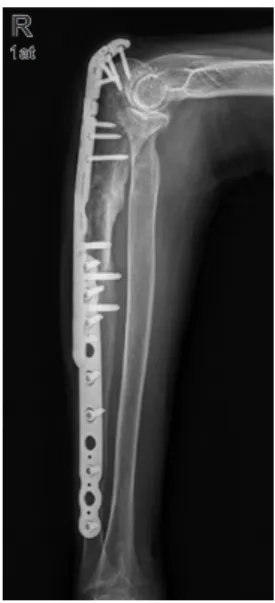
Fig. 4. Nine months after the second operation, radiological nonunion and implant loosening was observed.
Atypical Ulnar Fracture Associated with Bisphosphonate Therapy Dong-Soo Kim, et al.
103
갑상선 호르몬 상승(201.6 pg/ml)이 보였으며, 골밀도 검사상 요추부 T점수 –2.3점, 대퇴부 T점수 –3.1점이었다. 골주사 검사를 시행하였으나 다른 부위의 골절 양상은 관찰되지 않 았다.
수술은 전신 마취하에 앙와위 자세로 진행하였다. 불유 합 부위에 일부 섬유성 조직이 관찰되어 모두 제거하였으 며, 이후 골소파술을 시행하였다. 동측 전상장골극에서 해면 골을 채취하였다. Bone chip, Edenfuse 및 자가 해면골을 이 용해 골 이식을 한 뒤 Olecranon Plate (Synthes, Oberdorf, Switzerland)를 이용해 고정하였다. 골절판 끝 부위는 피로 골절 예방을 위해 Anatomical Forearm Plate (Hankiltech, Hwaseong, Korea)를 이용해 추가 고정하였다(Fig. 5).
술 후 3개월 단순 방사선 사진상 가골 형성이 관찰되었으 며, 술 후 1년 단순 방사선 사진상 완전한 골유합이 관찰되었 다. 술 후 1년 10개월까지 외래 추시를 하였으며, 전완부 불편 감 및 통증은 없었고 주관절 운동 범위 또한 건측과 같았다 (Fig. 6).
고 찰
비전형 대퇴골 골절의 경우 Mahjoub 등3)의 보고에 의하 면 92.6%에서 비스포스포네이트 복용력이 있으며, 특히 3년 이상 복용할 경우 그 위험성이 더 높은 것으로 알려져 있다.4) Ito 등5)의 보고에 따르면 이전 발표되었던 비전형 척골 골절 14명의 환자 모두 비스포스포네이트를 복용하였으며, Tan 등6)
이 보고한 7예에서는 모두 7년 이상 장기간 복용을 하였다. 본 증례에서는 5년간 비스포스포네이트를 복용하였으며, 위의 문헌과 종합해 보았을 때 비스포스포네이트를 장기간 복용 시 척골의 비전형 골절 발생 위험이 있음을 알 수 있었다.
Ito 등5)에 따르면 총 14예의 비전형 척골 골절 환자 중 보 존적 치료를 받은 5명에서 3명은 불유합, 1명은 부분적으로 골유합을 얻었으며, 1명은 언급이 없었다. 수술적 치료를 받 은 9명에서 5명은 내고정술을 받았으며, 나머지 4명은 골이 식과 함께 내고정술을 시행 받았다. 골이식을 받은 4명은 모 두 골유합을 얻은 반면, 내고정술만 시행 받은 환자 중 2명 은 불유합이 발생해 골이식을 통한 재수술을 받아야 했다.
본 증례에서는 내고정술 시행 후에 불유합이 발생하였다. 추 가적인 연구 및 보고가 필요할 것으로 보이나 비전형 척골 골 절에 대한 수술적 치료 시 금속 내고정에 추가하여 골이식을 하는 것이 도움이 될 것으로 보인다.
일반적으로 대퇴골의 비전형 골절 시 수술적 치료로 골수 강내정을 이용한 내고정이 가장 추천된다.7) 대퇴골의 비전형 골절의 경우 골수강내정이 금속판을 사용한 내고정 시보다 골유합까지의 시간이 더 짧으며, 재수술의 비율도 더 낮기 때 문이다. 이를 적용하여 척골의 경우에도 골수강내정의 치료 를 생각해 볼 수 있다. 하지만 Kong 등8)의 증례보고에서 척 골의 비정형 골절에서 골수강내정을 이용해 내고정을 하였지 만 실패하였다. 실패의 원인으로 척골에서는 대퇴골에서와는 다르게 피질골이 얇고 골수강이 좁아 확공이 어려우며, 적은 확공으로 충분한 가골형성을 통한 골화를 얻기 못했기 때문 R
R
ap ap
Fig. 5. The operation was performed with an olecranon plate and fore- arm plate.
Fig. 6. Radiographs 1 year and 10 months after surgery show main- tained bone union.
R R
1at 1at
Journal of the Korean Fracture Society Vol. 33, No. 2, April 2020
104
으로 보인다. 골수강내정을 이용한 치료는 제한점이 있으며 더 많은 증례와 추가적인 연구가 필요할 것으로 보인다.
대퇴골의 비전형 골절 시 골수강내정을 이용한 치료 후 불 유합 혹은 지연유합 등의 비율은 일반적인 전형적인 대퇴골 골절 시보다 높다.9) 하지만 척골의 비전형 골절 시 치료 후 불 유합 등 추가적인 수술적 치료가 필요한 비율에 대한 연구는 없으나, Ito 등5)의 연구에서는 총 14명에서 결과를 알 수 없 는 1명을 제외한 13명 중 완전한 골유합을 얻은 환자는 6명이 었고, 수술을 받은 환자 9명 중 2명에서 재수술을 하였다. 이 는 일반적인 근위부 척골 골절 시 불유합이 약 6%로 보고 되 고 있는 것10)과 비교해 볼 때 높은 비율이다. 본 증례에서도 첫 수술적 치료 후 불유합이 있었다. 이를 종합해 보면 비전 형 척골 골절에서 일반적인 척골 골절보다 불유합이 발생할 가능성이 더 높고 수술적 치료 시에도 재수술을 할 가능성이 높음을 예측해 볼 수 있다. 비전형 척골 골절에 대한 수술적 치료 추시 시 이에 대하여 주의 깊게 관찰해야 한다.
저자들은 척골의 비전형 골절 및 금속 내고정 수술 후 발 생한 불유합에 대해 경험하였다. 척골의 비전형 골절은 비스 포스포네이트를 장기간 복용한 환자에서 발생 가능한 합병 증으로, 주의해서 관찰해야 한다. 보존적 치료보다는 수술적 치료가 더 추천되며, 수술적 치료 시 금속 내고정에 추가하여 골이식을 처음부터 시행하는 것이 골유합을 얻는 데 유리할 것으로 생각된다. 또한 일반적인 골절보다 불유합 비율이 높 아 수술 후 추시 시 주의를 요한다.
결론적으로, 근위 척골의 비전형 골절의 치료로 금속내고 정술 및 골이식이 추천되며, 불유합 가능성이 높으므로 주의 하여 추시하여야 한다.
요 약
비스포스포네이트는 골다공증 치료제로 장기간 복용 시 비전형 골절이 나타날 수 있다. 비전형 골절은 주로 대퇴골 전 자하 혹은 간부 골절로 나타나며 많은 보고를 통해 치료에 대 한 가이드라인이 제시되고 있는 상황이다. 하지만 척골 근위 부의 비전형 골절에 대한 보고는 비교적 드물며 보고 사례들 중 불유합 비율이 높은 편이다. 저자들은 척골 근위부 비전형 골절에 대해 금속 고정 후 발생한 불유합에 대해 성공적으로 치료한 사례를 보고하고자 한다.
색인 단어: 척골 비전형 골절, 불유합, 비스포스포네이트, 골
이식ORCID
김동수, https://orcid.org/0000-0001-6666-7359 박지강, https://orcid.org/0000-0002-8653-549X 최의성, https://orcid.org/0000-0003-4978-3256 정호승, https://orcid.org/0000-0001-7297-5534 홍석현, https://orcid.org/0000-0002-5274-5376 안병현, https://orcid.org/0000-0001-9906-022X
References
1. Schilcher J, Koeppen V, Aspenberg P, Michaëlsson K: Risk of atypical femoral fracture during and after bisphosphonate use.
Acta Orthop, 86: 100-107, 2015.
2. Silverman S, Kupperman E, Bukata S: Bisphosphonate-related atypical femoral fracture: managing a rare but serious complica- tion. Cleve Clin J Med, 85: 885-893, 2018.
3. Mahjoub Z, Jean S, Leclerc JT, et al: Incidence and characteris- tics of atypical femoral fractures: clinical and geometrical data. J Bone Miner Res, 31: 767-776, 2016.
4. Khow KS, Shibu P, Yu SC, Chehade MJ, Visvanathan R: Epi- demiology and postoperative outcomes of atypical femoral frac- tures in older adults: a systematic review. J Nutr Health Aging, 21: 83-91, 2017.
5. Ito H, Miyakoshi N, Kasukawa Y, et al: Treatment of atypical fracture of the ulnar diaphysis by open reduction and internal fixation with teriparatide. Case Rep Orthop, 2019: 9103412, 2019.
6. Tan SH, Saseendar S, Tan BH, Pawaskar A, Kumar VP: Ulnar fractures with bisphosphonate therapy: a systematic review of published case reports. Osteoporos Int, 26: 421-429, 2015.
7. Koh A, Guerado E, Giannoudis PV: Atypical femoral fractures related to bisphosphonate treatment: issues and controversies related to their surgical management. Bone Joint J, 99: 295-302, 2017.
8. Kong KM, Kwon YU, Min YK, Kim DY: Ulnar insufficiency fractures in patients on prolonged bisphosphonate therapy: a case report. J Korean Fract Soc, 32: 143-147, 2019.
9. Egol KA, Park JH, Prensky C, Rosenberg ZS, Peck V, Tejwani NC: Surgical treatment improves clinical and functional out- comes for patients who sustain incomplete bisphosphonate- related femur fractures. J Orthop Trauma, 27: 331-335, 2013.
10. Niéto H, Billaud A, Rochet S, et al: Proximal ulnar fractures in adults: a review of 163 cases. Injury, 46 Suppl 1: S18-S23, 2015.