SURFACE CHARACTERISTICS OF ANODIC OXIDIZED TITANIUM ACCORDING TO THE PORE SIZE
Heon-Seok Ha, D.D.S., Chang-Whe Kim, D.D.S., M.S., Ph.D.,
Young-Jun Lim, D.D.S., M.S.D., Ph.D., Myung-Joo Kim, D.D.S., M.S., Ph.D.
Department of Prosthodontics, Graduate School, Seoul National University
Statement of problem.The success of osseointegration can be enhanced with an implant that has improved surface characteristics. Anodic oxidation is one of the surface modifying method to achieve osseointegration. Voltage of anodic oxidation can change surface charac- teristics and cell activity.
Purpose. This study was performed to evaluate MG63 cell responses such as affinity, prolif- eration and to compare surface characteristics of anodic oxidized titanium in various voltage.
Material and method.The disks for cell culture were fabricated from grade 3 commercial- ly pure titanium, 1 mm in thickness and 12 mm in diameter. Surfaces of 4 different roughness were prepared. Group 1 had a machined surface, used as control. Group 2 was anodized under 220 V, group 3 was anodized under 300 V and group 4 was anodized under 320 V.
The microtopography of specimens was observed by scanning electron microscope (JSM-840A, JEOL, Japan) and atomic force microscope(Autoprobe CP, Park Scientific Instrument, USA).
The surface roughness was measured by confocal laser scanning microscope(Pascal, LSM5, Zeiss, Germany). The crystal structure of the titanium surface was analyzed with x-ray diffractometer(D8 advanced, Bruker, Germany). MG63 osteoblast-like cells were cultured on these specimens. The cell morpholgy was observed by field emission electron microscope(Hitachi S-4700, Japan). The cell metabolic and proliferative activity was evaluated by MTT assay.
Results and conclusion. With in limitations of this in vitro study, the following conclusions were drawn.
1. In anodizing titanium surface, we could see pores which did not show in contol group. In higher anodizing voltage, pore size was increased.
2. In anodizing titanium surface, we could see anatase. In higher anodizing voltage, thick- er oxide layer increased crystallinity(anatase, anatase and rutile mixed).
3. MG63 cells showed more irregular, polarized and polygonal shape and developed more lamellipodi in anodizing group as voltage increased.
4. The activity of cells in MTT assay increased significantly in group 3 and 4 in comparison with group 1 and 2. However, there was no difference between group 3 and 4 at P<0.05.
Proliferation of MG63 cells increased significantly in pore size(3-5.5 ㎛) of group 3 and 4 in comparison with in pore size(0.2-1 ㎛) of group 2.
Key Words
Anodic oxidation, pore size, anatase, MG63 cell, MTT assay
J Korean Acad Prosthodont : Volume 44, Number 3, 2006
※Supported by grant no 4-2005-0003 from the Seoul National University Dental Hospital Research Fund.
C
ellular behaviors such as adhesion, mor- phologic change, functional alteration, and pro- liferation are greatly affected by surface proper- ties, including hydrophilicity, roughness, tex- ture, and morphology.1Osteoblast-like cells demonstrate significantly higher levels of cell attachment on rough surfaces than they do on smooth surfaces.2It is well known that titani- um is one of the best materials for inducing osseointegration, while a number of other mate- rials, such as stainless steel, tend to promote fibrous tissue formation.3,4In the air at room temperature, the surface of titanium is covered spontaneously by oxide layer which is 1.5-10 nm in thickness.5It was defined that the oxide lay- er has low level of electronic conductivity, great thermo-dynamical stability, and low ion-forma- tion tendency in aqueous environments.6The excellent biocompatibility of titanium implants is related to the TiO2surface. Albrektsson et al.7 proposed six factors which have been generally accepted as especially important for the estab- lishment of reliable osseointegration : implant mate- rial, implant design, surface quality, status of the bone, surgical technique, and implant loading condition. Further modifications of the surface oxide properties of an implant have potential to ensure clinically favorable performance.8A prospective randomized clinical study by Rocci et al.9found a 10% higher survival rate following immediate loading of oxidized implants in the pos- terior mandible compared with the outcome of machined implants(95.5% and 85.5%, respec- tively; p=0.0575). Glauser et al. performed two prospective clinical studies on immediate loading using oxidized and machined implants.10,11 They reported a failure rate of 3% for oxidized implants and 17.3% for machined implants. Although these were two separate studies, the results indi- cate a difference.The success of osseointegration can be enhanced with an implant that has improved surface char- acteristics. Although the range of biomechanical properties which promote an optimal bone- implant interface are not all known, surface roughness is thought to be one of the more important considerations for investigation. Clearly, the optimal degree and type of surface roughness has not been well defined. The pattern, size and distribution of peaks and valleys that compose the surface roughness may significantly influence the overall intimacy and mechanical interlocking of the bone-implant interface.12 Larsson13 con- cluded that an increased oxide thickness and roughness on the sub-micrometer scale were advantageous surface properties for early bone tis- sue response. Knowledge is still lacking about the role of surface oxide thickness during the dynam- ic build-up of an osseointegration process. Very few in vivo studies have investigated bone tissue responses to surface oxide thickness of C.P. tita- nium implants. Anodic oxidation of the elec- tropolished surfaces, which produced areas of increased roughness and a thicker surface oxide, had an enhancing effect on the rate of bone for- mation. Increasing the oxide thickness of rough machined implants only had no significant effect on the bone response. The results show that both surface topography on the submicrometer scale and oxide thickness influence the bone response to titanium.14Sul et al.15showed that implants with oxide thickness of approximately 600, 800, and 1000 nm demonstrated significantly stronger bone responses in the evaluation of removal torque val- ue than did implants that had an oxide thickness of approximately17 and 200 nm. The surface topography relates to the degree of surface rough- ness and orientation of surface irregularities.
Buser et al.16reported that increased bone to metal contact correlated to increased surface roughness.
There are basically 2 ways to modify the surface layer, ie, creation of a convex texture or a concave texture. Additive treatments such as plasma spray-coating of hydroxyapatite particles or tita- nium beads, or physical or chemical vapor depo- sition, are performed to create convex surface morphology. It is possible that deposited particles can fracture from the convex surface. In con- trast, mechanical treatment such as sandblasting or chemical treatment with acid or alkaline can cre- ate concave surface texture.17Anodic oxidation also create concave surface texture.
Anodic oxidation is an electrochemical process that increase the TiO2surface layer and roughness.
The implant is immersed in a suitable electrolyte and becomes an anode in an electrochemical cell. When a potential is applied to the sample, ion- ic transport of charge transfers through the cell, and an electrolytic reaction takes place at the anode, resulting in the growth of an oxide film.18 Niki et al.19and Ishizawa et al.20reported strong bone response to a new implant surface anodized in a sulfuric acid and phosphoric acid(H2SO4+ H3PO4) mixed electrolyte system, resulting in an oxide thickness of about 4 ㎛. Hall and Lausmaa introduced the TiUnite implant(Nobel Biocare, Go¨teborg, Sweden), which was also anodized in a mixed electrolyte system contain- ing H2SO4+ H3PO4. The oxide thickness of the TiUnite implant is claimed to be 1 to 2 ㎛ at the coronal end, including the first threads, and 7 to 10 ㎛ at the apical ends.21
Electrochemically oxidized implants are currently being used clinically, and improved experimen- tal and clinical performance has been reported.
Significantly more bone-implant contact and bone inside the thread area were found for the oxi- dized implants than for the machined ones. The reasons for the stronger bone reaction to the oxi- dized implants compared to the machined controls might be single or multiple. The thicker oxide lay-
er itself might lead to a stronger bone response.
The change in the morphology of the oxidized implants(size and distribution of pores) might be another reason, whereas the machined surface lacks such features. The surface enlargement and increased surface roughness for the oxidized implants may be a relevant factor for the strong bone reaction.22Anodic oxidation is efficient to con- trol the thickness, composition and topography of oxide film on titanium.23With such an increase of the oxide thickness, the microstructural prop- erties and crystallinity of the titanium oxide var- ied substantially with the oxide thickness. In oxides thicker than 600 nm, porous microstructures appeared due to voltage surge and micro arc- ing(breakdown phenomenon). The crystal struc- tures of the titanium oxide revealed different oxide structures at different thickness: thermal oxide was amorphous while anodic oxidation produced mainly the anatase phase.24The thick- ened titanium oxide layer is highly crystalline con- taining anatase and rutile, which are the common crystalline forms of titanium oxide and phos- phates.17Titanium dioxide occurs in four forms:
rutile, tetragonal mineral usually of prismatic habit, often twinned; anatase or octahedrite, a tetragonal mineral of octahedral habit; and brookite, an orthorhombic mineral. Titanium dioxide(B) or TiO2(B), a monoclinic allotrope of tita- nium dioxide has a density lower than that of the other three allotropes. A certain amount of titania of anatase and/or rutile structures on the oxidized titanium surfaces was required for the apatite for- mation. The structure of rutile is matching to the structure of apatite.25It was reported that the matching structure could be the nuclei for crys- tal growth.26During implantation titanium releas- es corrosion products into the surrounding tissue fluid, even though it is covered by a thermody- namically stable oxide film.27
The purpose of this study was to evaluate
MG63 cell morphology, proliferatve response and viability and to compare surface characteristics of anodic oxidized titanium in various condi- tions.
MATERIALS AND METHODS 1. Preparation of titanium disk
The disks for cell culture were fabricated from grade 3 commercially pure titanium, 1 mm in thick- ness and 12 mm in diameter. Surfaces of 4 different roughness were prepared.
Group 1 : machined surface, used as control Group 2 : anodized under 220 V
Group 3 : anodized under 300 V Group 4 : anodized under 320 V
Disks were anodized with the pulse power.
The Electrolyte solution contained 0.25M H2SO4 and 0.25M H3PO4. After anodizing, they were rinsed with distilled water and sterilized with eth- ylene oxide gas(130℃, 10psi, 3hours).
2. Surface characterization
The microtopography of specimens was observed by scanning electron microscope(JSM-840A, JEOL, Japan) and atomic force microscope (Autoprobe CP, Park Scientific Instrument, USA).
The surface roughness was measured by con- focal laser scanning microscope(Pascal, Zeiss, Germany). The crystal structure of the titanium surface was analyzed with x-ray diffractome- ter(D8 advanced, Bruker, Germany).
3. Cell culture
MG63 osteoblast-like cells were used for these studies because of resemblances to human osteoblast cell. MG63 cells were originally isolated from an osteosarcoma and they display numer-
ous osteoblastic traits that are typical of immature osteoblasts, including stimulation of alkaline phosphatase specific activity and osteocalcin production in response to treatment with 1,25- (OH)2D3.28,29
MG63 cells were purchased from the American type culture collection(Rockvill, MD) and cul- tured in Dulbeccco’s modified eagle medi- um(DMEM) containing 10% fetal bovine serum(FBS) and 1% antibiotic-antimycotic(10,000 unit/ml penicillin, 10 mg/ml streptomycin, 25 ㎍ /ml amphotericin B) at 37℃, 5% CO2for 12 hour, 24 hour, 48 hour for MTT assay on each group samples.
4. Scanning electron microscopy
For the observation of cell morphology, the samples were fixed with 4% paraformaldehyde 1 ml and reserved at 4℃.
The cell morpholgy was observed by field emission electron microscope(Hitachi S-4700, Japan).
5. Cell proliferation assay
Examination of cell viability and proliferation forms the basis for numerous in vitro assay of a cell population’s response to external factor.
The reduction of tetrazolium salts is a reliable way to examine cell proliferation. The yellow tetra- zolium MTT (3-(4, 5-dimethylthiazolyl-2)-2, 5- diphenyltetrazolium bromide) is reduced by metabolically active cells, in part by the action of dehydrogenase enzymes, to generate reducing equivalents such as NADH and NADPH. The resulting intracellular purple formazan can be sol- ubilized and quantified by spectrophotometric means.
The plated cells with medium were left 1 ml per well and 250 ㎕ of 2 mg/ml MTT solution(M2128,
Sigma-Aldrich, USA) was added. After incubation at 37℃ for 4 hours, the media were removed with needle and syringe and detergent agent (dimethylsulfoxide) 300 ㎕ were added to each well and pipette up and down to dissolve crystals for 10 minutes. 200 ㎕ was transferred to 96 well and absorbance was measured micro reader(Eliza 540 nm).
6. Statistics
SPSS 12.0 for windows was used to carry out sta- tistical analysis. One-way analysis of variance (ANOVA) was used for statistical analysis of the MTT assay data.
RESULTS
1. Surface morphology and roughness
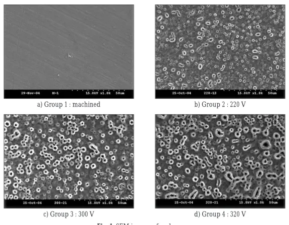
The machined sample showed the appearance of machined grooves and ridges. In the anodized samples showed the porous oxide layer, the pore size increased with change of voltage. Pore size were 0.2 - 1.0 ㎛ in 220 V, 3.0 - 5.5 ㎛ in 300 V and 320 V.(Fig. 1)
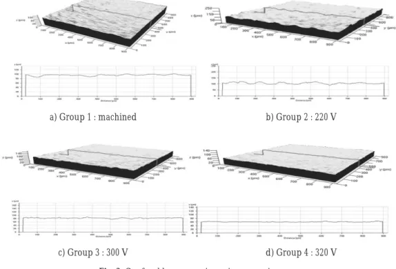
Confocal laser scanning microscopy images are shown in Fig. 2. The roughness of the oxidized titanium surface was characterized by average roughness(Ra). As the applied voltage increased, Ra values decreased. Ra value were 2.84(±0.89)
㎛ in machined surface, 3.29(±0.37) ㎛ in 220 V, 1.78(±0.14) ㎛ in 300 V, and 1.74(±0.14) ㎛ in
Fig. 1. SEM images of each group.
a) Group 1 : machined b) Group 2 : 220 V
c) Group 3 : 300 V d) Group 4 : 320 V
Fig. 2.Confocal laser scanning microscopy images.
a) Group 1 : machined b) Group 2 : 220 V
c) Group 3 : 300 V d) Group 4 : 320 V
Fig. 3. The graph of Ra and Sa values.
Group1 Group2 Group3 Group4
5 4.5 4 3.5 3 2.5 2 1.5 1 0.5 0
Sa Ra
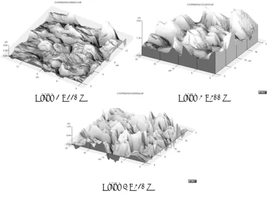
320 V. The graph of Ra and Sa values of each group are shown in Fig. 3. AFM imaging demonstrated the discontinuity of pores and the distance between the peak and the valley.(Fig. 4)
2. Crystal structure of oxide layer
XRD patterns indicated that the anodic oxide films containing anatase, rutile, and amorphous oxides. The amount of anatase increased with change of voltage. Rutile observed at 320 voltage group. The absence of rutile in other groups might be due to the sensitivity of XRD. The results are shown in Fig. 5.
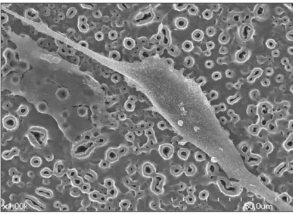
3. Cell morphology
Three kinds of fully spread cell morphology were shown, a polygonal shape, a polarized shape and a round shape. Flat morphology of MG63 cells were adhered to machined titanium surface. As the surface changed more roughly, MG63 cells Fig. 4.Atomic force microscopy images.
Group 2 : 220 V
Group 4 : 320 V
Group 3 : 300 V
Fig. 5.XRD profiles of each group.
XRD profiles Ti
anatase rutile
1200
1000
800
600
400
200
0
30 35 40 45 50
2 theta
320V
300V
200V
Ti
Fig. 6.Group 1, 24 hour. Fig. 7.Group 2, 24 hour.
Fig. 8.Group 3, 24 hour. Fig. 9.Group 4, 24 hour.
Fig. 10.MTT assay.
group1 group2 group3 group4 glass
12hr 0.8
0.7 0.6 0.5 0.4 0.3 0.2 0.1 0.0
24hr 48hr
O. D.
showed more irregular, polarized, and polygonal growth and developed more lamellipodi con- nected to the surface and stretched to the pores.(Fig.
7-9)
4. MTT assay
The disks of machined surface(Group 1) were used as control whereas glass disks were used for comparison. Statistical analysis revealed that the proliferation of MG63 cells was increased in anodized groups, there was significant differ- ence compared with machined and glass, but no significant difference was shown between the group 3 and 4 at P<0.05.(Fig. 10. and Table I) Also, there was no significant difference between Group 1 and glass.(P<0.05) The same results were shown in each time of observations.
DISCUSSION
Anodic oxidation of titanium implants demon- strates changes of various oxide properties, not only oxide thickness, but also surface morphology, pore configuration, crystallinity, chemical com- position, and surface roughness.20Anodized tita- nium surface had porous oxide layer, and there were increase in both size and number of pores, as the anodizing voltage became higher.30
Cell attachment, spreading and subsequent proliferation are closely related to the surface
properties of the substrate, e.g. composition, roughness, wettability and morphology.20 The absorption concentration of complement C3, which is related to the cellular attachment, increas- es with an increase in the thickness and/or crys- tallinity of the titanium oxide. Oxide crystallini- ty seems to be a more significant factor than oxide thickness.31After the spherical cells attach on the surfaces, the following event for cell-sub- strate interaction is cell spreading. The substratum surface topography alters cell shape and modu- lates fibronectin at the transcriptional and post- transcriptional levels, as well as the amount of fibronectin assembly into the extracellular matrix.32 It was reported that the surface texture of the Ti substrates can also affect the expression of fibronectin and vitronectin integrin receptor, modify their clustering or aggregation, and there- fore determine variations in shape and spreading of cells.33,34 Fibronectin, a cell-surface protein, enables cells to interact with the extracellular matrix. Fibronectin consists of two 250-kd polypep- tide chains that are linked by a disulfide near their carboxyl termini. This highly elongated protein, 600 Å long and 25 Å wide, contains a linear array of domains, each able to specifically bind cer- tain molecules outside the cell, such as fibrin, collagen, and heparin. In addition, fibronectin has a cell-binding domain. This cell-surface protein is important for cell migration in development and for wound healing. Vitronectin is an adhesive gly- Table I. Data of MTT assay
culture time group1 group2 group3 group4 glass
(hours)
12 average 0.275 0.371 0.375 0.409 0.207
SD 0.037 0.065 0.043 0.071 0.058
24 average 0.237 0.341 0.361 0.356 0.200
SD 0.032 0.072 0.045 0.074 0.047
48 average 0.427 0.598 0.737 0.725 0.408
SD 0.080 0.098 0.166 0.089 0.068
coprotein with a molecular weight of 75-kd found in human plasma, tissue and extracellular matri- ces. It tends to promotes the attachment and spreading of cells. It has implications for throm- bosis since it binds to heparin, protecting throm- bin and factor Xa from heparin-dependent inac- tivation by antithrombin. Vitronectin is also involved in inflammation and has been identified as the carrier protein of beta-endorphin and has also been found to protect bystander cells from lysis by complement. We could see in our experiment that cell proliferation was enhanced by oxide surfaces leading to more irregular and distinct polygonal spreading of the cells. Many more lamellipodia involved in cell migration were observed in cells on the more intensively anodized titanium than on the machined titanium. These phenomena indicated that the ability of cell migration on anodic oxidized titanium could be higher than that on machined titanium and increased as anodizing voltage increased. Focal contacts on the machined titanium were more intensive than on the oxidized groups. Focal contacts act as a special structure of cell adhesion on the substrates, but in general, cells that form strong focal adhesions are less migratory. More stress fibers but fewer lamellipodia were formed on the control or weakly anodized surfaces. As a highly organized cytoskeleton with stress fibers is often associated with strong cell adhesion, the reorganization of the actin cytoskeleton, togeth- er with the results of the amount and distribution of focal contacts, further confirm the assump- tion that the cells on anodic oxides may have higher motility in comparison with those on the control.23,35Although the initial cell response to dif- ferent surface topographies is unclear, it were known that an increase in calcium and phos- phorus deposition in physiological fluids and increase in protein production and calcium uptake by osteoblast-like cells.36The titanium
oxide itself could have promoted mineralization owing to its ability to bind calcium and thereby stimulate bone formation.37
Oxides, formed on surfaces and behaved in a hydrophobic nature were identified as rutile-type titanium oxide only, while oxides formed on surfaces behaving in a hydrophilic character were identified as a mixture of dominant rutile and anatase oxides. It is not clear that this structural difference between rutile and anatase oxides will contribute to the observed differences between hydrophilic and hydrophobic behaviors in terms of the relationship between surface roughness and contact angle.17Sul et al.8suggested that surface properties of implants directly influence bone responses. Based on the bone response in their study, which was expressed as a function of quantitative changes in the surface oxide properties.
TiO2 in a crystalline phase, ie, a mixture of anatase and rutile phase rather than amorphous, seemed to be optimal. The optimal oxide thickness of a porous surface structure appeared to be in the range of 1,000 to 5,000 nm. The optimum surface chemistry of magnesium incoporated, oxidized implants consisted of approximately 9% mag- nesium at relative atomic concentration in TiO2
matrix. An optimum porosity of open pores was in the range of 19% to 30%, ie, approximately 24%;
with a pore size of ≤ 2.0 ㎛. Surface roughness val- ues of 0.7 to 1.0 ㎛ for Sa, 0.9 to 1.4 ㎛ for Sq, and 27% to 46% for Sdr seemed to be optimal. Because roughness of the surface plays a predominant role in cell adhesion during the implant healing phas- es, this factor should be considered in the man- ufacturing of endosseous implants. However, surface roughness is not as important as other sur- face properties in biologic responses. Furthermore, primary stability could be negatively influenced by an increase in surface roughness, which could counteract the stabilization of the implant known to be essential for implant fixation.38Other surface
properties should be also considered important in the biologic response and may be more critical parameters of biocompatibility than surface roughness.17
The presence of porous surfaces on the anodic oxide was suggested to increase the surface roughness and energy and may cause micro- scopic tissue-cell ingrowth, thereby improving implant fixation.39
The size of the pores, which originate from sparks on the interface of the oxide and elec- trolyte, was related to the nature and concen- tration of ions in the electrolyte.40 Previously published studies had suggested an optimal pore size for bone ingrowth in the range of 50 to around 400 ㎛.41,42Schupbach et al.43suggested that the bone can be formed into smaller pores with diameters of less than 2 ㎛. It is not necessary for osteoblasts to enter into the pores to form bone.
Osteoblasts are polarized cells, and the findings of the investigation indicate that these cells stay at the surface and deposit bone matrix into the pores of oxidized surface. Wong et al.44reported the push out tests revealed small deposits of bone in these 1-2 ㎛ pores. One explanation for the increased pushout strength of this group is like- ly the increased mechanical interlocking that occurs between bine matrix and these small pores. Their study described the fixation of implants by direct apposition of bone during the early healing stages in trabecular bone. Porous structures supply positive guidance cues for anchorage-dependent cells to attach, leading to enhance cell attachment. In contrast, the cells attached to a smooth titanium surface by focal con- tacts around their periphery as predominant adhesion structures, since repulsive signals from the environment led to retraction of the filopodia back to the cell bodies. These cells showed well- organized stress fibers, which exert tension across the cell body, resulting in flattened cells.45In our
study, The pore sizes of group 3 and 4 which were good results in MTT assay were various from 3.0 to 5.5 ㎛. The shapes of pores were various from round to oval. The difference in diameter may be due to the method of measurement in oval shape pore.
The surface modification of the titanium with anodic oxidation enhances cellular adhesion with minor change in the gene expression of osteoblast cells. Thus the enhanced cell adhe- sion produced by anodic oxidation might result in increased bone growth, and contribute to the achievement of a tight fixation within a shorter peri- od of time after surgery.6The cell reaction to an implant surface is a very complex situation. This study was aim to find adequate surface charac- teristics of initial cell attachment and proliferation.
CONCLUSIONS
Surface characteristics of anodic oxidized tita- nium influence osteoblast response. The good response of osteoblast can result in reinforce- ment of osseointegration.
There are various factors which affect the sur- face characteristics in anodic oxidization of tita- nium such as electrolyte, time of anodizing, and applied voltage. The changing voltage is one of the easy way to control the anodic oxidation.
We inspect the surface characteristics of anod- ic oxidized titanium and the response of MG63 cell.
The following conclusions were drawn.
1. In anodizing titanium surface, we could see pores which did not show in contol group. In higher anodizing voltage, pore size was increased.
2. In anodizing titanium surface, we could see anatase. In higher anodizing voltage, thicker oxide layer increased crystallinity(anatase, anatase and rutile mixed).
3. MG63 cells showed more irregular, polarized,
polygonal shape and developed more lamel- lipodi in anodizing group as voltage increased.
4. The activity of cells in MTT assay increased sig- nificantly in group 3 and 4 in comparison with group 1 and 2. However, there was no dif- ference between group 3 and 4 at P<0.05.
Proliferation of MG63 cells increased signifi- cantly in pore size (3-5.5 ㎛) of group 3 and 4 in comparison with in pore size (0.2-1 ㎛) of group 2.
REFERENCES
1. Lampin M, Warocquire-Clerout R, Legris C, Degrange M, Sigot-Luizard MF. Correlation between substratum roughness and wettability, cell ad- hesion, and cell migration. J Biomed Mater Res 1997;36:99-108.
2. Bowers KT, Keller JC, Randolph BA, Wick DC, Michaels CM. Optimization of surface micro- morphology for enhanced osteoblast responses in vitro. Int J Oral Maxillofac Implants 1992;7:302- 310.
3. Schwartz Z, Swain LD, Marshall TS, Sela J, Gross U Amir D, Muller-Mai C, Boyan BD. Modulation of matrix vesicle enzyme activity and phos- phatidylserine content by ceramic implant ma- terials during endosteal bone healing. Calcif Tissue Int 1992;51:429-437.
4. Sela J, Shani J, Kohavi D, Soskolne WA, Itzhak K, Schwartz Z, Boyan BD. Uptake and biodistribution of 99mtechnetium methylene~[32P]diphosphonate during endosteal healing around titanium, stain- less steel and hydroxyapatite implants in rat tibial bone. Biomaterials 1995;16:1373-1380.
5. Kasemo B, Lausmaa J. Aspect of surface physics on titanium implants. Swed Dent J 1983;28(Suppl.):19- 36.
6. Kim Y. Microarray-based expression analysis of hu- man osteoblast-like cell response to anodized ti- tanium surface. thesis, Seoul National University 2004.
7. Albektsson T, Brenemark PI, Hanson HA, Lindstorm J. Osseointegrated titanium implants. Acta Orthop Scand 1981;52:155-170.
8. Sul YT, Johansson CB, Wennerberg A, Cho LR, Chang BS, Albrektsson T. Optimum surface prop- erties of oxidized implants for reinforcement of os- seointegration: surface chemistry, oxide thick- ness, porosity, roughness, and crystal structure. Int J Oral Maxillofac implants 2005;20:349-359.
9. Rocci A, Martignoni M, Gottlow J. Immediate loading of Branemark System with TiUnite and ma-
chined surfaces in the posterior mandible: a ran- domized, open-ended trial. Clin Implant Dent Relat Res 2003;5(suppl 1):57-63.
10. Glauser R, Lundgren A, Gottlow J, et al. Immediate occlusal loading of branemark MkIV TiUnite im- plants placed predominantly in soft bone: 1-year results of a prospective, clinical study. Clin Implant Dent Relat Res 2003;5(suppl 1):47-56.
11. Glauser R, Ree A, Lundgren A, Gottlow J, Hammerle CH, Scharer P. Immediate occlusal loading of Branemark implants applied in various jawbone regions: a prospective, 1-year clinical study. Clin Implant Dent Relat Res 2001;3:204- 213.
12. Klokkevold PR, Nishimura RD, Adachi M, Caputo A. Osseointegration enhanced by chemical etching of the titanium surface. Clin Oral Impl Res 1997;8:442-447.
13. Larsson C. The Interface between bone and metals with different surface properties. Light micro- scopic and Ultra-structural studies. thesis, Go¨teborg:Go¨teborg university, 1997.
14. Larsson C, Emanuelsson L. Bone response to sur- face modified titanium implants - studies on the tis- sue response after 1 year to machined and elec- tropolished implants with different oxide thickness.
J Mater Sci Mater Med 1997;8:721-729.
15. Sul YT, Johansson CB, Jeong YS, Wennerberg A, Albrektsson T. Resonance frequency and removal torque analysis of implants with turned and an- odized surface oxides. Clin Oral Impl Res 2002;
13:252-259.
16. Buser D, Schenk RK, Steinemann S, Fiorellini JP, Fox CH, Stich H. Influence of surface characteristics on bone integration of titanium implants. A histo- morphometric study in miniature pigs. J Biomed Mater Res 1991;29:889-902.
17. Lim YJ, Oshida Y, Andres C, Barco MT. Surface characterizations of variously treated titanium materials. Int J Oral Maxillofac Implants 2001;16:333- 342.
18. Aalam AA, Nowzari H. Clinical evaluation of dental implants with surfaces roughened by anodic oxidation, dual acid-etched implants, and ma- chined implants. Int J Oral Maxillofac Implants 2005;20:793-798.
19. Niki M, Ito G, Matsuda T, Orgino M. Comparative push-out data of bioactive and non-bioactive ma- terials interface. Toronto, Canada: University of Toronto Press, 1991:350-356.
20. Ishizawa H, Fujino M, Ogino M. Histomorphometric evauation of the thin hydroxyapatite layer formed through anodization followed by hydrothermal treatment. J Biomed Mater Res 1997;35:199-206.
21. Hall J, Lausmaa J. Properties of a new porous ox- ide surface on titanium implants. Appl Osseointegration Res 2001;1:5-8.
22. Ivanoff CJ, Widmark G, Johansson C, Wennerberg
A. Histologic evaluation of bone response to oxi- dized and turned titanium micro-implants in hu- man jawbone. Int J Oral Maxillofac implants 2003;18:341-348.
23. Zhu X, Chen J, Scheideler L, Reichl R, Geis- Gerstorfer J. Effect of topography and composition of titanium surface oxides on osteoblast respons- es. Biomaterials 2004;25:4087-4103.
24. Sul YT, Johansson CB, Petronis S, Krozer A, Jeong Y, Wennerberg A, Albrektsson T. Characteristics of the surface oxides on turned and electrochem- ically oxidized pure titanium implants up to dielectic breakdown: the oxide thickness, micropore con- figurations, surface roughness, crystal structure and chemical composition. Biomaterials 2002;23:491-501.
25. Yang B, Uchida M, Kim HM, Zhang X, Kokubo T.
Preparation of bioactive titanium metal via anodic oxidation treatment. Biomaterials 2004;25:1003- 1010.
26. Bryan MC. Crystal structure of cholesterol mono- hydrate. Nature 1976;260:727-729.
27. Meachim G, Williams DF, Changes in nonosseous tissue adjacent to titanium implants. J Biomed Mater Res 1973;7:555-572.
28. Franceschi RT, James WM, Zerlauth G. 1 alpha, 25- dihydroxyvitamin D3 specific regulation of growth, morphology, and fibronectin in a human os- teosarcoma cell line. J Cell Physiol 1985;123:401-409.
29. Boyan BD, Schwartz Z, Bonewald LF, Swain LD.
Localization of 1,25-(OH)2D3-responsive alkaline phosphatase in osteoblast-like cells (ROS 17/2.8, MG 63, and MC 3T3) and growth cartilage cells in culture. J Biol Chem 1989 Jul 15;264:11879-11886.
30. Park KH. Osseointegration of anodized titanium implants. MSD thesis, Seoul National University 2003.
31. McAlarney ME, Oshiro MA, McAlarney CV. Effect of titanium oxide passive film crystal structure, thick- ness, and crystallinity on C3 adsorption. Int J Oral Maxillofac Implants 1996;11:73-80.
32. Chou L, Firth J, Uitto V, Brunette D. Substratum sur- face topography alters cell shape and regulates fi- bronectin mRNA level, mRNA stability, secre- tion and assembly in human fibroblasts. J Cell Sci 1995;108:1563-1573.
33. Degasne I, Basle, Demais ME, Hure V, Lesourd G, Grolleau M, Mercier B, Hormia M, Kononen M.
Immunolocalization of fibronectin and vitronectin receptors in human gingival fibroblasts spreading on titanium surfaces. J Periodontal Res 1994;29:146- 152.
34. Chappard L. Effect of roughness, fibronectin and vitronectin on attachment, spreading, and pro- liferation of human osteoblast-like cells(Saos-2) on titanium surface. Calcif Tissue Int 1999;64:499- 507.
35. Badley RA, Woods A, Carruthers L, Rees DA.
Cytoskeletal changes in fibroblast adhesion and de- tachment. J Cell Sci 1980;43:379-390.
36. Martin JY, Schwartz Z, Hummert TW, Schraub DM, Simpson J, Lankford J Jr, Dean DD, Cochran DL, Boyan BD. Effect of titanium surface roughness on proliferation, differentiation, and protein synthe- sis of human osteoblast-like cells(MG63). J Biomed Mater Res 1995;29:389-401.
37. Kokubo T, Kim HM, Kawashita M, Nakamura T. Bioactive metals:preparation and properties. J Mater Sci Mater med 2004;15:99-107.
38. Carlsson L, Ro¨stlund T, Albrektsson B, Albrektsson T. Implant fixation improved by close fit. Acta Orthop Scand 1988;59(3):272-275.
39. Groessner-Schreiber B, Tuan RS. Enhanced ex- tracellular matrix production and mineralization by osteoblasts cultured on titanium surfaces in vit- ro. J Cell Sci 1992;101:209-217.
40. Zhu X, Ong JL, Kim S, Kim K. Surface character- istics and structure of anodic oxide films con- taining Ca and P on a titanium implant material.
J Biomed Mater Res 2002;60:333-338.
41. Bobyn JD, Pilliar RM, Cameron HU, Weatherly GC.
The optimum pore size for the fixation of porous surfaced metal implants by the ingrowth of bone.
Clin Orthop Relat Res 1980;150:263-270.
42. Clemow AJT, Weistein AM, Klawitter J, Koeneman J, Andresson J. Interface mechanics of porous ti- tanium implants. J Biomed Mater Res 1981;15:73- 82.
43. Schupbach P, Glauser R, Rocci A, Martignoni M, Sennerby L, Lundgren A, Gottlow J. The human bone-oxidized titanium implant interface: A light microscopic, scanning electron microscopic, and en- ergy-dispersive X-ray study of clinically retrieved dental implants. Clin Implant Dent Relat Res 2005;7(suppl1):36-43.
44. Wong M, Eulenberger J, Schenk R, Hunziker E.
Effect of surface topology on the osseointegra- tion of implant materials in trabecular bone. J Biomed Mater Res 1995;29:1567-1575.
45. Zhu X, Chen J, Scheideler L, Altebaeumer T, Geis- Gerstorfer J, Kern D. Cellular reactions of os- teoblasts to micron- and submicron-scale porous structures of titanium surfaces. Cell Tissues Organs.
2004;178(1):13-22.
Reprint request to:
CHANG-WHEKIM, D.D.S., M.S.D. Ph.D.
DEPARTMENT OFPROSTHODONTICS,GRADUATESCHOOL, SEOULNATIONALUNIVERSITY
28-1, YEONGUN-DONG,CHONGNO-GU,SEOUL, 110-749, KOREA [email protected]