- 118 - Pheochromocytoma is a rare catecholamine pro
ducing tumor and its occurrence during pregnancy is extremely rare (reported incidence of one in 54,000 pregnancies).1 Because of its rarity and confusion with the much more common forms of pregnancyrelated hypertension, the diagnosis of pheochromocytoma in pregnancy may be overlooked.
If unrecognized, maternal and fetal mortality amounts to 4050%.1 However, after antenatal diagnosis of pheochromocytoma, maternal mortality was reduced substantially to <2% and fetal mortality to <14%.2 Early diagnosis of pheochromocytoma in pregnancy needs high index of suspicion due to the wide variability and nonspecificity of its clinical signs and symptoms.
대 한 주 산 회 지 제27권 제2호, 2016 Korean J Perinatol Vol.27, No.2, June, 2016 http://dx.doi.org/10.14734/kjp.2016.27.2.118
� Case report �
We report a case of a 28yearold woman who was diagnosed with pheochromocytoma early on the basis of detailed history taking and underwent laparoscopic adrenalectomy at advanced gestational age compared with those of previous reported cases.
Case report
A 28yearold primigravida presented with a blood pressure of 173/104 mm Hg at 22 weeks of gestation.
She complained of headache and was otherwise asymptomatic. A urine dipstick test showed 2+ for glucose and trace for protein. A presumptive diagnosis of severe preeclampsia and gestational diabetes were made. However, on physical examination, there was no evidence of peripheral edema. Fetal sonographic examination was unremarkable. Laboratory investi
gations were all within normal limits except for trace proteinuria and 4+ glycosuria. With these findings, she was considered to be inconsistent with a diagnosis of preeclampsia.
An evaluation of secondary hypertension was carried out. Biochemical investigation showed that
Early Detection and Successful Laparoscopic Adrenalectomy for Pheochromocytoma in Pregnancy;
A Case Report
Jonghyun Kim, M.D., Ph.D.
Department of Obstetrics and Gynecology, Research Institute of Clinical Medicine, Chonbuk National University Medical School, Chonbuk, Korea
Pheochromocytoma is an extremely rare tumor in pregnant women with potentially fatal consequences. We report a case of pregnant woman at 22 weeks of gestation with pheochromocytoma. A correct diagnosis on the basis of differential clues from severe preeclampsia was obtained and laparoscopic adrenalectomy was performed.
Key Words: Pheochromocytoma, Pregnancy, Laparoscopy, Adrenalectomy
Received: 13 May 2016, Revised: 9 June 2016 Accepted: 10 June 2016
Correspondence to: Jonghyun Kim, M.D., Ph.D.
Department of Obstetrics and Gynecology, Research Institute of Clinical Medicine, Chonbuk National University Medical School, 20, Geonjiro Deokjin-gu, Jeonju, 54907 Korea
Tel: +82. 63-250-2290, Fax: +82. 63-254-4833 E-mail: [email protected]
Copyrightⓒ 2016 by The Korean Society of Perinatology
This is an Open Access article distributed under the terms of the Creative Commons Attribution Non-Commercial License (http://creativecommons.org/license/
by-nc/4.0/), which permits unrestricted non-commercial use, distribution, and reproduction in any medium, provided that the original work is properly cited.
The Korean Journal of Perinatology · pISSN 1229-2605 eISSN 2289-0432 · e-kjp.org
김종현 : - 임신 중 진단된 갈색세포종의 진단 및 치료 -
- 119 - 24hour urinary excretion of vanillylmandelic acid (VMA) and metanephrine was 18.2 mg (normal range 18 mg/24 hours) and 2 mg (normal range 00.8 mg/24 hours), respectively. With a high suspicion of a pheochromocytoma, additional biochemical tests and magnetic resonance imaging (MRI) of the abdomen were performed. Urine norepinephrine was 1,011.6 µg/24 hours (normal range <80 µg/24 hours) and epinephrine was 21.9 µg/24 hours (normal range <20 µg/24 hours). Plasma norepinephrine was 3,053.8 pg/
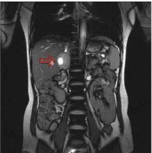
mL (normal range <80 pg/mL) and epinephrine was 10.5 pg/mL (normal range <20 pg/mL). MRI of the abdomen confirmed a right adrenal mass measuring 4 cm, with internal cystic and solid component (Fig. 1).
At 25+6 weeks of gestation after 14 days of pre
paration with the αblocking drug doxazosin, a laparoscopic right adrenalectomy was successfully carried out. Histopathologic examination confirmed pheochromocytoma. The postoperative course was uneventful. At the 14th day postoperation, 24hour urinary excretion of VMA and metanephrine was normalized to 2.4 mg (normal range 18 mg/24
hours) and 0.3 mg (normal range 00.8 mg/24 hours), respectively. Plasma norepinephrine and epinephrine was decreased to 311.0 pg/mL (normal range <80 pg/mL) and 22.6 pg/mL (normal range
<20 pg/mL), respectively. The remaining weeks of the pregnancy progressed normally and a normal baby of 3.2 kg weight was delivered at 39 weeks of gestation by vaginal delivery. Apgar scores were 9 at 1minute and 10 at 5minute. The mother and the newborn recovered well and were discharged on the 3rd postpartum day.
Discussion
The diagnosis of pheochromocytoma during pregnancy is often overlooked due to the similarity of usual signs and symptoms, such as nausea and hypertension in patients with pregnancyrelated hypertension. In a review of the literature from 1980 to 1987, the overall maternal mortality was found to be 17% with a fetal loss of 26%, although antenatal diagnosis of pheochromocytoma has reduced the maternal mortality to <1% and the fetal loss to 15%.3,
8 However, only 26% of pheochromocytomas were diagnosed antenatally.
Based on history and clinical exam, sustained epi
sodes of hypertension occurring after the 20th week of gestation, edema and weight gain are all important clues for preeclampsia, whereas par oxysmal epi
sodes of hypertension occurring throughout the entire pregnancy, severe headaches, sweating, palpitation and orthostatic hypotension are clues for pheochromocytoma.3 The presence of proteinuria, oliguria or anuria, hyperglycemia, elevated liver enzymes and thrombocytopenia are laboratory clues for preeclampsia.4
Paroxysmal attacks occurring in pheochromo
Fig. 1. Magnetic resonance imaging of the abdomen.
Arrow indicates right adrenal tumor.
Jonghyun Kim : - Pheochromocytoma in Pregnancy -
- 120 - cytoma during pregnancy as potentially fatal hy
pertensive crisis can be precipitated by general anesthesia, vaginal delivery, the mechanical effects of the gravid uterus, uterine contractions and increas
ed fetal movements.5 The fetal risks are mainly determined by the vasoconstrictive effects of cate
cholamines on the uteroplacental circulation and by the severity and duration of hypertensive spells.1
Urinary levels of catecholamines do not change during normal pregnancy, therefore, the diagnosis of pheochromocytoma can be confirmed by mea
surement of 24hour urine VMA, metanephrines, or catecholamines. Following biochemical confirmation, the next step is to localize the pheochromocytoma.
Abdominal ultrasound as an initial imaging test is safe but less reliable, particularly with the gravid uterus in the third trimester.1 Computed tomogra
phy or MRI can be used, but MRI is the preferred modality in pregnancy as it is free from ionizing radiation and produces high quality images.6 If other imaging methods are unhelpful, the Iodine 123
metaiodoenzylguanidine scan is mainly indi cated for detecting multiple tumors or extraadrenal tumors and metastasis.7
Definitive treatment of pheochromocytoma is sur
gical removal. Appropriate preoperative medical management to block the effects of released cate
cholamine is essential, therefore, αadrenergic blockade using phenoxybenzamine or doxazosin should be initiated about 1014 days preoperatively.1 βblockade may be required to treat tachycardia, but it should never be prescribed before αblockade, because βblockade alone can precipitate a hyper
tensive crisis attributed to unopposed αadrenergic effects by catecholamines.4
In pregnant patients, the optimal time of surgical tumor removal is a challenging and controversial
is sue depending on the gestational age, clinical re
sponse to treatment, the accessibility of the tumor, and the presence or absence of fetal distress.4 Har
per et al8 have recommended that tumor resection should be performed in the first and second trime
sters, and elective cesarean delivery followed by tumor resection in later pregnancy. During the first trimester, surgical tumor removal may lead to increased risk of spontaneous abortion.1, 8 Some au
thors have advocated that the tumor diagnosed before 24 weeks of gestation should be removed as soon as possible after hypertension is controlled medically, but after 24 weeks of gestation, tumor removal should be deferred until the fetus is viable and removed simultaneously at the time of cesarean section or shortly after delivery.1, 4 However, a case who received conservative treatment until fetal ma
turity is attained, showed the difficulty in managing pheochromocytoma in pregnancy.4
Although midpregnancy laparoscopic surgery is difficult to perform this because it is difficult to secure visual field exposure, we undertook a laparoscopic surgery since our institution has had experience in laparoscopic adrenalectomy in the last decades and we expect to have a better prognosis in patients with fetal surgical intervention. In case of present case, compared to the most of previously presented cases, we made an early diagnosis and successfully undertook adrenalectomy through subsequent laparo scopic surgery. Keeping the wellbeing states of the mother and fetus to the full term and it was possible to have the childbirth through vaginal de
livery. This could be the great significance this case has.
In conclusion, despite its low incidence, pheochro
mocytoma should be kept in mind as one of the causes of hypertension in pregnant patients, owing to its high
김종현 : - 임신 중 진단된 갈색세포종의 진단 및 치료 -
- 121 - mortality. Here we present a hypertensive pregnant patient presenting with pheochromocytoma and illu
strate the importance of careful history taking and physical examination in preventing such serious maternal and fetal complications.
References
1) Lenders JW. Pheochromocytoma and pregnancy: a deceptive connection. Eur J Endocrinol 2012;166:143-50.
2) Ahlawat SK, Jain S, Kumari S, Varma S, Sharma BK.
Pheochromocytoma associated with pregnancy: case report and review of the literature. Obstet Gynecol Surv 1999;54:
728-37.
3) Nakajima Y, Masaoka N, Sodeyama M, Tsuduki Y, Sakai M.
Pheochromocytoma-related cardiomyopathy during the
antepartum period in a preterm pregnant woman. J Obstet Gynecol Res 2011;37:908-11.
4) Oliva R, Angelos P, Kaplan E, Bakris G. Pheochromocytoma in pregnancy: a case series and review. Hypertension 2010;
55:600-6.
5) Bullough A, Karadia S, Watters M. Phaeochromocytoma:
an unusual cause of hypertension in pregnancy. Anesthesia 2001;56:43-6.
6) Almog B, Kupferminc MJ, Many A, Lessinq JB. Pheochro- mocytoma in pregnancy – a case report and review of the literature. Acta Obstet Gynecol Scand 2000;79:709-11.
7) Grodski S, Jung C, Kertes P, Davies M, Bantinq S. Pheo- chromocytoma in pregnancy. Intern Med J 2006;36:604-6.
8) Harper MA, Murnaghan GA, Kennedy L, Hadden DR, Atkinson AB. Phaeochromocytoma in pregnancy. Five cases and a review of the literature. Br J Obstet Gynaecol 1989;96:594-606.