접수일:2008년 12월 3일, 승인일:2008년 12월 12일
기증 제대혈 검체의 Ureaplasma 오염률
노은연1,2ㆍ신 수1,2ㆍ윤종현1,2ㆍ장지영2
서울대학교 보라매병원 진단검사의학과1, 서울특별시립 보라매병원 공여제대혈은행2
= Abstract =
Ureaplasma Contamination Rate in Donated Cord Blood Units
Eun Youn Roh1,2, Sue Shin1,2, Jong Hyun Yoon1,2, Jee Young Chang2
Department of Laboratory Medicine, Seoul National University Boramae Hospital1, Seoul Metropolitan Public Cord Blood Bank2, Seoul, Korea
Background: Ureaplasma is one of the most common microorganisms in the genital tract, and also one of the most important contaminants in cell culture laboratories and cell banks. We analyzed the Ureaplasma contamination rate in donated cord blood units (CBUs) before cell processing.
Methods: One hundred fifty-one donated CBUs with informed consent collected between 3 November and 28 December 2006 were randomly selected and enrolled for study. All of the CBUs were obtained from full-term (37∼42 weeks gestation) deliveries. We performed blood cultures and Ureaplasma DNA PCR assays using samples from the collection bags before processing.
Results: Two CBUs had positive blood culture results; however, Ureaplasma DNA was not found in the samples tested.
Conclusion: The contamination rate of Ureaplasma in the donated CBUs from full-term deliveries without gestational and/or perinatal complications was extremely low. With the results of our previous Mycoplasma contamination assay (0%), donated CBUs can be used in culture and expansion processes without concerns for pre-processing mollicute contamination. (Korean J Blood Transfus 2008;19:239-244)
Key words: Donated cord blood, Ureaplasma, Contamination
서 론
인간의 비뇨생식기에는 7종의 mollicute (Myco- plasma hominis, Mycoplasma fermentans, Myco- plasma primatum, Mycoplasma genitalium, Myco- plasma spermatophilum, Mycoplasma penetrans
Ureaplasma. urealyticum)가 상재하며, 이 중 하나
인 Ureaplasma urealyticum은 만삭 및 조산분만시
양수에서 가장 흔히 분리되는 미생물로 융모양막
염(chorioamnionitis), 산욕기 자궁내막염(puerperal
endometritis), 수술 후 창상감염, 신생아 패혈증,
뇌수막염 및 기관지폐이형성증(bronchopulmonary
dysplasia) 등의 질환을 유발하는 것으로 알려져 있다.
1-4)Ureaplasma를 비롯한 mollicute는 임상적인 중 요성 외에 세포배양시 오염의 주요 원인으로 세 포배양 실험실이나 세포은행에서 배양되는 세포 의 15∼80% 정도가 mollicute에 오염된다고 보고 되었으며, 배양 세포에 mollicute 오염이 있는 경 우 성장속도 저하, 형태학적 변화, 염색체 변이, 싸이토카인 발현의 유도 또는 억제, 막 구성의 변 화, 아미노산 및 핵산대사의 변이 등을 유발하여 실험 결과의 신뢰성을 떨어뜨리고 생물학적 산물 을 폐기하는 경우를 초래한다. 하지만 오염시 변 화가 잘 드러나지 않고 성장조건이 까다로워 일 상적인 미생물 배양검사로는 검출되지 않기 때문 에 다른 세균 및 진균 오염과 달리 간과하는 경우 가 흔하다.
5-8)Ureaplasma의 검출에는 배양, 혈청학적 검사, 간접 DNA 염색(Hoechst 33258), Gen-Probe, PCR 등의 방법이 이용되는데 배양은 Ureaplasma의 확 진법이지만 영양 요구성이 까다롭고 1∼2주 이상 의 기간이 소요되며 집락을 현미경으로 관찰해야 하는 등 어려운 점이 많아서, 최근에는 배양법에 비해 민감하고 1일 이내에 결과를 확인할 수 있 는 PCR 방법이 널리 이용되고 있으며 자체 제작 한(in-house) 검사법이나 상용화된 PCR 키트가 많 이 소개되고 있다.
8-10)제대혈은 태아가 만출된 후 태반이 배출되기 직전(in-utero) 탯줄에서 채취하므로 비뇨생식기, 위장관, 피부의 상재균 오염이 드물지 않으며, 제 대혈 검체의 미생물 오염은 5%에서 많게는 15%
까지 보고되고 있다.
11,12)제대혈에는 골, 신경, 내 피세포 등으로 분화 가능한 간엽줄기세포가 풍부 하여 조혈모세포이식 외에 각종 난치성 질환의 세포치료제로 주목 받고 있으며, 제대혈 줄기세 포의 체외 증식 및 분화에 대한 연구가 활발히 진
행되는 추세이다.
13,14)이에 본 연구자들은 비뇨생 식기 상재균 오염의 가능성이 있는 제대혈 검체 에 대하여 가장 흔한 mollicute인 Ureaplasma 오염 률을 살펴 저장 제대혈의 안전성을 다시 한 번 확 인하고 조혈모세포의 체외 증식과 세포치료제 개 발을 위한 세포배양 및 분화연구의 줄기세포 공 급원으로서의 적합성을 평가하고자 하였다.
대상 및 방법
1. 대상
2006년 11월 3일부터 12월 28일까지 제대혈 기 증에 관해 서면으로 동의한 산모에게서 채취한 제대혈 350단위 중 151단위를 무작위로 선택하였 다. 보건복지부의 표준지침에 따라 제대혈 기증 은 임신 37주에서 42주 사이의 만삭분만이며 임 신관련 합병증이 없는 경우로 한정하였다. 이 중 76단위에 대해서는 저장처리가 실시되었고 75단 위는 채취된 양이 기준에 미치지 못하여 접수가 거절된 검체였다.
2. 방법
1) 채취
서울특별시립 보라매병원 의학연구윤리심의
위원회(Institutional review board, IRB)의 심의를
통과한(IRB No. 06-2006-2) 기증 동의서에 자발적
으로 동의 및 서명한 산모에게서 분만을 담당한
산부인과 의사가 태아 만출 직후 탯줄을 결찰하
고 항응고제(citrate phosphate dextrose adenine,
CPDA-1)가 포함된 채취백에 제대혈을 채취하였
으며, 운송 담당자가 이를 실온에서 24시간 이내
에 제대혈은행으로 운반하였다.
2) 미생물 배양 검사
가공처리를 시작하기 전 채취백에서 무균적으 로 취한 제대혈 10 mL를 BacT/Alert SA (호기성 세균 혈액배양병)과 BacT/Alert SN (혐기성세균 혈액배양병)에 각 5 mL씩 접종하였으며, 통상적 인 혈액배양법에 따라 BacT/Alert 자동화 시스템 (bioMerieux, Durham, NC, USA)에서 5일간 배양 하였다. 균이 증식한 경우 그람염색을 실시하였 고, BacT/Alert SA는 BAP와 MAC배지에, BacT/
Alert SN는 Brucellar agar와 chocolate agar에 계대 배양을 실시한 후 단일 집락을 선택하여 Vitek 카 드 시스템(bioMerieux, Durham, NC, USA)으로 균 동정을 실시하였다.
3) Ureaplasma PCR
(1) 검체 DNA 추출: 제대혈 채취백에서 검체 300μL를 취한 후 Puregene
ⓇDNA purification Kit (Gentra Systems, Minneapolis, MN, USA)를 이용하 여 제조사의 지침에 따라 DNA를 추출하였다.
(2) 양성대조균주 DNA 추출: ATCC 27813 Ureaplasma urealyticum 표준균주를 액체배지 ATCC 1331 Urea broth 10B에 접종하고 5% CO
2, 37
oC에 배양하였다. pH 변화에 의해 배지의 색이 노란색 (yellow)에서 주황색(orange)으로 바뀌면(약 9∼12 시간 배양) 증균된 액체배지 30 mL를 취하여 80,000 rpm에서 20분간 원침한 후 침사를 15분간 75∼95
oC 수조에서 가온하여 DNA를 추출하였 다.
(3) PCR: Urease 유전자의 염기서열을 이용한 다음의 시발체(5'-CCA GGA AAA GTA GTA CCA GGA GC-3'; 5'-CTC CTA ATC TAA CGC TAT CAC C-3')로 PCR 증폭을 시행하였다.
9)Taq polymerase 1U, dNTP 각각 0.2 mM, Tris-HCl 10 mM, KCl 50 mM, MgCl
21.75 mM이 포함된 amfiSure PCR premix (Genedepot, Seoul, Korea)에 Ureaplasma 시발체 세트(primer set) 각각 10 pmol
및 증류수를 혼합하여 총반응량을 25μL에 맞추 고 DNA 100 ng을 첨가한 다음 PTC 0200 (Bio- RAD, CA, USA) thermocycler로 94
oC에서 1분간 변성한 후, 94
oC에서 30초, 55
oC에서 30초, 68
oC에 서 90초씩 35 cycle 증폭하고, 72
oC에서 5분간 다 시 증폭하였다. 양성대조와 음성대조를 함께 증 폭하였는데, 양성대조 주형(template)은 ATCC 27813 표준균주 Ureaplasma genomic DNA를 이용 하였고 음성대조에는 DNA를 첨가하지 않았다.
증폭 산물은 ethidium bromide를 포함한 agarose gel에 50 V, 35분간 전기영동 한 후 429∼450 bp 사이에서 밴드 여부를 확인하였다.
결 과
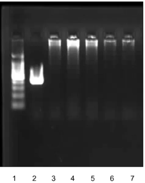
산모의 연령 분포는 30.4±3.1세였고, 신생아의 성별은 남아 73명, 여아 78명, 자연분만이 108건, 제왕절개를 통한 분만이 43건이었다. 분만시 문 제가 있었던 경우로는 태변착색의 예가 1건이었 으며 본 제대혈은행의 운영지침에 따라 시행하는 3개월 후의 2차 문진결과 산모 및 신생아의 주산 기 감염, 염증 등 임상적인 이상소견은 확인되지 않았다. 전체 151단위의 제대혈 중 2건은 산전 진 단에서 기형이 발견되어 접수가 거절되었으며, 1 건은 임신초기 양수 파수 의심하여 항생제 치료 를 받았으나 이후 이상소견 없어 정상분만을 한 예였다. 제대혈에 대한 미생물 배양 검사에서는 2단위의 제대혈에서 각각 citrobacter spp.와 알 수 없는 그람 양성 간균이 동정되었으나 Ureaplasma PCR 검사는 모두 음성이었다(Fig. 1).
고 찰
제대혈은 분만 후 태반과 탯줄에 남아 있는 혈
액으로 원래는 적출물로 폐기되어 왔으나 1990년
Fig. 1. Electrophoretic analysis by PCR to detect Ureaplasma. Lane 1, 100 bp DNA marker; Lane 2, positive control; Lane 3, negative control; Lane 4∼
7, cord blood samples.
대 이후 조혈모세포 공급원으로 주목을 받기 시 작하였으며, 최근에는 조혈모세포의 체외 증폭에 관한 시도와 함께 제대혈 간엽줄기세포를 이용한 세포치료제 개발에 관한 연구가 이어지면서 조혈 모세포이식 및 각종 악성 및 양성 질환의 치료에 제대혈 이용이 시도되고 있다. 이에 따라 제대혈 은행뿐 아니라 이와 연계된 연구기관 및 생명공 학 회사들이 활발히 연구에 참여하고 있으며
15)가까운 미래에 제대혈이 세포치료와 재생의학 분 야에서 중요한 줄기세포 공급원이 될 것으로 예 상된다. 분만과정에서 부수적으로 획득되는 제대 혈은 채취과정이 골수와 달리 공여자(신생아, 산 모)에게 의학적인 위험을 증가시키지 않으며, 개 체로 분화할 수 있는 배아와 달리 윤리적인 문제 는 최소화하면서 많은 잠재적 공여자를 확보할 수 있는 장점이 있지만, 분만이라는 검체 채취 환
경의 특수성 때문에 세포배양 및 가공 이전에 이 미 미생물에 오염될 가능성을 배제할 수 없다.
U. urealyticum은 임산부의 비뇨생식기에서 가 장 흔히 발견되는 미생물로 40∼80%까지 그 빈 도가 보고되고 있으며,
3,16)Ureaplasma를 비롯한 mollicute 오염은 세포배양시 중요하고 흔한 문제 로 검출이 용이하지 않기 때문에 오염이 확산되 는 것을 막지 못하여 피해가 커질 수 있고, 특히 줄기세포를 이용한 세포치료제 개발 연구 등에서 는 경제적으로 큰 손실을 야기할 수 있어서 세포 은행이나 세포치료제 관련 GMP 시설에서는 매 우 주의를 기울이는 부분이다.
제대혈의 Ureaplasma 오염률은 임신관련 합병 증이 없는 건강한 만삭분만 산모에서는 보고된 바가 없었지만, 임신 23∼31주의 조산아를 대상 으로 시행한 연구에 의하면 제대 조직의 4.7%에 서 U. urealyticum이 분리되었으며,
17)조산아의 제 대혈 배양시 23%에서 U. urealyticum 또는 M.
hominis가 검출되었고, 배양 양성인 조산아에서
음성인 조산아에 비해 자궁내 감염, 염증소견 및
신생아 전신성 염증반응 증후군(neonatal systemic
inflammatory syndrome)의 빈도가 높았다.
18)비뇨생식기 상재균으로 흔히 임산부에서 분리
되는 mollicute 오염은 발견이 쉽지 않고 피해가
커서 세포배양을 위한 줄기세포 공급시에 간과할
수 없는 부분인데, 본 연구에서 PCR 방법을 이용
하여 검사한 결과 Ureaplasma PCR 양성을 보이
는 제대혈 검체가 없어 세포배양 등의 실험적 조
작을 행하기 전 채취상태의 제대혈 검체에서
Ureaplasma 오염률은 극히 낮은 것을 확인하였
다. 이는 Astori 등이 제대혈 체외 증폭 연구에서
Mycoplasma 오염이 관찰되지 않았음을 보고한
것과 일치하는 소견이며,
19)또한 동일한 대상군
에서 Mycoplasma 오염이 관찰되지 않았던 저자
들의 이전 연구 결과와도 일치하는 소견이다.
20)분만장에서 태아 만출 후 채취되는 제대혈은 비뇨기, 생식기, 소화기 및 피부의 상재균 오염 가능성이 적지 않을 것으로 예상되었으나, 본 연 구와 동일한 방법으로 채취하고 처리한 제대혈 895 단위를 분석한 결과 제대혈의 미생물 오염률 은 2.3%로 다른 연구자들이 보고한 5∼15%
11,12)보다 현저히 낮은 값을 보여 분만 및 제대혈 채취 과정 중 미생물 오염의 빈도가 낮음을 알 수 있었 고, 또한 Ureaplasma는 조산, 사산 등의 합병증과 관련이 알려져 있고 본 연구의 대상군에는 조기 양막파수, 조산 등의 임신 관련 합병증의 예가 거 의 포함되지 않았으므로 임신 주수 37∼42주의 만삭분만으로 대상자를 한정하는 기증제대혈의 Ureaplasma 오염률은 매우 낮은 것으로 생각된 다.
저자들은 본 연구 및 관련 연구 결과를 바탕으
로
19,20)분만과정과 연관된 검체 채취의 특수성에
도 불구하고 임신 주수와 합병증 여부에 따라 대 상자가 한정되는 기증제대혈에서는 Ureaplasma 및 Mycoplasma 오염의 가능성이 희박함을 확인 하였다. 따라서 기증제대혈 검체에 대해서 Myco- plasma 및 Ureaplasma 오염을 평가할 선별검사를 추가로 시행할 필요는 없으며, 기증제대혈은 체 외 조혈모세포증식과 세포배양 및 세포치료제 개 발 연구에 이용하기에 적합한 줄기세포 공급원이 다.
요 약
배경: Ureaplasma는 흔한 생식기 상재균인 동 시에 세포배양이나 세포은행에서 매우 중요한 오 염원의 하나로 저자들은 본 연구를 통해 가공처 리를 거치지 않은 기증제대혈의 Ureaplasma 오염 률을 확인하고자 하였다.
방법: 2006년 11월부터 12월까지 임신 주수 37 주에서 42주 사이의 만삭분만 산모로부터 기증된
제대혈 중 무작위로 선정한 151단위에 대하여, 채취백에서 취한 제대혈 검체로 Ureaplasma DNA PCR 검사 및 혈액배양검사를 실시하였다.
결과: 151단위의 제대혈 중 2 검체가 혈액배양 검사에서 양성을 보였으나 Ureaplasma DNA는 검출되지 않았다.
결론: 임신관련 합병증 및 주산기 합병증이 없 는 만삭분만 산모로부터 기증된 제대혈은 Urea- plasma 오염이 극히 드물고 저자들이 이전에 확 인한 Mycoplasma 오염률(0%)과 함께 판단할 때, 기증제대혈은 채취시 mollicute 오염의 가능성을 배제할 수 있어 체외증식이나 배양 등의 연구에 적합한 줄기세포 공급원이다.
참고문헌