Korean J Gastroenterol Vol. 58 No. 4, 226-228 http://dx.doi.org/10.4166/kjg.2011.58.4.226
IMAGE OF THE MONTH
Korean J Gastroenterol, Vol. 58 No. 4, October 2011 www.kjg.or.kr
개회충에 의한 호산구성 간농양
이상호, 신운건
한림대학교 의과대학 내과학교실
Eosinophilic Liver Abscess Caused by Toxocara Canis
Sang Ho Lee and Woon Geon Shin
Department of Internal Medicine, Hallym University College of Medicine, Seoul, Korea
CC This is an open access article distributed under the terms of the Creative Commons Attribution Non-Commercial License (http://creativecommons.org/licenses/
by-nc/3.0) which permits unrestricted non-commercial use, distribution, and reproduction in any medium, provided the original work is properly cited.
교신저자: 신운건, 134-701, 서울시 강동구 길동 445번지, 한림대학교의료원 강동성심병원 소화기내과
Correspondence to: Woon Geon Shin, Department of Internal Medicine, Kangdong Sacred Heart Hospital of Hallym University Medical Center, 445, Gil-dong, Gang- dong-gu, Seoul 134-701, Korea. Tel: +82-2-2225-2814, Fax: +82-2-478-6925, E-mail: [email protected]
Financial support: None. Conflict of interest: None.
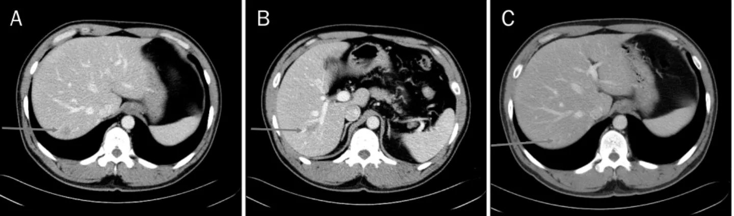
Fig. 1. (A) CT showed 20 mm sized, well-defined, and faintly low attenuated mass in the S8 of the liver. (B) CT showed 12 mm sized, ill-defined, and faintly low attenuated lesion in the S7 of the liver. (C) Follow up CT (after 8 weeks) showed decreased size of (20 mm → 13 mm) well-defined, and faintly low attenuated mass in the S8 of the liver.
증례: 상지의 근력 감소로 신경과에서 추적 중인 45세 남자 환자가 복부전산화단층촬영에서 간에 이상 소견을 보여 혈액 종양내과를 방문하였다. 내원 당시 복통, 황달 및 체중감소 등의 특이증상은 호소하지 않았다. 과거력으로는 고혈압이 있 었으며, 내원 당시의 활력징후는 정상이었다. 진찰소견에서 간비대는 없었고 흉부청진에서도 특이소견은 없었다. 당시 시 행한 말초혈액검사에서 백혈구 15,540/mm3, 혈색소 15.2 g/dL, 혈소판이 250,000/mm3, 호산구 8.2% (정상치 <7%)였 으며, 다른 혈액검사에서 이상소견은 보이지 않았다. 복부전
산화단층촬영에서 간의 7번, 8번 분절에 20 mm, 12 mm 크 기의 저음영 종양이 관찰되었다(Fig. 1A, B).
8주 후 추적한 복부전산화단층촬영에서 간의 8번 분절에서 관찰되던 종괴의 크기는 감소하였고(Fig. 1C), 간의 7번 분절 에서 관찰되던 종괴는 보이지 않았다. 당시 시행한 말초혈액 검사에서 백혈구 12,150/mm3, 호산구 7.3%였으며 기생충 검 사에서 간흡충, 폐흡충, 유구낭미충, 스파르가눔의 항원에 대 한 IgG 항체 검사결과 모두 음성이었다.
10주 후 추적한 복부전산화단층촬영에서 간의 7번 분절에
Lee SH and Shin WG. Eosinophilic Liver Abscess Caused by Toxocara Canis
227
Vol. 58 No. 4, October 2011
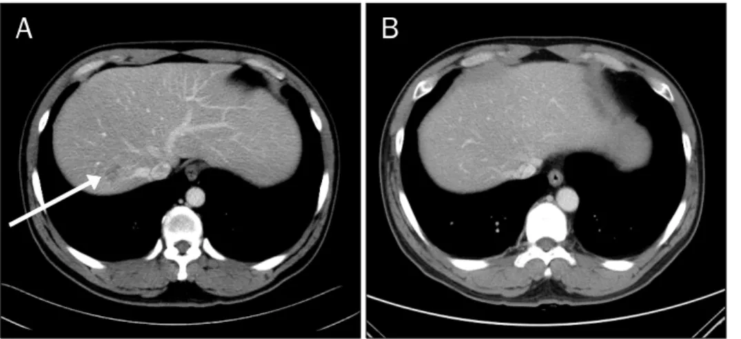
Fig. 2. (A) Follow up CT (after 10 weeks) showed 23 mm sized low attenuated lesion with irregular shape in the S7 of the liver. (B) Follow up CT (after 12 weeks) showed disappear- ance of low attenuated lesion in the S7 of the liver.
23 mm의 새로운 종괴가 발견되었다(Fig. 2A). 말초혈액검사 에서 백혈구 11,780/mm3, 호산구 9.1%였다. 호산구 증다증 의 평가를 위하여 골수검사를 권유하였지만, 보호자가 거부하 여 우선 스테로이드 치료를 시작하였다. 치료기간 중 환자는 속쓰림과 역류증상으로 소화기내과를 방문하였다. 환자의 개 를 기르고 있는 경력과 호산구 증다증 그리고 복부전산화단층 촬영에서 크기와 위치가 변하는 저음영 병변 등을 고려하여 개회충(toxocara canis)에 대한 ELISA 검사를 시행하였고 양 성소견을 보였다. 알벤다졸(albendazole) 400 mg을 투약하 였고, 3개월 후 추적한 복부전산화단층촬영에서 간의 7번 분 절에서 보이던 종괴는 관찰되지 않았고(Fig. 2B), 호산구도 정 상범위로 관찰되었다. 현재 특별한 증상 없이 추적관찰 중이 다.
진단: 개회충에 의한 호산구성 간농양
톡소카라증(Toxocariasis)은 개나 고양이를 숙주로 하는 개 회충(Toxocara canis)과 고양이회충(Toxocara catis)에 의한 인간 감염이다. 개회충에 의한 인간 감염은 두 가지 경로로 이루어진다. 개의 소장에 있던 개회충의 충란이 배설물을 통 하여 배출되고, 그것에 의해 오염된 흙 또는 채소를 인간이 섭취함으로서 감염된다. 그러면 그 유충이 소장으로 배출되고 소장벽으로 침투한 후에 간문맥을 침범한다. 다른 경로로는 인간이 캡슐화된 유충을 가지고 있는 감염된 연장동물을 생식 하는 경우가 있다. 간에 도달한 유충들은 간문맥류를 통하여 폐, 눈, 심장 그리고 뇌 등의 조직에 분포하여 출혈, 괴사 그리 고 호산구성 염증을 일으키며 육아종을 형성하기도 한다.1-4 이번 증례의 환자는 기르던 개의 충란이 배설물을 통해 배출 된 경로에 의해 감염되었다고 추정하였다.
임상적으로 톡소카라증 환자의 대부분은 무증상이어서, 이 질병은 주로 말초 호산구 증다증을 평가하는 과정에서 진단이 된다. 가능한 임상적 특징으로는 백혈구 및 호산구의 증가, 복부 불편감 및 복통, 그리고 발열과 전신 무력감 등으로 알려
져 있다.5,6
개회충에 의한 간병변을 진단하기 위해서는 혈청학적 검사 와 영상검사 그리고 조직검사가 필요하다. 특히 복부전산화단 층촬영에서 다발성의 저음영 종양으로 나타나는 경우가 많아 전이성 종양과 감별이 필요하다. 이번 증례에서도 확진을 위 해서는 간조직검사가 필요하였지만 조직검사의 침습성과 가 능한 부작용을 고려할 때 ELISA를 이용한 혈청학적 검사와 변화하는 저음영의 종양을 특징으로 하는 복부전산화단층촬 영 소견으로 임상적인 진단을 내릴 수 있다고 판단하였다.7 톡소카라증의 치료에는 주로 벤지미다졸(benzimidazole) 유도체가 사용된다. 이번 증례에서 투약한 알벤다졸(alben- dazole)은 이전 연구에서 10 mg/kg를 5일간 투여하였을 때 평균 30주 간의 추적관찰기간 중 환자의 47%에서 치료 반응 을 보였다.8
톡소카라증에 의한 간농양의 치료 후 추적에는 말초혈액 호산구 수와 영상검사가 필요하다. 이번 증례의 경우 개회충 에 의한 간농양을 진단하여 알벤다졸 400 mg을 투약하고 3 개월 후에 추적한 결과 말초 호산구 수가 정상화되고 복부전 산화단층촬영에서 저음영의 종괴가 관찰되지 않았다.
REFERENCES
1. Chang S, Lim JH, Choi D, et al. Hepatic visceral larva migrans of Toxocara canis: CT and sonographic findings. AJR Am J Roentgenol 2006;187:W622-629.
2. Beaver PC, Jung RC, Cupp EW. Clinical parasitology. 9th ed.
Philadelphia: Lea & Febiger, 1984:320-329.
3. Kaplan KJ, Goodman ZD, Ishak KG. Eosinophilic granuloma of the liver: a characteristic lesion with relationship to visceral larva migrans. Am J Surg Pathol 2001;25:1316-1321.
4. Beaver PC. The nature of visceral larva migrans. J Parasitol 1969;55:3-12.
5. Kwon NH, Oh MJ, Lee SP, Lee BJ, Choi DC. The prevalence and diagnostic value of toxocariasis in unknown eosinophilia. Ann
228
이상호, 신운건. 개회충에 의한 호산구성 간농양The Korean Journal of Gastroenterology Hematol 2006;85:233-238.
6. Despommier D. Toxocariasis: clinical aspects, epidemiology, medical ecology, and molecular aspects. Clin Microbiol Rev 2003;16:265-272.
7. Jacquier P, Gottstein B, Stingelin Y, Eckert J. Immunodiagnosis
of toxocarosis in humans: evaluation of a new enzyme-linked im- munosorbent assay kit. J Clin Microbiol 1991;29:1831-1835.
8. Magnaval JF. Comparative efficacy of diethylcarbamazine and mebendazole for the treatment of human toxocariasis.
Parasitology 1995;110:529-533.