대한소화기학회지 2009;54:205-211 □ REVIEW □ DOI: 10.4166/kjg.2009.54.4.205
연락처: 류지곤, 110-744, 서울시 종로구 대학로 101 서울대학교 의과대학 내과학교실, 간연구소 Tel: (02) 2072-1962, Fax: (02) 762-9662 E-mail: [email protected]
Correspondence to: Ji Kon Ryu, M.D.
Department of Internal Medicine and Liver Research Institute, Seoul National University College of Medicine, 101, Daehagro, Jongno-gu, Seoul 110-744, Korea
Tel: +82-2-2072-1962, Fax: +82-2-762-9662 E-mail: [email protected]
급성 췌장염의 중증도 평가
서울대학교 의과대학 내과학교실, 간연구소
류 지 곤
Evaluation of Severity in Acute Pancreatitis
Ji Kon Ryu, M.D.
Department of Internal Medicine and Liver Research Institute, Seoul National University College of Medicine, Seoul, Korea
Acute pancreatitis has a variable etiology and natural history, and some patients have severe complications with a significant risk of death. The prediction of severe disease should be achieved by careful ongoing clinical assess- ment coupled with the use of a multiple factor scoring system and imaging studies. Over the past 30 years sev- eral scoring systems have been developed to predict the severity of acute pancreatitis. However, there are no complete scoring index with high sensitivity and specificity till now. The interest in new biological markers and predictive models for identifying severe acute pancreatitis testifies to the continued clinical importance of early se- verity prediction. Among them, IL-6, IL-10, procalcitonin, and trypsinogen activation peptide are most likely to be used in clinical practice as predictors of severity. Even if contrast-enhanced CT has been considered the gold standard for diagnosing pancreatic necrosis, early scanning for the prediction of severity is limited because the full extent of pancreatic necrosis may not develop within the first 48 hour of presentation. (Korean J Gastroenterol 2009;54:205-211)
Key Words: Acute pancreatitis; Severity; Scoring system
서 론
급성 췌장염은 췌장의 급성 염증 과정이며 흔히 췌장 주 변조직과 다른 원격 장기의 이상이 동반된다. 급성 췌장염 의 중증도는 매우 다양하여 췌장에만 염증이 발생하는 경증 의 형태에서부터 다발 장기부전 및 사망이 동반되는 중증의 형태까지 발생할 수 있다. 급성 췌장염의 평균 사망률은 5%
정도이나,1 경증 췌장염에서는 사망률이 1% 미만인데 반해,2 중증 췌장염에서는 매우 높아져서 무균 괴사 췌장염에서는 10%, 감염 괴사 췌장염의 경우는 25-30%에 이른다.3 급성
췌장염 환자에서 사망은 약 50%에서 발병 2주 내에 발생하 므로 초기에 중증 경과를 보일 것으로 예측되는 환자를 선 별하여 집중 치료하는 것이 매우 중요하다. 지금까지 알려 진 급성 췌장염의 중증도 판별 기준은 다양하고 복잡하며 예민도가 충분히 높지 않아 실제 임상적으로 적용하는 데 어려움이 있다. 이 글에서는 지금까지 알려진 중증도 판정 기준의 유용성과 한계점을 알아보고 최근 새롭게 제시되고 있는 판정 기준을 소개하고자 한다.
206 대한소화기학회지: 제54권 제4호, 2009
Table 1. Atlanta Criteria for Severity of Acute Pancreatitis
Severity criteria Definition
Organ failure with 1 or more
Shock Systolic blood pressure <90 mmHg Pulmonary insufficiency PaO2<60 mmHg
Renal failure Serum creatinine level >2 mg/dL after rehydration Gastrointestinal tract bleeding 500 mL in 24 hours
Local complications
Pancreatic necrosis More than 30% of the parenchyma or more than 3 cm Pseudocyst Collection of pancreatic juice enclosed by a wall
Abscess Circumscribed collection of pus containing little or no pancreatic necrosis
Ranson score >3
APACHE II score >8
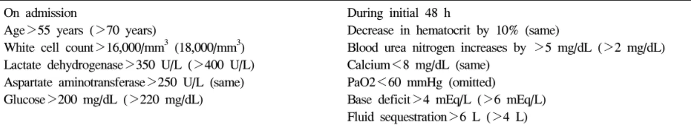
Table 2. Ranson’s Criteria for the Prediction of Severity of Acute Pancreatitis On admission During initial 48 h
Age>55 years (>70 years) Decrease in hematocrit by 10% (same)
White cell count>16,000/mm3 (18,000/mm3) Blood urea nitrogen increases by >5 mg/dL (>2 mg/dL) Lactate dehydrogenase>350 U/L (>400 U/L) Calcium<8 mg/dL (same)
Aspartate aminotransferase>250 U/L (same) PaO2<60 mmHg (omitted)
Glucose>200 mg/dL (>220 mg/dL) Base deficit>4 mEq/L (>6 mEq/L) Fluid sequestration>6 L (>4 L)
The criteria for nongallstone (alcoholic) acute pancreatitis are listed first; the changes (if any) in the criteria for gallstone pancreatitis are in parentheses.
본 론
1. 중증 급성 췌장염(Atlanta 분류)
1992년 Atlanta에서 급성 췌장염을 임상, 검사실 소견 및 국소 합병증에 따라 경증과 중증으로 구분하였다. 즉 4가지 기준 중 1가지만 만족하면 중증 췌장염으로 분류하였다. 첫 째, 장기 부전이 있을 때, 둘째, 국소 합병증이 동반될 때, 셋째, Ranson 지표가 3점 이상일 때, 넷째, APACHE (Acute Physiology and Chronic Health Evaluation) II 점수가 8점 이상 일 때이다(Table 1). Atlanta 기준이 세계적으로 가장 많이 사 용되고 있지만 실제 임상에서는 여러 문제점이 있다. 일시 적인 장기 부전과 지속적인 장기 부전이 구분되어 있지 않 아 일부 환자에서 과대평가될 가능성이 있으며 중증 췌장염 으로 진단된 10-15%만이 실제로 중증으로 판정된다.4 또한 실제 대부분의 사망은 국소 합병증 자체보다는 장기 부전에 기인하는데 Atlanta 분류는 장기 부전과 국소 합병증 사이에 중증도 차이가 없다는 문제가 있다. 중증 급성 췌장염 환자 207명을 분석한 최근 연구에 의하면 장기 부전 유무가 사망 률과 재원 기간을 예측하는 가장 중요한 인자이며, 장기 부 전이 동반되지 않은 중증 췌장염 환자에서는 치사율이 0%
였다.5 그러므로 급성 췌장염을 경증, 중등도 중증, 중증 3가 지로 분류할 것을 제안하였다. 한편 장기 부전 중 소화관 출 혈은 매우 드물고 다른 장기 부전과 유사하게 중증도를 반 영하지 못하므로 장기 부전 항목에서 삭제해야 한다는 주장 도 있다.1 최근에는 국소 합병증의 정의에 대한 개정 필요성 이 제시되고 새로운 용어가 등장하고 있어 향후 개정이 필 요할 것으로 보인다.6-8
2. Ranson 지표
1974년 Ranson이 발표한 임상 지표법으로 다변수 평가법 중 가장 많이 알려진 것이다. 43개의 임상, 생화학 지표를 분석한 결과 11개의 항목이 예후와 관련 있음이 밝혀졌다.9 입원 시에 5개 항목, 입원 후 48시간 이내에 6개 항목을 측 정하여 3가지 이상 관찰되는 경우 중증 췌장염으로 정의하 였다(Table 2). 처음에는 알코올 췌장염을 대상으로 하였다 가 이후 담석 췌장염 환자를 대상으로 한 지표가 추가되어 발표되었다. Ranson 지표의 주된 문제점은 11개의 지표를 모두 측정해야 하며 완전한 평가를 위해 48시간이 필요하므 로 중증도의 판정이 늦어질 수 있고 48시간 이후 지속적인 판정이 불가능하다는 점이다. 또한 3점 이하이거나 6점 이 상일 경우에는 비교적 정확하지만 중간 점수일 때는 예후와
류지곤. 급성 췌장염의 중증도 평가 207
Table 3. Glasgow Severity Scoring System for Acute Pan- creatitis
Age>55 years
White cell count>15,000/mm3 PaO2<60 mmHg
Serum lactate dehydrogenase>600 U/L Serum aspartate aminotransferase>200 U/L Serum albumin<3.2 g/dL
Serum calcium<8 mg/dL Serum glucose>180 mg/dL Serum urea>45 mg/dL
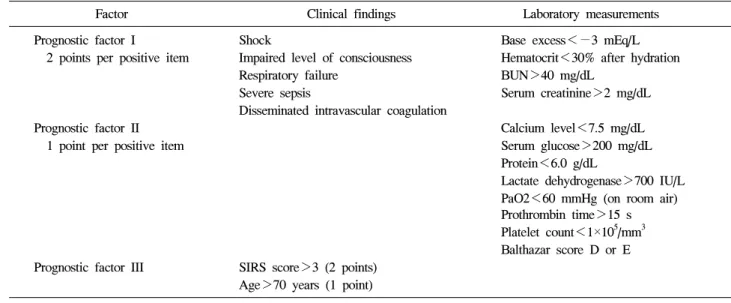
Table 4. Japanese Severity Score for Acute Pancreatitis
Factor Clinical findings Laboratory measurements Prognostic factor I Shock Base excess<−3 mEq/L 2 points per positive item Impaired level of consciousness Hematocrit<30% after hydration
Respiratory failure BUN>40 mg/dL
Severe sepsis Serum creatinine>2 mg/dL Disseminated intravascular coagulation
Prognostic factor II Calcium level<7.5 mg/dL
1 point per positive item Serum glucose>200 mg/dL Protein<6.0 g/dL
Lactate dehydrogenase>700 IU/L PaO2<60 mmHg (on room air) Prothrombin time>15 s Platelet count<1×10⁵/mm3 Balthazar score D or E Prognostic factor III SIRS score>3 (2 points)
Age>70 years (1 point) 상관도가 떨어진다.10
3. Glasgow 지표
Imrie 등은 알코올과 담석 췌장염에 모두 사용할 수 있는 Ranson 지표와 유사한 다변수 평가법을 발표하였다. Ranson 지표 중 3개 지표를 삭제하고 알부민을 첨가하여 총 9개의 지표로 단순화하였다(Table 3). 이 지표는 Ranson 지표와 유 사한 정확도를 보이지만 역시 판정에 48시간이 필요하다는 단점이 있다.
4. APACHE II 지표
APACHE II 지표는 특정 질환에 대한 임상 평가가 아니 라 중환자실에서 이용되어온 지표로 12가지의 생리적인 측 정치와 나이, 5개의 장기에 기초한 만성 건강 상태를 평가 하고 이를 점수화하여 전체 점수를 합산하는 방법으로 산출 된다. 체온, 심박수, 호흡수, 동맥혈 산소, 동맥혈 pH, 혈청 나트륨, 혈청 크레아티닌, hematocrit, 백혈구수, Glasgow coma scale, 연령 점수, 질환 점수 등이 포함된다. APACHE
II 지표는 입원 수시간 내에 급성 췌장염의 중증도를 판정 할 수 있고 수시로 반복 측정할 수 있어 진행 여부를 평가 할 수 있다는 장점이 있다. Atlanta 분류에 의하면 8점 이상 을 중증 췌장염으로 정의하였으며 Ranson 지표, Glasgow 지 표에 비해 중증도 예측에 우수성을 보이나 점수 산정과 계 산이 복잡하고 급성 췌장염에 적합한 적절한 기준이 불명확 하며 고연령에서 점수가 지나치게 높다는 문제점이 있다.
비만이 중증 급성 췌장염의 발병과 관련이 있고 사망의 독 립적인 예측인자로 인식되면서 APACHE II 지표에 body mass index (BMI)를 더하여 새로운 APACHE-O 지표가 만들 어졌다.11 즉 BMI가 26-30인 경우 1점, 30 이상인 경우 2점 으로 계산한다.
5. 일본 중증도 지표
1998년 일본 췌장학회에서는 임상 징후, 혈액 검사, 영상 소견에 기초하여 일본 중증도 지표를 만들었고 이후 2002년 개정된 지표를 사용하고 있다(Table 4).12 점수를 바탕으로 Stage 0: mild acute pancreatitis, Stage 1: moderate acute pancreatitis, Stage 2: severe acute pancreatitis I (severity score, 2-8 points), Stage 3: severe acute pancreatitis II (severity score, 9-14 points), Stage 4: extremely severe acute pancreatitis (severity score, 15-27 points)로 분류하였다. 사망 및 중증도 예측의 예민도는 Ranson 지표와 유사하나 역시 복잡하다는 문제가 있다.
6. Artificial neural network (ANN)
ANN은 정보 분석 알고리즘으로 생물학적인 신경계와 유 사하게 고안되었다.13 ANN은 복잡한 임상 자료를 분석하여 여러 다양한 임상 상황에서 사용할 수 있도록 고안되어 진
208 The Korean Journal of Gastroenterology: Vol. 54, No. 4, 2009
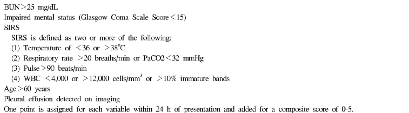
Table 5. BISAP (Bedside Index for Severity in Acute Pancreatitis) Scoring System BUN>25 mg/dL
Impaired mental status (Glasgow Coma Scale Score<15) SIRS
SIRS is defined as two or more of the following:
(1) Temperature of <36 or >38oC
(2) Respiratory rate >20 breaths/min or PaCO2<32 mmHg (3) Pulse>90 beats/min
(4) WBC <4,000 or >12,000 cells/mm3 or >10% immature bands Age>60 years
Pleural effusion detected on imaging
One point is assigned for each variable within 24 h of presentation and added for a composite score of 0-5.
단 및 생존율 분석 등 의학에서도 사용되고 있다.14,15 급성 췌장염의 중증도 예측에도 ANN을 사용한 연구가 있는데 APACHE II, Ranson 지표보다 더 정확하다는 연구 결과가
있다.16,17 Mofidi 등은 최근 연구에서 나이, shock, systemic
inflammatory response syndrome (SIRS), 동맥혈 산소, LDH, 혈당, blood urea nitrogen (BUN), Ca, hematocrit, 백혈구수 등 10개의 변수를 갖고 ANN를 이용하여 분석한 결과 중증 경 과로의 진행 예측, 다발성 장기부전의 발생, 사망 예측 등에 있어서 모두 유의하게 정확하였다.17
7. Bedside index for severity in acute pancrea- titis (BISAP) 지표
212개 병원에서 급성 췌장염으로 진단 받은 17,922명의 환자를 자료 분석하여 2008년 BISAP 지표가 발표되었다 (Table 5).18 입원 24시간 동안 BUN>25 mg/dL, impaired mental status, SIRS, age>60, pleural effusion 5개 항목을 갖 고 각각 1점을 주어 점수가 높아짐에 따라 사망률이 높아짐 을 보고하였다. 5점일 때는 20% 이상의 사망률을 보인 반면 1점 이하에서는 1% 미만의 사망률을 보였다. 18,256명의 환 자를 갖고 검증한 결과에서도 APACHE II 지표와 유사한 정확도를 보여 매우 간편하면서 정확한 지표라고 하였다.
이후 최근 발표된 전향 연구에서도 BISAP 지표는 사망률과 유의한 상관관계를 보였고, 3점 이상인 경우 장기부전의 발 생, 장기부전의 지속, 췌장 괴사의 위험도가 각각 7.4, 12.7, 3.8로 간단하면서도 유용한 검사로 보고했다.19
8. 기타 다변수 평가법
2007년 Ueda는 3개의 변수를 이용한 simple prognostic score를 발표하였다.20 BUN>25 mg/dL, LDH>900 IU/L, pan- creatic necrosis in enhanced computed tomography (CT)로 0점 에서 3점으로 분류하였을 때 사망률과 상관관계가 매우 높 으며 기존의 다변수 평가법과 유사한 정확도를 보고하였다.
국내에서 Lee 등은 109명의 환자를 대상으로 한 연구에서
크레아티닌, CT index, 혈청 교정 칼슘 농도를 이용하여 새 로운 평가법을 고안했는데(R=2.512 loge (creatinine mg/dL)+
1.729 loge (CT index)−4.780 loge (corrected calcium mg/dL)) 사망률을 예측하는 예민도 및 특이도가 각각 83.3%, 89.5%
였다.21
2009년 독일에서는 3개의 변수를 이용하여(반발통 또는 guarding이 없음, 정상 hematocrit, 정상 크레아티닌) 중증이 아닌 급성 췌장염을 예측하는 강력한 지표라고 하였고 harmless acute pancreatitis score (HAPS)라고 명명하였다.22 이는 처음 394명의 환자를 대상으로 조사하였고 이후 전향 다기관 연구로 452명의 환자를 통해 검증한 결과 98%의 예 민도로 중증이 아닌 경과를 예측하였다.
9. 단일 검사 지표
1) 혈액 농축 및 hematocrit
심각한 급성 췌장염이 발생하면 복강 내 체액 손실로 인 해 혈액이 농축되어 높은 적혈구 용적률을 보인다. Brown 등은 입원 시 hematocrit이 44% 이상이고 24시간 이내에 감 소하지 않을 경우 94%의 예민도로 췌장 괴사와 장기부전 발생을 예측한다고 하였다.23 그러나 이후 다른 연구에서는 유사한 예민도를 보여주지 못했다.24 한편 이후 Brown 등은 hematocrit 44% 이상, BMI 30 이상, pleural effusion 3가지를 종합한 Panc 3 지표를 발표하였고, 중증 급성 췌장염을 예 측하는 간편하고 정확한 검사라고 하였다.25 최근 연구에서 는 24시간 이내에 hematocrit이 44% 이상 또는 Hb이 14.6 mg/dL이면 사망률 증가와 관련이 높았다.26
2) C-reactive protein (CRP)
CRP는 혈중 interleukin (IL)-1과 IL-6에 의해 자극을 받아 간세포에서 합성되는 급성기 단백이다. 현재 급성 췌장염의 중증도 평가에서 가장 널리 알려져 있고 이용되는 단일 지 표이다. 증상 발생 후 48시간이 지나서 측정해야 유용하며 이때 측정하면 APACHE II 지표와 유사한 정확도를 보인다
Ryu JK. Evaluation of Severity in Acute Pancreatitis 209
Table 6. Balthazar CT Score
Grade CT findings
A Normal
B Focal or diffuse enlargement of the pancreas, including irregularities of contour and inhomogeneous attenuation C Pancreatic gland abnormalities in grade B plus per pancreatic inflammation
D Grade C plus a single fluid collection
E Grade C plus 2 or more fluid collections and/or the presence of gas in or adjacent to the pancreas
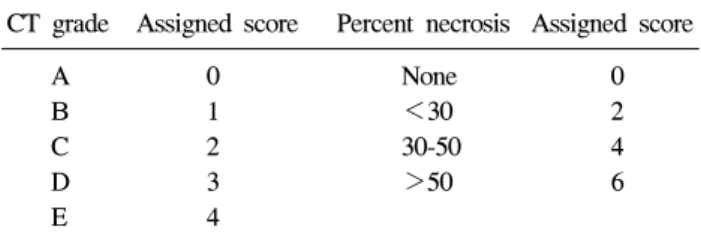
Table 7. CT Severity Index
CT grade Assigned score Percent necrosis Assigned score
A 0 None 0
B 1 <30 2
C 2 30-50 4
D 3 >50 6
E 4
CT grade based on Balthazar score plus pancreatic necrosis with a maximum score of 10 points.
는 보고도 있다.27 중증과 경증을 구분하는 CRP의 cut-off 값 은 120-210 mg/L로 알려져 있다.28,29 가장 큰 단점은 발병 초 기 48시간 내에는 정확도가 떨어진다는 점과 혈중 사이토카 인의 매개로 간세포에서 합성되므로 직접적인 중증도 반영 에 한계가 있다.
3) Blood urea nitrogen (BUN)과 크레아티닌 BUN과 크레아티닌은 대부분의 다변수 평가법에서 중요 한 인자로 포함되어 있으나 단일 인자로서의 역할에 관한 연구는 거의 없었다. 그러나 최근 미국의 69개 병원에서 5,819명의 환자를 대상으로 시행한 후향 연구에 의하면 48 시간 동안 BUN의 지속적 상승이 사망률과 관련성이 있어 서 사망을 예측하는 가장 중요한 단일 인자였다.30 또한 129 명의 환자를 대상으로 한 다른 전향 연구에서는 첫 48시간 동안 크레아티닌 1.8 mg/dL 이상의 상승은 췌장 괴사의 발 생과 높은 관련성이 있었다.31
4) 사이토카인(Cytokine)
급성 췌장염의 발생 및 다발 장기부전에 여러 사이토카인 이 관여한다고 알려져 있어 이를 측정하여 췌장염의 중증도 판정에 이용하려는 연구들이 있었다. IL-6는 급성기 단백 생 성의 주요 매개체로 24시간 내에 경증과 중증을 판정하는 데 비교적 정확하다는 보고가 있다.32 IL-8, IL-10, TNF-α에 대한 연구도 있는데,28,32,33 아직까지는 소규모 연구이고 특 히 실제로 쉽게 측정할 수 있는 방법이 개발되어 있지 않아 임상적으로 응용하기에는 한계가 있다. 한편 IL-10과 혈청 칼슘 두 가지를 병합하였을 때 예민도 88%, 특이도 93%로 장기부전 발생을 비교적 정확하게 예측하였다는 연구도 있 다.34
5) 기타
Procalcitonin은 칼시토닌의 불활 propeptide로 중증 세균 감염 또는 진균 감염, 환자의 혈청에서 검출되며 다발 장기 부전 환자에서 발견되는 것으로 알려져 있다. 몇몇 연구 결 과에서 입원 당시의 procalcitonin이 CRP, APACHE II 지수 보다 더 정확히 급성 췌장염의 중증도를 예측한다는 결과가
있으나35,36 아직 더 연구가 필요한 상태이다. 소변의 trypsi-
nogen-2의 정량 검사가 중증 급성 췌장염 환자에서 의미 있 게 높다는 연구들도 있다.37,38
Trypsinogen activation peptide (TAP)는 trypsinogen이 활성 화되는 과정에서 분해되어 나오는 작은 펩티드인데 246명 의 환자를 대상으로 한 연구에서 소변 TAP의 측정이 급성 췌장염 발생 24시간에 중증도를 예측할 수 있는 정확한 인 자라고 알려졌으며 또한 CRP보다 더 예민도가 높았다.29
10. 방사선 검사
조영 증강 복부 CT는 급성 췌장염의 중증도 판정뿐만 아 니라 합병증 진단에 널리 사용되고 있고 A등급부터 E등급 까지 분류한 Balthazar 등급 분류가 널리 이용되고 있다 (Table 6).39
조영제에 의해 췌장 실질이 증강되지 않으면 췌장 괴사를 의미하며 이는 췌장 괴사의 진단에 gold standard이며 100%
의 예민도와 양성 예측도를 보인다고 알려져 있어,40 급성 췌장염의 중증도 판정에 매우 중요하다. 1990년 Balthazar 등급과 췌장 괴사의 정도를 합쳐서 점수화한 CT 중증도 지 수가 제안되었고,41 매우 중요한 예후 평가 방법이다(Table 7). 당시 연구에 의하면 CT 중증도 지수가 7-10이면 17%의 사망률인 반면 4-6인 경우 6%, 0-3일 때는 3%의 사망률을 보고하였다. 이후 268명의 환자를 대상으로 한 연구에서 CT 중증도 지수가 5 이상인 경우 사망률 8배, 장기 입원 경과 17배, 수술을 받을 가능성이 10배 더 높았다.42 한편 Mortele 등은 췌장 염증의 등급을 3등급으로(0, 2, 4) 줄이고, 췌장 괴사 정도도 30%를 기준으로(0, 2, 4) 간소화하고 췌장외 합 병증 유무를(0, 2) 추가한 CT 중증도 지수를 제안하였는데,43
210 대한소화기학회지: 제54권 제4호, 2009
기존의 중증도 지수보다 재원 기간, 수술 필요성, 감염 발 생, 장기부전의 발생 등을 예측하는 데 더 높은 정확도를 보 였다. 조영 증강 CT가 중증 급성 췌장염의 진단 및 예후 판 정에서 매우 중요한 검사인 것은 사실이나 몇 가지 제한점 이 있다. CT 중증도 지수는 급성 췌장염 발병 6-10일 때 가 장 정확하다고 알려져 있으며 특히 발병 48시간 이내에는 췌장 괴사가 잘 나타나지 않으므로 초기에 중증도를 판정하 기에는 한계가 있다. 또한 조영제가 췌장의 미세혈관 순환 장애를 유발하여 췌장염을 악화시킨다는 일부 보고가 있어 모든 급성 췌장염 환자를 대상으로 조영 증강 CT를 시행할 필요는 없다. 그러므로 조영 증강 CT는 다른 진단을 감별하 기 위한 경우가 아니면 다른 중증도 지표에서 중증 췌장염 이 의심될 때 검사하는 것이 추천되며 또한 췌장 괴사의 정 도를 정확히 판정하기 위해서는 48시간 이후에 권장되고 있 다.
결 론
Ranson 지표가 처음 발표된 이후 지난 30여 년 동안 급성 췌장염의 중증도를 발병 2-3일 내에 예측할 수 있는 여러 다중 평가법이 고안되었다. 가장 이상적인 중증도 평가법은 조기에 예측이 가능하고 비침습적이고 간편한 방법이어야 한다. 그러나 APACHE II 지표와 일본 중증도 지표는 너무 복잡하다는 문제가 있고 BISAP 지표는 비교적 간단하나 향 후 대규모 연구가 필요하며 여러 연구자들 간에 학회를 통 한 합의가 필요하다. 또한 컴퓨터 소프트웨어의 발달로 ANN의 개발을 통한 새로운 지표의 개발을 기대할 수 있다.
단일 생화학 지표로는 현재 CRP가 임상적으로 많이 이용되 고 있으나 발병 48시간 이내에는 정확도가 떨어진다는 문제 가 있으며 향후 IL-6, IL-10, procalcitonin, 소변 TAP 등이 유 용한 지표로 사용될 가능성이 있다. 조영 증강 CT는 췌장 괴사 여부를 유무 및 정도를 판정할 수 있는 매우 중요한 검사법이며 중증 급성 췌장염의 예후 판정에도 중요한 역할 을 하지만 발병 48시간 이내에는 괴사 여부가 잘 나타나지 않는다는 한계점을 알고 있어야 한다. 1992년 Atlanta 분류 가 제정된 이후 급성 췌장염의 병태 생리에 관한 기초 연구 의 발전과 임상 의학의 눈부신 발전으로 더 이상 조기 수술 은 시행되지 않고 있으며 비수술 치료의 역할이 더 중요해 지고 있고 합병증의 정의에 관한 새로운 용어 제정 필요성 이 대두되고 있는 실정이다. 그러므로 향후 학회를 통한 합 의가 도출되어 새롭고 이상적인 급성 췌장염의 중증도 평가 법이 개발될 것으로 생각한다.
참고문헌
1. Banks PA, Freeman ML. Practice guidelines in acute pan- creatitis. Am J Gastroenterol 2006;101:2379-2400.
2. Uhl W, Warshaw A, Imrie C, et al. IAP guidelines for the surgical management of acute pancreatitis. Pancreatology 2002;2:565-573.
3. Pandol SJ, Saluja AK, Imrie CW, Banks PA. Acute pan- creatitis: bench to the bedside. Gastroenterology 2007;132:
1127-1151.
4. Bradley EL III. Atlanta redux. Pancreas 2003;26:105-106.
5. Vege SS, Gardner TB, Chari ST, et al. Low mortality and high morbidity in severe acute pancreatitis without organ fail- ure: a case for revising the Atlanta classification to include
“moderately severe acute pancreatitis”. Am J Gastroenterol 2009;104:710-715.
6. Bollen TL, Besselink MG, van Santvoort HC, et al. Toward an update of the Atlanta classi. cation on acute pancreatitis:
review of new and abandoned terms. Pancreas 2007;35:
107-113.
7. Bollen TL, van Santvoort HC, Besselink MG, et al. The Atlanta Classification of acute pancreatitis revisited. Br J Surg 2008;95:6-21.
8. Morgan DE. Imaging of acute pancreatitis and its compli- cations. Clin Gastroenterol Hepatol 2008;6:1077-1085.
9. Ranson JH, Rifkind KM, RosesDF, Fink SD, Eng K, Spencer FC. Prognostic signs and the role of operative management in acute pancreatitis. Surg Gynecol Obstet 1974;139:69-81.
10. Imrie CW. Classification of acute pancreatitis and the role of prognostic factors in assessing severity of disease. Schweiz Med Wochenschr 1997;127:798-804.
11. Johnson CD, Toh SK, Campbell MJ. Combination of APACHE-II score and obesity score (APACHE-O) for the prediction of severe acute pancreatitis. Pancreatology 2004;
4:1-6.
12. Ogawa M, Hirota M, Hayakawa T, et al. Development and use of a new staging system for severe acute pancreatitis based on a nationwide survey in Japan. Pancreas 2002;25:
325-330.
13. Drew PJ, Monson JR. Artificial neural networks. Surgery 2000;127:3-11.
14. Mofidi R, Powell TI, Brabazon A, et al. Prediction of the ex- act degree of internal carotid artery stenosis using an artificial neural network based on duplex velocity measurements. Ann Vasc Surg 2005;19:829-837.
15. Mofidi R, Deans C, Duff MD, de Beaux AC, Paterson Brown S. Prediction of survival from carcinoma of oesophagus and oe-
류지곤. 급성 췌장염의 중증도 평가 211
sophago-gastric junction following surgical resection using an artificial neural network. Eur J Surg Oncol 2006;32:533-539.
16. Halonen KI, Leppäniemi AK, Lundin JE, Puolakkainen PA, Kemppainen EA, Haapiainen RK. Predicting fatal outcome in the early phase of severe acute pancreatitis by using novel prognostic models. Pancreatology 2003;3:309-315.
17. Mofidi R, Duff MD, Madhavan KK, Garden OJ, Parks RW.
Identification of severe acute pancreatitis using an artificial neural network. Surgery 2007;141:59-66.
18. Wu BU, Johannes RS, Sun X, et al. The early prediction of mortality in acute pancreatitis: a large population-based study.
Gut 2008;57:1698-1703.
19. Singh VK, Wu BU, Bollen TL, et al. A prospective evaluation of the bedside index for severity in acute pancreatitis score in assessing mortality and intermediate markers of severity in acute pancreatitis. Am J Gastroenterol 2009;104:966-971.
20. Ueda T, Takeyama Y, Yasuda T, et al. Simple scoring sys- tem for the prediction of the prognosis of severe acute pancreatitis. Surgery 2007;141:51-58.
21. Lee BJ, Kim CD, Jung SW, et al. Analysis of the factors that affect the mortality rate in severe acute pancreatitis. Korean J Gastroenterol 2008;51:25-33.
22. Lankisch PG, Weber-Dany B, Hebel K, Maisonneuve P, Lowenfels AB. The harmless acute pancreatitis score: a clin- ical algorithm for rapid initial stratification of nonsevere disease. Clin Gastroenterol Hepatol 2009;7:702-705.
23. Brown A, Orav J, Banks PA. Hemoconcentration is an early marker for organ failure and necrotizing pancreatitis. Pancreas 2000;20:367-372.
24. Remes-Troche JM, Duarte-Rojo A, Morales G, Robles-Diaz G. Hemoconcentration is a poor predictor of severity in acute pancreatitis. World J Gastroenterol 2005;11:7018-7023.
25. Brown A, James-Stevenson T, Dyson T, Grunkenmeier D.
The Panc 3 score: a rapid and accurate test for predicting se- verity on presentation in acute pancreatitis. J Clin Gastroen- terol 2007;41:855-858.
26. Wu BU, Johannes RS, Conwell DL, Banks PA. Early hemo- concentration predicts increased mortality only among trans- ferred patients with acute pancreatitis. Pancreatology 2009;9:
639-643.
27. Wilson C, Heath DI, Imrie CW. Prediction of outcome in acute pancreatitis: a comparative study of APACHE II, clin- ical assessment and multiple factor scoring systems. Br J Surg 1990;77:1260-1264.
28. Sandberg AA, Borgstrom A. Early prediction of severity in acute pancreatitis. Is this possible? JOP 2002;3:116-125.
29. Neoptolemos JP, Kemppainen EA, Mayer JM, et al. Early
prediction of severity in acute pancreatitis by urinary trypsi- nogen activation peptide: a multicentre study. Lancet 2000;
355:1955-1960.
30. Wu BU, Johannes RS, Sun X, Conwell DL, Banks PA. Early changes in blood urea nitrogen predict mortality in acute pancreatitis. Gastroenterology 2009;137:129-135.
31. Muddana V, Whitcomb DC, Khalid A, Slivka A, Papachristou GI. Elevated serum creatinine as a marker of pancreatic ne- crosis in acute pancreatitis. Am J Gastroenterol 2009;104:
164-170.
32. Mayer J, Rau B, Gansauge F, Beger HG. Inflammatory medi- ators in human acute pancreatitis: clinical and pathophysio- logical implications. Gut 2000;47:546-552.
33. McKay CJ, Gallagher G, Brooks B, Imrie CW, Baxter JN.
Increased monocyte cytokine production in association with systemic complications in acute pancreatitis. Br J Surg 1996;
83:919-923.
34. Mentula P, Kylänpää ML, Kemppainen E, et al. Early pre- diction of organ failure by combined markers in patients with acute pancreatitis. Br J Surg 2005;92:68-75.
35. Kylänpää-Bäck ML, Takala A, Kemppainen E, Puolakkainen P, Haapiainen K, Repo H. Procalcitonin strip test in the early detection of severe acute pancreatitis. Br J Surg 2001;88:222- 227.
36. Modrau IS, Floyd AK, Thorlacius-Ussing O. The clinical val- ue of procalcitonin in early assessment of acute pancreatitis.
Am J Gastroenterol 2005;100:1593-1597.
37. Hedström J, Korvuo A, Kenkimäki P, et al. Urinary trypsi- nogen-2 test strip for acute pancreatitis. Lancet 1996;347:729- 730.
38. Lempinen M, Kylänpää-Bäck ML, Stenman UH, et al. Pre- dicting the severity of acute pancreatitis by rapid measurement of trypsinogen-2 in urine. Clin Chem 2001;47:2103-2107.
39. Balthazar EJ. CT diagnosis and staging of acute pancreatitis.
Radiol Clin North Am 1989;27:19-37.
40. Balthazar EJ. Staging of acute pancreatitis. Radiol Clin North Am 2002;40:1199-1209.
41. Balthazar EJ, Robinson DL, Megibow AJ, Ranson JH. Acute pancreatitis: value of CT in establishing prognosis. Radiology 1990;174:331-336.
42. Simchuk EJ, Traverso LW, Nukui YM, Kozarek RA. Com- puted tomography severity index is a predictor of outcomes for severe pancreatitis. Am J Surg 2000;179:352-355.
43. Mortele KJ, Wiesner W, Intriere L, et al. A modified CT se- verity index for evaluating acute pancreatitis: improved corre- lation with patient outcome. AJR Am J Roentgenol 2004;183:
1261-1265.