서 론
급 성기의 뇌졸중 환자에서는 이차적으로 여러 가지 합 병증이 발생할 수 있다. 이러한 합병증은 사망률을 증가시키거나 뇌졸중 회복을 저해할 수 있어 환자의 재활
및 예후에 나쁜 영향을 미친다(1, 2). 한 다기관 연구에서는 급성기 뇌졸중 환자의 85%에서 내과적 합병증이 발생하는 것으로 보고하였는데 빈도 순으로 보면 통증(34%), 낙상 (25%), 요로 감염(24%), 폐렴(22%), 욕창(21%) 순이었다 (3). 뇌졸중의 급성기 합병증은 연구에 따라서 다양한 빈도
급성기 합병증 관리
Management of Acute Stroke Complication
이 종 민 | 건국의대 재활의학과 | Jongmin Lee, MD
Department of Rehabilitation Medicine, Konkuk University School of Medicine E - mail : leej@kku.ac.kr
J Korean Med Assoc 2009; 52(4): 365 -374
A
cute complications following stroke may increase mortality and impede functional recovery.Most of the complications are treatable and preventable. Close monitoring of the complications and proper management are necessary for the better outcome of the patients.
Common complications include aspiration pneumonia, dysphagia, urinary tract infection, incontinence, malnutrition, deep vein thrombosis, pressure sore, fall, pain, seizure, and depression. Proper positioning and early mobilization are recommended to prevent major complications. Aspiration pneumonia is one of the frequent causes of death in acute stroke setting. Dysphagia screening should be done to evaluate the risk of aspiration. If oral feeding is not safe, nasogastric tube feeding should be considered. The majority of urinary tract infections in acute stroke are associated with the use of indwelling catheters, therefore prolonged indwelling catheterization should be avoided. Nutritional assessment and supplements are necessary in acute stroke patients. Low dose subcutaneous heparin or low molecular weight heparin should be considered for patients with high risk of deep vein thrombosis. If heparin is contraindicated, compressive stockings are an alternative. Regular assessment for skin breakdown and fall risk is recommended for all patients. Shoulder pain is also one of the frequent complications in stroke patients. Proper handling and mobilization in acute stage may prevent shoulder pain.
Administration of anticonvulsants may prevent recurrent post-stroke seizures. Depression may limit functional outcome by inhibiting patient motivation and treatment with antidepressants should be considered. Proper management of acute complications needs multidisciplinary team approach that consists of physicians, nurses, therapists, and nutritionists. Adequate prevention and management of complications may improve functional outcome of acute stroke.
Keywords: Complications; Acute stroke; Management 핵 심 용 어: 합병증; 급성기 뇌졸중; 관리
Abstract
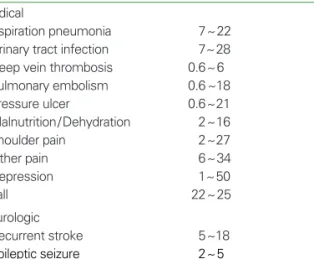
로 발생한다고 보고되고 있으며(Tablel 1), 예후에 대한 영 향도 다양하게 보고되고 있다(3~13). 이는 연구마다 합병증 의 정의, 조사 시기 및 대상이 다르기 때문인데 일부 연구는 발병 후 급성기 입원치료 시기의 환자들을 대상으로 하였고 어떤 연구들은 재활 치료 시기까지의 환자를 대상으로 하여 합병증의 양상에 차이가 있다. 그러나 대부분의 급성기 합 병증은 예방이나 치료가 가능한 것으로 뇌졸중 발생 초기부 터 적극적인 합병증의 발견 및 관리가 필요하며, 이는 최근 도입되고 있는 뇌졸중 유니트 치료에서도 필수적인 치료 내 용이다(14).
합병증 관리에 대한 임상진료지침
최근 미국과 유럽 등에서 근거중심의 진료를 위해 뇌졸중 에 대한 임상진료지침(clinical practice guideline)이 개발 되었는데 뇌졸중의 급성기 합병증 예방과 관리에 대한 진료 지침들이 제시되어 있다. 미국 American Heart Associa- tion (AHA)/American Stroke Association (ASA) 진료지침 에서는 근거 수준이 높은 Class I 권고사항으로 중증 환자가 아닌 경우 조기에 환자를 움직이게(mobilization)하고 흡 인, 영양실조, 폐렴, 심부정맥혈전증, 폐동맥색전증, 욕창, 관절 구축 등의 합병증을 예방하기 위한 조치들을 취할 것
을 권고하고 있으며, 식사나 물을 마시게 하기 전에 연하 장 애에 대한 평가를 하도록 권고하고 있다(15). 또한 폐렴이 나 요로 감염이 의심되는 환자는 항생제를 투여하며 움직이 지 못하는 부동 환자는 심부정맥혈전증을 예방하기 위해 항 응고제를 피하 투여할 것을 권고하고 있다(15). 이 외에도 경구로 식이나 수분 공급이 되지 않는 환자는 비위관(naso- gastric tube)이나 경피적 위루술(percutaneous endo- scopic gastrostomy, PEG)을 이용한 식이가 필요하고, 항 응고제보다 효과적이지는 못하지만 심부정맥혈전증의 예방 을 위해 아스피린을 사용할 수 있으며, 항응고제를 투여할 수 없는 환자는 간헐적 공기 압박 기기(intermittent pneu- matic compression device)를 사용할 것을 권고하고 있다 (Class II)(15). 그러나 예방적 목적의 항생제 투여는 바람 직하지 않으며 요로 감염의 위험이 높은 유치 도뇨관 (indwelling catheter)의 사용은 가능한 한 피하도록 권고 하고 있다(Class III)(15).
한편 유럽 European Stroke Organizaton (ESO)의 뇌졸 중 진료지침에서는 Class I 권고사항으로 심부정맥 혈전증 이나 폐동맥색전증의 위험이 높은 환자에서 저용량의 헤파 린이나 저분자량 헤파린의 피하 투여를 고려해야 하며, 뇌 졸중 후 간질 발작의 재발을 예방하기 위해 항경련제를 투 여해야 한다고 권고하고 있다(16). 연하 장애가 있는 환자 에서는 조기에 비위관을 이용한 경관 식이를 시작해야 하며 2주 이내에는 위루술을 이용한 식이는 고려하지 말라고 권 고하고 있으며, 영양 실조가 있는 경우 연하 곤란이 없으면 경구로 영양 보조 식이를 하도록 권고하고 있다(Class II) (16). 낙상위험이 있는 환자에서 칼슘과 Vitamin D의 보충 이 고려되며 골절의 과거력이 있는 여자 환자에서는 bisphos-phonate 투여가 필요하다(Class II)(16). 요실금 과 연하 곤란에 대한 평가가 필요하고(Class III), 감염이 있 을 경우에는 적절한 항생제를 사용하여 치료하는 것이 바람 직하며 조기에 환자를 움직이도록 함으로써 흡인성 폐렴, 심부정맥 혈전증, 욕창 등을 예방할 수 있다(Class IV)(16).
조기에 수분을 공급하고 압박 스타킹을 착용하여 정맥 혈전 증을 예방해야 하며 모든 뇌졸중 환자에서 낙상 위험에 대 한 평가를 권고하고 있다(Class IV)(16).
Table 1.Frequencies of acute complications following stroke Complication Range of frequencies* (%) Medical
Aspiration pneumonia 7~22
Urinary tract infection 7~28 Deep vein thrombosis 0.6~6 Pulmonary embolism 0.6 ~18
Pressure ulcer 0.6~21
Malnutrition/Dehydration 2~16
Shoulder pain 2~27
Other pain 6~34
Depression 1~50
Fall 22~25
Neurologic
Recurrent stroke 5~18
Epileptic seizure 2~5
* Data taken from (3~13).
합병증 예방을 위한 일반 처치
급성기 합병증 예방을 위해서는 생체 징후 안정을 위한 내 과적 치료 외에도 적절한 자세 유지와 관절 운동, 체위 변화, 기도 분비물 제거, 수분 및 영양 공급, 배뇨 및 배변 관리 등 의 일반적인 처치가 필요하다. 욕창을 방지하기 위해 매 2시 간마다 체위를 바꿔 주어야 하며 관절 구축 예방을 위해 침 상에서 관절 구축이 잘 발생하는 자세를 피해야 한다(17).
흔히 무릎 아래에 베개를 넣어 고관절과 슬관절이 굴곡된 자세로 환자를 유지하고 있는 경우가 있는데 이는 고관절과 슬관절의 관절 구축을 유발할 수 있는 잘못된 자세이다. 하 지의 올바른 침상 자세는 슬관절과 고관절을 신전시킨 자세 로 고관절이 외전되지 않도록 하며 발받침대로 족관절을 중 립 위치로 유지시켜야 한다. 상지는 어깨를 외전, 팔꿈치를 굴곡시킨 후 전완부를 약간 회내전시켜 베개에 올려놓거나 팔꿈치를 신전, 전완부를 회외전시켜 유지하고 손에 핸드 롤(hand roll)을 쥐어 경직의 발생을 줄여준다(18). 옆으로
누운 자세로 유지하기도 하는데 이때는 마비 측 하지가 위 로 오도록 하고 다리 사이에 베개를 넣어준다. 관절 운동은 관절 구축뿐 아니라 심부정맥혈전증, 흡인성 폐렴, 기립성 저혈압, 통증 등의 발생을 줄여줄 수 있으며 수동 운동으로 시작하여 점차 능동 운동을 시행하게 된다(17). 급성기에는 마비측 상지의 극상근과 삼각근 등의 근위약으로 견관절 아 탈구(subluxation)가 발생할 수 있는데 환자를 움직일 때 팔을 당기지 않도록 하고 앉아 있을 때는 베개나 랩보드 (lapboard)로 팔을 받쳐서 관절 및 상완신경총의 손상이 일 어나지 않도록 해야 한다(5, 17).
주요 급성기 합병증의 관리
1. 흡인성 폐렴
급성기 뇌졸중 환자의 약 37~45%의 환자에서 연하 곤란 이 있으며 20%에서 흡인성 폐렴이 발생한다(19, 20). 연하 곤란이 없는 뇌졸중 환자의 10%에서도 흡인성 폐렴이 발생
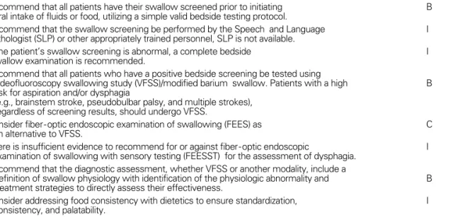
Table 2. AHA/ASA -endorsed guidelines for assessment of swallowing
Recommendation Recommendation level*
Recommend that all patients have their swallow screened prior to initiating B oral intake of fluids or food, utilizing a simple valid bedside testing protocol.
Recommend that the swallow screening be performed by the Speech and Language I Pathologist (SLP) or other appropriately trained personnel, SLP is not available.
If the patient’s swallow screening is abnormal, a complete bedside I
swallow examination is recommended.
Recommend that all patients who have a positive bedside screening be tested using
videofluoroscopy swallowing study (VFSS)/modified barium swallow. Patients with a high B risk for aspiration and/or dysphagia
(e.g., brainstem stroke, pseudobulbar palsy, and multiple strokes), regardless of screening results, should undergo VFSS.
Consider fiber-optic endoscopic examination of swallowing (FEES) as C
an alternative to VFSS.
There is insufficient evidence to recommend for or against fiber-optic endoscopic I examination of swallowing with sensory testing (FEESST) for the assessment of dysphagia.
Recommend that the diagnostic assessment, whether VFSS or another modality, include a
definition of swallow physiology with identification of the physiologic abnormality and B treatment strategies to directly assess their effectiveness.
Consider addressing food consistency with dietetics to ensure standardization, I consistency, and palatability.
* Recommendation level
A: a strong recommendation that the intervention is always indicated and acceptable, B: a recommendation that the intervention may be useful/effective,
C: a recommendation that the intervention may be considered,
D: a recommendation that a procedure may be considered not useful/effective, or may be harmful, I : Insufficient evidence to recommend for or against; clinical judgment should be used.
하는데 연하 곤란의 증상이 없이도 무증상 흡인이 발생할 수 있기 때문이다(19). 연하 곤란이 있을 경우 폐렴 발생의 상 대위험도는 3배 정도 증가하며 흡인이 있을 경우에는 12배 증 가하는 것으로 알려져 있다(20). 양측성 뇌졸중 환자나 뇌간 경색 환자에서 흡인의 위험이 높아 양측성 뇌졸중 환자의 54%에서 연하곤란으로 흡인이 발생하며 뇌간 경색환자에서 는 50%에서 흡인이 발생한다(21). 의식 수준이 저하되어 있 는 환자에서 연하 곤란과 흡인의 위험이 높으며 의식 저하 이 외에도 구음 장애나 발성 장애가 있는 경우, 자발적으로 기침 을 하지 못하는 경우, 침흘림이 있는 경우에 흡인의 위험이 높다(22). 그러나 단순히 구역반사 저하와 같은 임상 소견만 으로 흡인을 예측하기는 민감도가 떨어진다. 흡인의 선별검 사로 대표적인 것은 환자에게 직접 물을 삼키도록 해보는 것 으로 임상적으로 흡인 위험을 선별할 수 있는 방법이다(23).
연하 곤란은 구강기나 인두기에 주로 발생하는데 대부분 연하 반사가 지연되어 있거나 소실되어 있다. 환자의 평가 는 입술 및 혀의 운동, 저작근의 근력, 후두 거상(laryngeal elevation)의 정도 등의 평가가 포함되는데 이들 근육의 조 절이 충분하지 못하면 음식물 덩어리를 인두로 넘기지 못하 거나, 기도의 보호 작용이 충분히 일어나지 못하여 흡인의 위험이 증가하게 된다. 모든 급성기 뇌졸중 환자는 경구 식 이를 시작하기 전에 연하 곤란에 대한 침상 평가를 시행하 여야 하며, 기침 반사가 저하되어 있거나 연하 곤란이 의심 되는 환자는 비디오 투시 연하 검사나 광섬유 내시경 연하 검사를 시행하여 연하 기능을 평가하고 적절한 치료를 시행 하여야 한다(Table 2)(24 ~26). 연하 곤란의 치료는 식이 변형과 고개 숙이기와 같은 보상방법으로 식이를 직접 이용 하는 직접 훈련법과 행동 치료 기법 및 자극법과 같이 식이 를 이용하지 않는 간접 훈련법이 있다. 일반적으로 점도가 낮아 음식이 묽을수록 인두기의 조절이 어려워 흡인이 잘 일어나므로 비디오 투시 연하 검사의 결과에 따라 음식의 점 도를 조절하여 경구 식이를 시작한다(26).
연하 곤란이 있어 경구 식이가 안전하지 못한 환자의 경 우에는 비위관이나 경피 위루술을 이용한 경관 식이를 시행 한다(15, 16). 비위관 식이의 경우에도 식도 역류에 의해 흡 인의 위험이 있을 수 있으며 누워서 식이를 할 경우 위험이
크므로 경관 식이도 가능한 한 앉은 자세에서 시행한다. 비 위관을 장기간 거치해야 할 경우에는 경피 위루술을 이용한 경관 식이가 위장 출혈과 같은 합병증을 줄인다는 보고가 있어 고려해 볼 수 있다(27). 연하 곤란은 일측성 뇌졸중일 경우에는 대부분 1개월 이내에 빠르게 호전되어 2% 정도의 환자에서만 연하 곤란이 지속되게 된다(28). 그러나 뇌간 경색이나 양측성 뇌졸중 환자의 경우에는 잘 호전되지 않아 영양 공급을 위해 위루술이 필요할 수 있다.
2. 요로 감염
급성기 뇌졸중 환자는 신경인성 방광에 의한 배뇨곤란으 로 도뇨를 해야하는 경우가 많기 때문에 요로 감염의 위험 이 높다. 뇌졸중이 발생하면 배뇨를 관장하는 천추 반사가 저하되어 방광이 과도하게 팽창하게 되며, 소변을 배출 시 켜주지 않으면 요실금(overflow incontinence)이 발생하 거나 배뇨근의 손상이 발생할 수 있다(5). 따라서 급성기에 배뇨곤란이 있을 경우 4시간에서 6시간마다 간헐적 도뇨로 배뇨를 해주어야 한다. 자발적 배뇨가 돌아오면 도뇨의 횟 수를 줄이고 잔뇨가 100~150 ml 이하로 감소하면 도뇨를 중지한다. 수액 요법이 필요하거나 욕창이 있어 유치 도뇨 관(indwelling catheter)을 사용하여야 할 경우가 있는데 유치 도뇨관은 요로 감염의 주요 위험 요인이며 유치 기간 에 따라 매일 3~10%씩 요로 감염이 증가한다고 알려져 있 다(29). 따라서 유치 도뇨관의 사용을 가급적 피하는 것이 좋으며 사용해야 하는 경우에는 가능한 한 빨리 제거하고 간헐적 도뇨법(intermittent catherterization)이나 치골위 도뇨로 바꿔주는 것이 감염을 줄일 수 있다.
급성기가 지나면 점차 배뇨곤란 대신 배뇨근 과활동성에
따른 절박뇨(urgency)와 빈뇨 및 요실금 등의 증상이 나타
날 수 있는데 뇌졸중 환자의 20~50% 정도의 환자에서 배뇨
이상이 발생한다고 알려져 있다(30). 요실금의 원인이 불완
전 배뇨에 의한 것인지 아니면 배뇨량 자체가 적기 때문인
지에 따라 약물치료 방침이 달라지는데 tamsulosin과 같은
알파 차단제를 사용하여 완전 배뇨가 일어나게 하거나,
detrusitol과 같은 항콜린제를 투여하여 배뇨근을 이완시켜
주면 증상을 완화시킬 수 있다.
급성기 뇌졸중 환자에서 요로 감염은 40% 정도의 환자에 서 발생한다고 보고되고 있으며 적절한 항생제로 치료하여 야 하나 예방적 항생제 투여는 필요하지 않다(15, 16).
3. 영양 실조
급성기 뇌졸중 환자에서 영양 실조는 자주 동반되는 합병 증으로 22%의 환자에서 영양 실조가 발생하였다고 하며 (31), 이는 환자의 회복을 저해한다. 뇌졸중이 노인에서 많 이 발생하기 때문에 뇌졸중 발생시 이미 영양 상태가 좋지 않은 경우가 많으며 급성기 치료 시기에 칼로리 섭취가 적 기 때문에 영양 상태가 악화되기 쉽다. 또한 연하 곤란, 경 관 식이, 식욕 부진, 의식 장애, 실행증, 의사 소통의 장애, 우 울증 등으로 영양 상태가 더 악화될 수 있다. 입원시부터 영 양 실조 및 탈수의 위험에 대한 주의가 필요하여 수분 및 식 이섭취량을 매일 평가하고 정기적으로 체중을 측정하여 영 양 상태를 평가한다. 충분한 수분과 단백질, 칼로리 섭취가 되도록 해야 하며 경구 영양 공급이 충분하지 않으면 경관 식이를 통한 영양 공급이 필요하다(24).
4. 심부정맥혈전증
급성기 뇌졸중으로 부동 상태에 있는 환자는 심부정맥혈 전증의 발생 위험이 높으며, 특히 하지의 마비가 있는 경우 에 위험도가 더 높다. 혈전은 뇌졸중 발생 후 일주일 이내에 많이 발생하며 부동 상태가 지속되면 발병 후 수 주가 지난
후에도 발생할 수 있다(5). 급성기의 심부정맥혈전증 발생 률은 진단 방법에 따라 과거 23~75%까지 다양하게 보고되 었으나(32), 최근 적극적인 예방에 따라 발생률이 감소하는 추세에 있다.
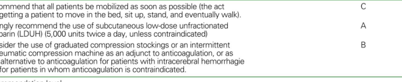
모든 급성기 뇌졸중 환자는 심부정맥혈전증에 대한 예방 적 처치를 시행해야 하는데 되도록 빨리 침상에서 움직이는 것이 중요하며, 저용량의 헤파린이나 저분자량 헤파린이 심 부정맥혈전증 예방에 효과적인 것으로 알려져 있다. 상부 위장관 출혈이나 뇌출혈 등으로 헤파린을 사용할 수 없을 경우에는 공기 압박 스타킹을 사용할 수 있다(Table 3)(15).
저분자량 헤파린과 헤파리노이드(heparinoid)가 통상적인 비분획 헤파린보다 심부정맥혈전증의 예방에 더 효과적이 나 출혈 등의 부작용에는 큰 차이를 보이지 않는 것으로 알 려져 있다(33). 간헐적 공기 압박 치료를 헤파린 치료와 같 이 병행하면 더 효과적이다(34). 심부정맥혈전증의 예방 치 료는 급성기 이후에도 지속하여 환자가 걸을 때까지는 하는 것이 좋으나 적절한 기간에 대해서는 아직까지 알려져 있지 않다.
심부정맥혈전증 환자의 증상은 잘 나타나지 않는 경우가 많아 50% 정도의 환자에서만 통증, 종창, 발적 등의 임상 증 상이 나타난다. 조기 발견을 위해 매일 부종과 통증을 관찰 하여야 하며 혈전이 의심되면 확진을 위한 검사를 시행하여 야 한다(32). 진단에는 CT 혈관조영술, 정맥 이중 조영술 (veous duplex scanning), 도플러 초음파 검사, d-dimer
Table 3. AHA/ASA- endorsed guidelines for prevention of deep vein thrombosis
Recommendation Recommendation level*
Recommend that all patients be mobilized as soon as possible (the act C of getting a patient to move in the bed, sit up, stand, and eventually walk).
Strongly recommend the use of subcutaneous low-dose unfractionated A
heparin (LDUH) (5,000 units twice a day, unless contraindicated)
Consider the use of graduated compression stockings or an intermittent B pneumatic compression machine as an adjunct to anticoagulation, or as
an alternative to anticoagulation for patients with intracerebral hemorrhagie or for patients in whom anticoagulation is contraindicated.
* Recommendation level
A: a strong recommendation that the intervention is always indicated and acceptable, B: a recommendation that the intervention may be useful/effective,
C: a recommendation that the intervention may be considered,
D: a recommendation that a procedure may be considered not useful/effective, or may be harmful, I : Insufficient evidence to recommend for or against; clinical judgment should be used.
assay 등이 사용되며 심부정맥혈전증으로 진단이 되면 항응 고제 치료를 시행한다(35). 출혈성 뇌졸중 환자와 같이 항응 고제 치료를 시행할 수 없는 환자는 폐동맥색전증을 예방하기 위해 대정맥 필터(vena cava filter)를 삽입하기도 한다(36).
5. 욕 창
욕창은 병원에 입원한 뇌졸중 환자의 9% 정도에서 발생 하며 통증 및 감염을 일으켜 입원 기간을 증가시킨다(24).
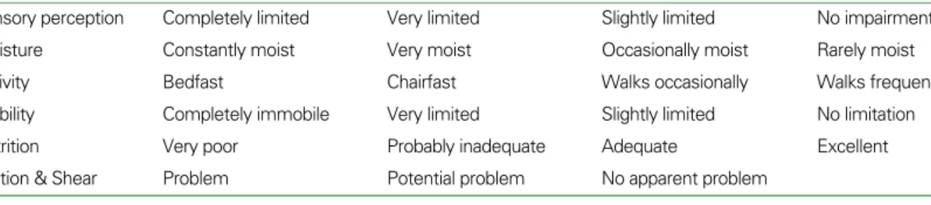
따라서 급성기에 욕창의 발생을 예방하는 것이 중요하며 특 히 움직이지 못하고 부동 상태에 있는 환자나 당뇨병이나 말초 혈관 질환이 동반된 환자, 요실금이나 변실금이 있는 환자, 체질량지수가 낮은 환자, 말기 환자 등이 위험도가 높 다(37). Braden scale과 같은 욕창 위험도 평가 도구를 사 용하여 욕창 발생을 예측하고 예방 조치를 취할 수 있다 (Table 4)(38). 욕창 예방을 위해서는 2시간 마다 체위를 바꿔주거나 돌려주어야 하며, 관절이나 체간을 움직여 주고 환자를 이동할 경우에는 피부에 압력이 가지 않도록 적절한 방법을 사용해야 한다. 마찰을 예방하기 위해 윤활제나 특 수 매트리스 등을 사용하며 요실금이나 변실금이 있을 경우 에는 피부를 청결하고 건조하게 관리해야 한다(39). 뇌졸중 의 중증도가 심하여 욕창 발생의 위험도가 높은 환자의 경 우에는 물 침대나 공기 침대와 같이 압력을 감소시켜주는 기구를 사용하기도 한다. 욕창은 골 융기 부위나 체중이 부 하되는 부위에 잘 발생하는데 자주 피부를 관찰하여 욕창을 조기에 발견해야 하며 신속히 치료를 시작하여야 한다 (Table 5). 욕창의 단계는 피부에 발적만 있는 1단계, 표피 또는 진피가 손상된 2단계, 피하조직까지 침범된 3단계와 근
막, 근육, 골조직까지 침범된 4단계로 분류할 수 있다. 욕창 의 치료는 비수술적 치료와 수술적 치료로 나눌 수 있는데 일 반적으로 1단계와 2단계까지는 드레싱과 같은 비수술적 치 료를 하고 3단계와 4단계는 수술적 치료를 필요로 한다(40).
6. 낙 상
낙상은 급성기에서도 흔히 발생하는 합병증으로 보고에 따라 25%까지 발생하며, 5%에서는 대퇴골 골절을 비롯한 심각한 손상을 유발한다(41). 뇌졸중 환자는 인지기능의 장 애, 하지 근위약, 평형 기능 및 이동 능력의 저하, 기립성 저 혈압, 우울증, 약물 부작용, 감각 장애 등에 의해 낙상이 일 어날 수 있다(42). 낙상을 예방하기 위해서는 급성기부터 위험 요인을 파악하고 예방적 치료를 시행하여야 한다. 적 절한 환자 이동, 균형 훈련, 운동치료, 보조기구 사용, calcium 및 Vitamin D 보조요법, bisphosphonate 치료 등이 균형기능을 호전시키고 골 강도를 증가시켜 낙상과 골 절을 예방할 수 있다(16). 필요한 경우에는 고관절 보호장 구를 착용시켜 대퇴골 골절을 예방할 수 있다(43).
7. 통 증
뇌졸중 환자는 두통, 견관절 통증, 중추성 통증과 같은 통 증이 발생할 수 있으며 이러한 통증은 부동(immobility), 근 위약, 경직 등에 의해 발생하는 근골격계 통증, 제1형 복합 부 위 통증 증후군(complex regional pain syndrome type 1), 뇌졸중 병변에 의한 중추성 통증 등 다양한 원인에 의해 발 생한다. 급성기부터 발생할 수 있으나 대부분 수 주 및 수 개월에 걸쳐 발생하며 급성기의 부적절한 자세 유지 및 관
Table 4.Braden scale for predicting pressure sore risk
1 2 3 4
Sensory perception Completely limited Very limited Slightly limited No impairment
Moisture Constantly moist Very moist Occasionally moist Rarely moist
Activity Bedfast Chairfast Walks occasionally Walks frequently
Mobility Completely immobile Very limited Slightly limited No limitation
Nutrition Very poor Probably inadequate Adequate Excellent
Friction & Shear Problem Potential problem No apparent problem
절 손상이 원인이 될 수 있다.
견관절 통증은 가장 흔한 통증으로 많게는 72%의 뇌졸중 환자에서 발생하는 것으로 알려져 있으며 특히 재활치료시 기에 치료의 제한을 가져와 환자의 기능 회복에 영향을 준 다(44, 45). 회전근개건염과 같은 기존 질환이 동반되어 있 는 경우도 있지만 대부분의 경우 견관절 아탈구, 경직, 관절 구축 중 하나 이상의 원인으로 발생하므로 급성기부터 팔걸 이나 스트랩을 이용한 견관절 보호, 부드러운 관절운동 및 적절한 관절 자세 유지가 견관절 통증의 예방을 위해 중요 하다(24, 46). 경직이 문제가 될 경우에는 관절의 스트레칭 운동이 필요하며 보툴리눔 독소를 이용하여 견갑하근의 근 긴장도를 완화시키는 방법이 시도되기도 한다(47).
뇌졸중 후 중추성 통증은 시상을 비롯한 척수시상로의 병 변에 의해 감각이상과 함께 신경성 통증이 발생하는 것으로 2~8%의 뇌졸중 환자에서 발생하며 대부분 발병후 1개월 이내에 발생한다(48). 마비측의 지속적 또는 발작적인 작열 통 및 난자통을 호소하며 이상감각, 이질통 및 통각과민 등 이 관찰된다. 치료의 목적은 통증을 경감시키는 것으로 비 스테로이드소염제나 아편유사진통제 등은 효과적이지 못하 며 amitryptiline과 같은 삼환계 항우울제나 lamotrigine, carbamazepine, gabapentine 등의 항경련제가 사용 된다(49).
8. 발 작
뇌졸중의 급성기 합병증으로 부분 발작이나 대발작 형태 의 간질 발작이 발생할 수 있다(15). 2주 이내의 급성기 뇌
졸중 환자의 5% 정도에서 발생하는 것으로 알려져 있으며 이들 중 40% 정도는 발병 후 24시간 이내에 발생한다(50).
출혈성 뇌졸중이나 대뇌 피질을 침범한 경우에 주로 발생하 는 것으로 알려져 있으며 뇌졸중의 중증도가 심할수록 발작 발생의 위험도가 높다(50). 피질하 또는 열공 뇌경색의 경 우에도 발생할 수 있다. Lorazepam (1~ 4 mg IV) 이나 diazepam (5 ~10 mg IV) 정맥 주사후 phenytoin 또는 carbamazepine 정맥주사로 치료하며 경우에 따라서는 뇌 파 모니터링이 필요하다(14).
뇌졸중 후 발작이 사망률을 높이거나 기능 회복을 저해하 는지는 아직까지 밝혀져 있지 않으며 급성기 발작 환자의 1/3 정도에서 간질 발작이 재발하는 것으로 알려져 있다 (51). 따라서 항간질제 치료의 시작 및 항간질제의 선택은 최초 발작이 환자의 기능 회복에 미쳤던 영향과 환자의 상 태를 고려하여 결정해야 하는데 대부분 단일 약물 치료로 조절이 된다. Phenytoin, phenobarbital, benzodiaze- pine 등의 약물들은 뇌졸중 후 회복을 저해하는 것으로 알 려져 있고(51), 특히 phenyoin은 골대사에 영향을 주고 항 응고제와도 상호작용이 있어 사용하지 않는 것이 좋다(52).
Lamotrigine과 gabapentine은 항응고제 및 항혈전제와 상 호작용이 없고 골대사에 영향을 주지 않아 뇌졸중 후 발생 하는 급성기 발작에 사용할 수 있다(52). 골대사에 문제가 없고 항응고제가 필요하지 않은 경우에는 비용을 고려할 때 저용량의 지속형 carbamazepine을 사용할 수 있다(52).
그러나 뇌졸중 후 간질 발작의 예방치료는 권고되고 있지 않다(16).
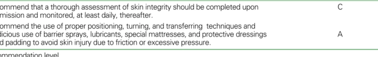
Table 5. AHA /ASA-endorsed guidelines for prevention of pressure ulcer
Recommendation Recommendation level*
Recommend that a thorough assessment of skin integrity should be completed upon C admission and monitored, at least daily, thereafter.
Recommend the use of proper positioning, turning, and transferring techniques and
judicious use of barrier sprays, lubricants, special mattresses, and protective dressings A and padding to avoid skin injury due to friction or excessive pressure.
* Recommendation level
A: a strong recommendation that the intervention is always indicated and acceptable, B: a recommendation that the intervention may be useful/effective,
C: a recommendation that the intervention may be considered,
D: a recommendation that a procedure may be considered not useful/effective, or may be harmful, I : Insufficient evidence to reco-mmend for or against; clinical judgment should be used.
9. 우 울 증
급성기 뇌졸중 환자의 50%에서 우울증이 발생하며 기능 회복 및 재활에 영향을 미친다(53). 좌측 전두엽 병변 환자 에서 잘 발생하는 것으로 알려져 있는데 전두엽의 노르아드 레날린, 도파민 및 세로토닌계의 손상에 의한 카테콜아민 고갈에 의한 것으로 생각되고 있다(54). 이 외에도 장애 발 생으로 인한 슬픔, 불안, 좌절과 같은 심리적 반응이 우울증 을 유발할 수 있는데 대부분의 뇌졸중 후 우울증 환자는 기 질적 원인과 심리적 원인이 동반되어 있다. 뇌졸중 환자에 서는 수면장애, 피로감, 정신운동지연과 같은 증상이 뇌졸 중 자체에 의한 증상으로 나타날 수 있어 우울증의 진단이 어려운 경우가 많다(55). 특히 실어증이나 인지장애가 있는 환자에서는 표준적인 진단도구가 도움이 되지 못하며 일반 적으로 우울한 기분과 치료 거부와 같은 관심의 소실이 우 울증의 진단에 도움이 된다(56).
뇌졸중 환자에서 우울증의 치료는 선택세로토닌재흡수 억제제(SSRI)나 삼환계 항우울제가 모두 효과적이나 삼환 계 항우울제는 부작용에 의해 중단해야 하는 경우가 많아 SSRI를 사용하는 것이 권장되고 있다(57, 58).
결 론
급성기 뇌졸중 합병증은 뇌졸중의 경과를 악화시켜 사망 률을 높이거나 뇌졸중 환자의 재활에 영향을 미친다. 주요 급성기 합병증으로는 흡인성 폐렴, 연하 곤란, 요로 감염, 영양실조, 심부정맥혈전증, 욕창, 낙상, 통증, 발작, 우울증 등이 있으며 이들 대부분은 예방 및 치료가 가능하다. 뇌졸 중 발병 초기부터 철저한 감시와 조기 발견을 통해 급성기 합병증을 관리하여야 하며, 이러한 급성기 합병증의 예방과 관리에는 의료진과 간호사, 치료사, 영양사 등 여러 분야 전 문가들의 팀 접근법이 필요하다.
참고문헌
11. Oppenheimer S, Hachinski V. Complications of acute stroke.
Lancet 1992; 339: 721- 724.
12. Worp HB, Kappelle LJ. Complications of acute ischemic stroke. Cerebrovasc Dis 1998; 28: 2180-2184.
13. Langhorne P, Stott DJ, Robertson L, MacDonald J, Jones L, McAline C, Dick F, Taylor GS, Murray G. Medical compli- cations after stroke: a multicenter study. Stroke 2000; 31:
1223-1229.
14. Bae HJ, Yoon DS, Lee J, Kim BK, Koo KJ, Kwon O, Park JM.
In - hospital medical complications and long-term mortality after ischemic stroke. Stroke 2005; 36: 2441- 2445.
15. Brandstater ME. Stroke rehabilitation. In: Delisa JA, ed.
Physical medicine and rehabilitation. 4th ed, Philadelphia:
Sauders, 2005: 1656 -1676.
16. Davenport RJ, Dennis MS, Welwood I, Warlow C. Compli- cations after acute stroke. Stroke 1996; 27: 415-420.
17. Dobkin BH. Neuromedical complications in stroke patients transferred for rehabilitation before and after diagnostic related groups. J Neurol Rehab 1987; 1: 3 -7.
18. Dromerick A, Reding M. Medical and neurological compli- cations during stroke rehabilitation. Stroke 1994; 25: 358 -361.
19. Indredavik B, Rohweder G, Naalsund E, Lydersen S. Medical complications in a comprehensive stroke unit and an early supported discharge service. Stroke 2008; 39: 414- 420.
10. Johnston KC, Li JY, Lyden PD, Hanson SK, Feasby TE, Adams RJ, Faught RE, Haley EC, for the RANTTAS Investigators.
Medical and neurological complications of ischemic stroke:
experience from the RANTTAS trial. Stroke 1998; 29: 447- 453.
11. Kalra L, Yu G, Wilson K, Roots P. Medical complications during stroke rehabilitation. Stroke 1995; 26: 990 - 994.
12. Roth JE, Lovell L, Harery RL, Heinemann AW, Semik P, Diaz S. Incidence of and risk factors for medical complications during stroke rehabilitation. Stroke 2001; 31: 523 - 529.
13. Weimer C, Ziegler A, Konig IR, Diener HC, on behalf of the German Stroke Date Collaborators. Prediction of functional outcome and survival after acute ischemic stroke. J Neurol 2002; 249: 888- 895.
14. Kaste M, Roine RO. General stroke management and stroke units. In: Mohr JP, Choi DW, et al. eds. Stroke: Pathophysi- ology, diagnosis, and management. 4th ed. Philadelphia:
Churchill Livingstone, 2004: 971-1024.
15. Adams HP, del Zoppo G, Alberts MJ, Bhatt DL, Brass L, Furlan A, Grubb RL, Higashida RT, Jauch EC, Kidwell C, Lyden PD, Morgenstern LB, Qureshi AI, Rosenwasser RH, Scott PA, Wijdicks EF. Guidelines for the early management of adults with ischemic stroke. Stroke 2007; 38: 1655 -1711.
16. European Stroke Organisation Executive Committee and the ESO Writing Committee. Guidelines of management of ischa- emic stroke and transient ischaemic attack 2008. Cerebro- vasc Dis 2008; 16: 311- 337.
17. Han TR, Kim YH, Paik NJ. Stroke rehabilitation. In: Han TR, Bang MS eds. Rehabilitation medicine. 3rd ed. Seoul: Koonja Publishing, 2008: 509- 548.
18. Ellwood PM. Bed positioning. In: Kottke FJ, Lehmann JF. eds.
Handbook of physical medicine and rehabilitation. 4th ed.
Philadelphia: W.B. Saunders, 1990: 520- 528.
19. Aviv JE, Sacco RL, Thomson J, Tandon R, Diamond B, Martin JH, Close LG. Silent laryngopharyngeal sensory deficits after stroke. Ann Otol Rhinol Laryngol 1997; 11: 609- 622.
20. Martino R, Foley N, Bhogal S, Diamant N, Speechley M, Teasell R. Dysphagia after stroke: incidence, diagnosis, and pulmonary complications. Stroke 2005; 36: 2756-2763 21. Daniels SK, Foundas AL. Lesion localization in acute stroke
patients with risk of aspiration. J Neuroimaging 1999; 9: 91- 98.
22. Hammond CA, Goldstein LB. Cough and aspiration of foods and liquids due to oral-pharyngeal dyshagia: ACCP evidence - based clinical practice guideline. Chest 2006; 129: 154-168.
23. DePippo KL, Holas MA, Reding MJ. Validation of the 3- oz water swallow test for aspiration following stroke. Arch Neurol 1992; 49: 1259 -1261.
24. Duncan PW, Zorowitz R, Bates B, Choi JY, Glasberg JJ, Graham GD, Katz RC, Lamberty K, Reker D. Management of adult stroke rehabilitation care: a clinical practice guideline.
Stroke 2005; 36: e100-e143.
25. Kim IS, Han TR. Evaluation and management of dysphagia.
Kor J Stroke 2006; 8: 40- 48.
26. Palmer JB, Drennan JC, Baba M. Evaluation and treatment of swallowing impairments. Am Fam Physician 2000; 61: 2453- 2462.
27. Norton B, Horner-Ward M, Donnely MT, Long RG, Holmes GK. A randomized prospective comparison of percutaneous endoscopic gastrostomy and nasogastric tube feeding after acute dysphagic stroke. BMJ 196; 312: 13 -16.
28. Blauer D. The natural history and functional consequences of dysphagia after hemispheric stroke. J Neurol Neurosurg Psychiatry 1989; 52: 236 - 241.
29. Warren JW. Catheter-associated urinary tract infections. Infect Dis Clin North Am 1997; 11: 609-622
30. Nakayama H, Jorgensen HS, Pedersen PM, Raaschou HO, Olsen TS. Prevalence and risk factors of incontinence after stroke. The Copenhagen Stroke Study. Stroke 1997; 28: 58- 62.
31. Axelsson K, Asplund K, Norberg A, Alafuzoff I. Nutritional status in patients with acute stroke. Acta Med Scand 1988;
224: 217- 224.
32. Brandstater ME, Roth EJ, Siebens HC. Venous thrombo- embolism in sroke: Literature review and implications for clinical practice. Arch Phys Med Rehabil 1992; 73: s379 -391.
33. Sandercock PA, Counsell C, Tseng MC.Low-molecular-weight
heparins or heparinoids versus standard unfractionated he- parin for acute ischaemic stroke. Cochrane Database Syst Rev 2008; 3: CD000119.
34. Kamran SI, Downey D, Ruff RL. Pneumatic sequential com- pression reduces the risk of deep vein thrombosis in stroke patients. Neurology 1998; 50: 1683 -1688.
35. Kelly J, Rudd A, Lewis R, Hung BJ. Venous thrombo-embo- lism after acute stroke. Stroke 2001; 32: 262-267.
36. Harvey RL, Green D. Deep venous thrombosis and pulmonary embolism in stroke. Top Stroke Rehabil 1996; 3: 54 -70.
37. Berlowitz Dr, Brandeis GH, Morris JN, Ash AS, Anderson JJ, Kader B, Moskowtz MA. Deriving a risk-adjustment model for pressure ulcer development using the Minum Data Set. J Am Geriatr Soc 2001; 49: 866- 871
38. Bergstrom N, Braden BJ, Laguzza A, Holman V. The Braden scale for predicting pressure sore risk. Nurs Res 1987; 36:
205-210.
39. Reddy M, Gill SS, rochon PA. Preventing pressure ulcers: a systematic review. JAMA 2006; 296: 974- 984.
40. Salcido R, Goldman R. Prevention and management of pre- ssure ulcers and other chronic wounds. In: Braddom RL, Busch- bacher RM, et al. eds. Physical medicine and rehabilitation.
2nd ed. Philadelphia: W.B. Saunders, 2000: 645 -664.
41. Forster A, Young J. Incidence and consequences of falls due to stroke: a systematic inquiry. BMJ 1995; 311: 83- 86.
42. Teasell R, McRae M, Foley N, Bhardwaj A. The incidence and consequences of falls in stroke patients during inpatient re- habilitation: factors associated with high risk. Arch Phys Med Rehabil 2002; 83: 329- 333.
43. Parker MJ, Gillespie LD, Gillespie WJ. Hip protectors for preventing hip fractures in the elderly. Cochrane Database Syst Rev 2001: CD001255.
44. Van Onwenaller C, LaPlace PM, Chartraine A. Painful shoulder in hemiplegia. Arch Phys Med Rehabil 1985; 67: 23-26.
45. Lindgren I, Jonsson AC, Norrving B, Lindgren A. Shoulder pain after stroke: a prospective population-based study. Stroke 2007; 38: 343-348.
46. Ada L, Foongchomcheay A, Canning C. Supportive devices for preventing and treating subluxation of the shoulder after stroke. Cochrane Database Syst Rev 2005: CD003863.
47. van Kuijk AA, Geurts AC, Bevaart BJ, van Limbeek J. Treat- ment of upper extremity spasticity in stroke patients by focal neuronal or neuromuscular blockade: a systematic review of the literature. J Rehabil Med 2002; 34: 51- 61.
48. Hansson P. Post-stroke pain case study: clinical characte- ristics, therapeutic options and long-term follow- up. Eur J Neurol 2004; 11: 22-30.
49. Frese A, Husstedt IW, Ringelstein EB, Evers S. Pharmacologic treatment of central post-stroke pain. Clin J Pain 2006; 22:
252- 260.
50. Olsen TS. Post- stroke epilepsy. Curr Atheroscler Rep 2001; 3:
340-344.
51. Camilo O, Goldstein LB. Seizures and epilepsy after ischemic stroke. Stroke 2004; 35: 1769 -1775.
52. Ryvlin P, Montavont A, Nighoghossian N. Optimizing therapy of seizures in stroke patients. Neurology 2006; 67: S3- S9.
53. Robinson RG, Starr LB, Kubos KL, Price TR. A two-year longi- tudinal study of post-stroke mood disorders: findings during the initial evaluation. Stroke 1983; 14: 736-741.
54. Robinson RG, Szetela B. Mood change following left hemi- sphere brain injury. Ann Neurol 1981; 9: 447- 453.
55. Thomas SA, Lincoln NB. Factors relating to depression after
stroke. Br J Clin Psychol 2006; 45: 49-61.
56. Kauhanen M, Korpelainen JT, Hiltunen P, Brusin E, Mononen H, Maatta R, Nieminen P, Sotaniemi KA, Myllyla VV. Post- stroke depression correlates with cognitive impairment and neurological deficits. Stroke 1999; 30: 1875-1880.
57. Hackett ML, Anderson CS, House AO. Management of de- pression after stroke: a systematic review of pharmacological therapies. Stroke 2005; 36: 1098-1103.
58. Bhogal SK, Teasell R, Foley N, Speechley M. Heterocyclics and selective serotonin reuptake inhibitors in the treatment and prevention of poststroke depression. J Am Geriatr Soc 2005; 53: 1051-1057.
Peer Reviewers’ Commentary
본 논문은 최근 급성기 뇌졸중 유니트의 필요성이 증대되면서 그 중요성이 재조명되고 있는 뇌졸중 발병 후 급성기의 합 병증 관리에 대하여 전반적으로 자세하게 기술하고 있다. 특히 뇌졸중 후 급성기 합병증의 관리에 대한 미국과 유럽의 임상진료지침(clinical practice guideline)들을 비교하여 제시하면서 근거중심의학적인 합병증 관리의 방향을 기술하고 있다. 다만 아쉬운 점은 국내의 역학적 자료가 아직 많이 부족하다는 점과 외국과의 발생 빈도가 다른 합병증들에 대한 임상적 접근 방법에서는 치료팀 사이에 논쟁이 있을 수 있으므로 향후 이를 반영한 독자적인 국내의 진료지침 개발이 필 요할 것으로 생각한다.
[정리:편집위원회]