Differentiation of gallbladder adenomyomatosis from early-stage gallbladder cancer before surgery
Jisum Moon, Yong Chan Shin, Tae-Gil Heo, Pyong Wha Choi, Jae Il Kim, Sung Won Jung, Heungman Jun, Sung Min Jung, and Eunhae Um
Department of Surgery, Ilsan Paik Hospital, Inje University College of Medicine, Goyang, Korea
Backgrounds/Aims: This study aimed to compare the perioperative and clinical outcomes in patients undergoing laparo- scopic cholecystectomy for gallbladder adenomyomatosis (GBA) or early-stage gallbladder cancer (GBC). Methods:
The perioperative and clinical outcomes of 194 patients diagnosed with GBA and 30 patients diagnosed with GBC who underwent laparoscopic cholecystectomy in our institution from January 2011 to December 2017 were retrospec- tively compared. Results: There were no significant differences between the GBA and GBC groups in sex (male:female ratio 1.0:0.8 vs. 1.0:0.7, p=0.734), BMI (23.9±3.4 vs. 24.0±3.8 kg/m2, p=0.916), or preoperative liver function tests.
Patients in the GBC group were significantly older (50.5±14.1 vs. 65.9±10.6 years, p<0.001) and had a higher ASA grade (40.3 vs. 63.4% grade II or III, p=0.043) than patients in the GBA group. Although there was no significant difference in preoperative diagnostic methods (p=0.442), the GBC group showed a significantly higher rate of mis- diagnosis on preoperative imaging compared with postoperative histopathologic findings (30.9% vs. 53.3%, p=0.011).
There were significantly more patients with gallstones in the GBA group than in the GBC group (68.6% vs. 40.0%, p=0.004). Conclusions: In older patients hospitalized for biliary colic without gallstones but with a thickened gallbladder wall with inflammation on preoperative diagnostic exam, the possibility of early-stage GBC should be considered. (Ann Hepatobiliary Pancreat Surg 2019;23:334-338)
Key Words: Gallbladder adenomyomatosis; Gallbladder cancer; Differential diagnosis
Received: September 17, 2019; Revised: October 1, 2019; Accepted: October 2, 2019 Corresponding author: Yong Chan Shin
Department of Surgery, Ilsan Paik Hospital, Inje University College of Medicine, 170 Juhwa-ro, Ilsanseo-gu, Goyang 10380, Korea Tel: +82-31-910-7743, Fax: +82-31-910-7319, E-mail: [email protected]
Copyright Ⓒ 2019 by The Korean Association of Hepato-Biliary-Pancreatic Surgery
This is an Open Access article distributed under the terms of the Creative Commons Attribution Non-Commercial License (http://creativecommons.org/
licenses/by-nc/4.0) which permits unrestricted non-commercial use, distribution, and reproduction in any medium, provided the original work is properly cited.
Annals of Hepato-Biliary-Pancreatic Surgery ∙ pISSN: 2508-5778ㆍeISSN: 2508-5859
INTRODUCTION
Various gallbladder (GB) diseases are characterized by generalized or localized wall thickening of the GB on computed tomography (CT) or ultrasound (US), including gallbladder adenomyomatosis (GBA), chronic cholecystitis, GB polyp, and early-stage, wall-thickening-type gall- bladder cancer (GBC).1,2 Among them, the differentiation GBA from GBC is still required because of the similarity in appearance, despite some reports being published con- cerning their imaging findings using US, CT, and mag- netic resonance imaging (MRI) since 1981.2,3 In this re- gard, Ching et al.2 reported that the differential diagnostic performance of contrast-enhanced CT for GBA and GBC showed 30% sensitivity and 93% specificity. MRI imag- ing is known to be useful because it can sensitively depict the pearl necklace sign, which is pathognomonic of ad-
enomyomatosis and directly indicates the presence of Rokitansky-Aschoff sinuses in the thickened wall.4,5 Re- cently, Joo et al.6 reported that high-resolution ultrasound (HRUS) and MRI with MR cholangiopancreatography have comparable sensitivity and accuracy.
Although there have been significant advances in diag- nostic imaging technology that can be helpful for dis- tinguishing GBA from early-stage GBC,7-9 some issues re- main unsolved. As mentioned above, MRI, one of the most useful diagnostic tools, is expensive and requires pa- tients to hold still for long periods of time. HRUS, while offering the ability to overcome the drawbacks of conven- tional US, is not yet widely available except in large gen- eral hospitals.
In order to accurately distinguish between the two con- ditions, it is necessary to analyze the perioperative demo- graphic and imaging data for diagnostic purposes. There-
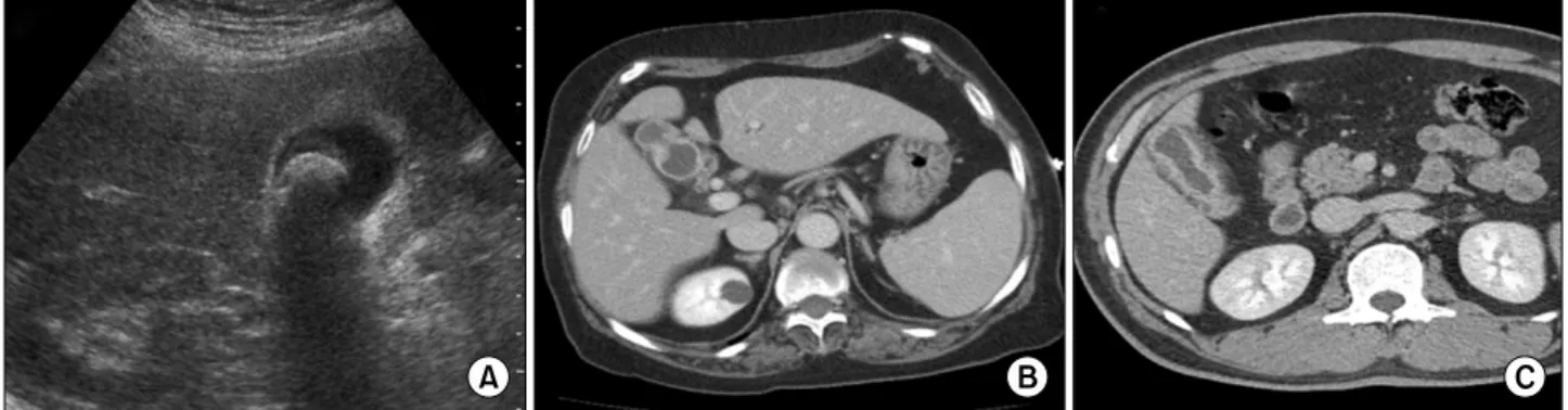
Fig. 1. Preoperative diagnostic images of gallbladder diseases. (A) Ultrasonographic finding of a patient with gallbladder cancer, considered as chronic calculous cholecystitis preoperatively. (B) Computed tomographic scan of a patient with gallbladder cancer, considered as gallbladder adenomyomatosis preoperatively. (C) Computed tomographic scan of a patient with gallbladder ad- enomyomatosis, considered as gallbladder cancer preoperatively.
fore, this study aimed to compare perioperative and clin- ical outcomes in patients undergoing laparoscopic chol- ecystectomy for GBA or early-stage GBC to evaluate the diagnostic performance of differences in preoperative demo- graphics and imaging findings between the two conditions.
MATERIALS AND METHODS
Patients
This retrospective study was approved by our institu- tional review board and the requirement for informed con- sent was waived. Between January 2011 and December 2017, a total of 2389 patients underwent laparoscopic cholecystectomy at a hospital, including 194 diagnosed with GBA (GBA group) and 30 diagnosed with GBC (GBC group). No patients had both adenomyomatosis and cancer and patients were assigned to separate groups ac- cording to the pathological result. We performed a retro- spective analysis of the perioperative and clinical out- comes of 224 consecutive patients.
The GBA group (n=194) consisted of 110 males and 84 females with a mean age of 50.5±14.0 years (range 17-86 years). The mean body mass index (BMI) was 23.9±3.4 kg/m2 (range 16.2-38.6 kg/m2). Laparoscopic cho- lecystectomy was performed because of gallstone-related symptoms in 122 patients and gallbladder wall thickening in 72 patients.
The GBC group (n=30) consisted of 18 male and 12 female patients with a mean age of 66.6±10.2 years (range 45-86 years). The mean BMI was 24.0±3.9 kg/m2 (range 15.8-32.8 kg/m2). In terms of TNM staging by the Ameri-
can Joint Committee on Cancer (8th edition), all tumors of the GBC group were classified as T1a or T1b. The tu- mor was located in the fundus in 9 patients, in the body in 12 patients, in the neck in 3 patients, and in the entire gallbladder in 5 patients.
Statistical analysis
Results are presented as mean and standard error of the mean. Patient demographics and clinical characteristics were compared using the 2 test or Fisher exact test for categorical variables and t-test or Mann-Whitney test for continuous variables, as appropriate. In assessing risk fac- tors associated with GBC, only variables statistically sig- nificant by univariate analysis were included in the multi- variate analysis, which was performed using logistic re- gression. All statistical analyses were performed using SPSS, version 21.0 (IBM, Armonk, NJ), with p-values < 0.05 considered statistically significant.
RESULTS
In most patients, preoperative diagnostic examination made it difficult to differentiate gallbladder diseases with wall thickening and the preoperative diagnostic images were inaccurate when compared to postoperative pathol- ogy reports (Fig. 1). The demographic and perioperative findings of both groups are listed in Table 1. There were no significant differences in sex (1.0:0.8 vs. 1.0:0.7, male:
female, p=0.734), BMI (23.9±3.4 vs. 24.0±3.8 kg/m2, p=
0.916), or preoperative liver function tests between the GBA and GBC groups. The GBC group was significantly
Table 1. Demographics and perioperative findings GBA group
(n=194)
GBC group (n=30) p-value
Age (years) <0.001
<60 142 (73.2%) 7 (23.3%)
≥60 52 (26.8%) 23 (76.7%)
Male sex 110 (56.7%) 18 (60.0%) 0.734 BMI (kg/m2) 23.9±3.4 24.0±3.8 0.916
ASA classification 0.043
1 116 (59.8%) 11 (36.7%)
2 68 (35.1%) 17 (56.7%)
3 10 (5.1%) 2 (6.6%)
Diagnostic method 0.442
US 61 (31.4%) 14 (46.7%)
CT 125 (64.5%) 16 (53.3%)
MRI 2 (1.0%) 0
EUS 6 (3.1%) 0
Misdiagnosis 60 (30.9%) 16 (53.3%) 0.011 Symptoms 122 (62.9%) 18 (60.0%) 0.761 Laboratory exam
WBC (/l) 6758.6±2313.8 7088.0±2481.1 0.473 AST (U/L) 33.6±33.4 28.9±14.2 0.442 ALT (U/L) 41.7±61.8 31.2±39.2 0.367 Total bilirubin
(mg/dl)
1.1±4.3 0.9±0.4 0.791
ALP (U/L) 79.0±42.5 87.8±54.2 0.360 GGT (U/L) 90.2±129.5 79.6±95.1 0.703
Operation time (min) 0.036
<60 89 (45.9%) 8 (26.7%)
≥60 105 (54.1%) 22 (73.3%)
Gallstones 133 (68.6%) 12 (40.0%) 0.004 GBA, gallbladder adenomyomatosis; GBC, gallbladder cancer;
BMI, body mass index; ASA, American society of anesthesiol- ogist; US, ultrasound; CT, computed tomography; MRI, mag- netic resonance imaging; EUS, endoscopic ultrasound; WBC, white blood cell; AST, aspartate transaminase; ALT, alanine transaminase; ALP, alkaline phosphatase; GGT, gamma-gluta- myltransferase
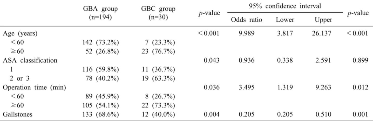
Table 2. Risk factors for gallbladder cancer GBA group
(n=194)
GBC group
(n=30) p-value 95% confidence interval
p-value Odds ratio Lower Upper
Age (years) <0.001 9.989 3.817 26.137 <0.001
<60 142 (73.2%) 7 (23.3%)
≥60 52 (26.8%) 23 (76.7%)
ASA classification 0.043 0.936 0.338 2.591 0.899
1 116 (59.8%) 11 (36.7%)
2 or 3 78 (40.2%) 19 (63.3%)
Operation time (min) 0.036 3.495 1.319 9.263 0.012
<60 89 (45.9%) 8 (26.7%)
≥60 105 (54.1%) 22 (73.3%)
Gallstones 133 (68.6%) 12 (40.0%) 0.004 0.205 0.205 0.510 0.001
GBA, gallbladder adenomyomatosis; GBC, gallbladder cancer; ASA, American Society of Anesthesiologists
older (50.5±14.1 vs. 65.9±10.6 years, p <0.001) and had a higher ASA grade (40.3 vs. 63.4, grade II or III (%), p=0.043) than the GBA group. The operation time of the GBC group was significantly longer than that of the GBA group (p=0.036) and there was a significant difference in the rate of cases with gallstones between 2 groups (68.6%
vs. 40.0%, p=0.004). Although there was no significant difference in preoperative diagnostic methods (p=0.442), the GBC group showed a significantly higher rate of dis- parity between preoperative imaging and postoperative histopathological findings (30.9% vs. 53.3%, p=0.011).
Risk factors for GBC were analyzed (Table 2). Multi- variate analysis revealed that old age (60 years or above), delayed operation time (60 min or above), and gallbladder without calculi were predictive factors for GBC (Table 2).
DISCUSSION
According to the Korea Central Cancer Registry’s an- nual report of 2016, as published by Korean Ministry of Health and Welfare, GBC and other biliary tract cancers account for 2.9% of all cancers in Korea.10 GBC is silent during the early-stage and remains asymptomatic until it gets to an advanced and unresectable stage. Therefore, early diagnosis and treatment of GBC is very important.
Inflammatory or obstructive GB changes may induce GBA at 2-5% of prevalence in any cholecystectomy speci- men.11,12 In surgery for pain aggravation or other symp- toms, accurate differential diagnosis between GBA and GBC is a major factor for choosing the adequate treat- ment. Although GBA has not been considered to have ma-
lignant potential, several reports have suggested a rela- tionship between GBA and GBC.2,13,14 Kai et al.11 reported GBC was associated with GBA in 25% of cases. Additio- nally, patients with GBC and GBA presented with a more advanced TNM stage than those without GBA. Given the differences in prognosis according to the TNM stage of GBC, preoperative differential diagnosis between GBA and GBC is indispensable to avoid nefarious consequences.
Despite the technical advances in imaging modalities (HRUS, multi-detector CT, and MRI), it is still difficult to distinguish between GBA and GBC before surgical resection. According to Ching et al. at 2007, the differ- ential diagnostic performance of CT for GBA and GBC was 30% sensitivity and 93% specificity.2 However, Bang et al. found improved values of 50% sensitivity and 98.2%
specificity.15 The improvement in diagnostic performance of US is most likely the result of the technological ad- vances which have been utilized since 2000, such as har- monics, compounding techniques, and speckle reduction.
In a previously published study, the diagnostic perform- ance of HRUS was equivalent to that of MRI for differ- entiating GBA from GBC.15 The presence of either intra- mural echogenic foci or cystic spaces, which indicate cho- lesterol crystals/stones or bile within the pathognomonic Rokitansky-Aschoff sinuses, respectively, had a sensitivity of 80.0%, specificity of 85.7% and accuracy of 82.2% for the diagnosis of GBA on HRUS.6,16 MRI may be superior to HRUS for the depiction of intramural cystic spaces. As shown above, multiple imaging modalities would be help- ful for evaluating and choosing treatment strategies since each modality has different advantages.
As with most other epithelial cancers, there is a strong relationship between age and gallbladder cancer.17 In this study, 76.7% of patients in the GBC group were over 60 years old; patients in this group were also significantly older than those in the GBA group. The absence of chol- elithiasis was an independent risk factor for GBC. The as- sociation between GBA and gallstones ranges from 36 to 95%, and gallstones have been also found to be associated with GBC in varying frequency.18 In this study, the GBA group showed a significantly higher rate of presence of gallstones compared to the GBC group (68.6 vs. 40.0%, p=0.004). If gallstones are absent in patients with an un- clear distinction between GBA and GBC on preoperative imaging, the presence of GBC may be considered.
The study has some limitations. First, this was a retro- spective study. Thus, it was difficult to determine the ex- act diagnosis of patients and surgical plan. However, this study included only patients who underwent laparoscopic cholecystectomy for early stage GBC; thus, we consider that the selection bias associated with retrospective studies was minimized. Second, patients enrolled in this study did not undergo a variety of diagnostic tests. More specifi- cally, no patients underwent preoperative EUS in the GBC group, no matter how few patients in that group (n=30).
Therefore, in many cases, the preoperative diagnosis was different from that after surgery.
In conclusion, this study suggests that the possibility of early-stage GBC should be considered in older patients hospitalized for biliary colic without gallstones but with a thickened gallbladder wall with inflammation on pre- operative examination.
REFERENCES
1. Gerard PS, Berman D, Zafaranloo S. CT and ultrasound of gall- bladder adenomyomatosis mimicking carcinoma. J Comput Assist Tomogr 1990;14:490-491.
2. Ching BH, Yeh BM, Westphalen AC, Joe BN, Qayyum A, Coakley FV. CT differentiation of adenomyomatosis and gall- bladder cancer. AJR Am J Roentgenol 2007;189:62-66.
3. Stunell H, Buckley O, Geoghegan T, O'Brien J, Ward E, Torreggiani W. Imaging of adenomyomatosis of the gall bladder.
J Med Imaging Radiat Oncol 2008;52:109-117.
4. Yoshimitsu K, Honda H, Jimi M, Kuroiwa T, Hanada K, Irie H, et al. MR diagnosis of adenomyomatosis of the gallbladder and differentiation from gallbladder carcinoma: importance of showing Rokitansky-Aschoff sinuses. AJR Am J Roentgenol 1999;172:1535-1540.
5. Haradome H, Ichikawa T, Sou H, Yoshikawa T, Nakamura A, Araki T, et al. The pearl necklace sign: an imaging sign of ad- enomyomatosis of the gallbladder at MR cholangiopancreato- graphy. Radiology 2003;227:80-88.
6. Joo I, Lee JY, Kim JH, Kim SJ, Kim MA, Han JK, et al.
Differentiation of adenomyomatosis of the gallbladder from ear- ly-stage, wall-thickening-type gallbladder cancer using high-reso- lution ultrasound. Eur Radiol 2013;23:730-738.
7. Oktar SO, Yücel C, Ozdemir H, Ulutürk A, Işik S. Comparison of conventional sonography, real-time compound sonography, tissue harmonic sonography, and tissue harmonic compound so- nography of abdominal and pelvic lesions. AJR Am J Roentgenol 2003;181:1341-1347.
8. Dahl JJ, Soo MS, Trahey GE. Clinical evaluation of combined spatial compounding and adaptive imaging in breast tissue. Ultrason Imaging 2004;26:203-216.
9. Yen CL, Jeng CM, Yang SS. The benefits of comparing conven- tional sonography, real-time spatial compound sonography, tissue harmonic sonography, and tissue harmonic compound sonog- raphy of hepatic lesions. Clin Imaging 2008;32:11-15.
10. Jung KW, Won YJ, Kong HJ, Lee ES. Cancer statistics in Korea: incidence, mortality, survival, and prevalence in 2016.
Cancer Res Treat 2019;51:417-430.
11. Kai K, Irie H, Ide T, Masuda M, Kitahara K, Miyoshi A, et al.
Actual status of clinical diagnosis in patients with primary gall- bladder cancer associated with adenomyomatosis. Indian J Gastro- enterol 2013;32:386-391.
12. Ozgonul A, Bitiren M, Guldur ME, Sogut O, Yilmaz LE. Fundal variant adenomyomatosis of the gallbladder: report of three cases and review of the literature. J Clin Med Res 2010;2:150-153.
13. Pang L, Zhang Y, Wang Y, Kong J. Pathogenesis of gallbladder adenomyomatosis and its relationship with early-stage gallbladder carcinoma: an overview. Braz J Med Biol Res 2018;51:e7411.
14. Kim BS, Oh JY, Nam KJ, Cho JH, Kwon HJ, Yoon SK, et al.
Focal thickening at the fundus of the gallbladder: computed to- mography differentiation of fundal type adenomyomatosis and localized chronic cholecystitis. Gut Liver 2014;8:219-223.
15. Bang SH, Lee JY, Woo H, Joo I, Lee ES, Han JK, et al.
Differentiating between adenomyomatosis and gallbladder can- cer: revisiting a comparative study of high-resolution ultrasound, multidetector CT, and MR imaging. Korean J Radiol 2014;15:
226-234.
16. Ootani T, Shirai Y, Tsukada K, Muto T. Relationship between gallbladder carcinoma and the segmental type of adenomyo- matosis of the gallbladder. Cancer 1992;69:2647-2652.
17. Gore RM, Yaghmai V, Newmark GM, Berlin JW, Miller FH.
Imaging benign and malignant disease of the gallbladder. Radiol Clin North Am 2002;40:1307-1323, vi.
18. Lowenfels AB, Maisonneuve P, Boyle P, Zatonski WA. Epide- miology of gallbladder cancer. Hepatogastroenterology 1999;46:
1529-1532.