TAA is a relatively rare skin appendage tumor which was first described by Landry and Winkel- mann in 19721. It is a distinctive benign neoplasm with apocrine differentiation in which epithelial elements mostly assume the shape of tubules and cysts. The neoplasm is difficult to differentiate his- tologically from papillary eccrine adenoma (PEA) in some cases. Falck and Jordaan2 proposed to in- clude TAA and PEA under a single heading, tubulopapillary hidradenoma because there are enough similarities between these two entities such as tubular structures with papillary projec- tions into lumina.
Herein, we report a case of TAA that occurred on nostril, which is a very unusual site and showed characteristic signs of apocrine secretion.
CASE REPORT
The patient was a 58-year old Korean woman who presented with a slow-growing nodule on the left nostril which had been present for 6 years. It was
asymptomatic and sometimes bled after irritation.
Examination revealed skin colored to erythema- tous, firm, 0.7x0.5 cm sized, lobulated nodule on the left nostril (Fig. 1). Family history was denied.
The lesion was completely excised. Histologic examination revealed a well-defined, uncapsulated dermal tumor consisting of numerous irregularly dilated tubules and cyst of varying sizes. The tubules were comprised of two rows of epithelial cells. Outer layer consisted of cuboidal or flattened cells, with the luminal columnar cells showing de- capitation secretion. Amorphous eosinophilic ma- terial was seen in the lumina (Fig. 2). The luminal border and the intraluminal substances reacted positively with the D-PAS (Fig. 3). The epithelial cells were strongly positive with anti-cytokeratin antibody (Fig. 4). Luminal cells of the tubules were stained with the anti-epithelial membrane antigen (EMA) antibody (Fig. 5). S-100 protein was detected in the peripheral layer of the tubular structures and stroma (Fig. 6). In following up for 12 months, there is no evidence of recurrence.
DISCUSSION
TAA is a benign sweat gland neoplasm comprising tubules and cysts showing signs of apocrine secretion and papillation into the lumina is commonly ob- served. Clinically it presents as a single, well cir- cumscribed nodule that displays a verrucous, sometimes eroded , surface usually found on the scalp3. However, involvement of perianal area,
A Case of Tubular Apocrine Adenoma
Tuk Woo Lim, M.D., Kyung Dal Kim, M.D., Nack In Kim, M.D.
Department of Dermatology, College of Medicine, Kyung Hee University, Seoul, Korea Tubular apocrine adenoma(TAA) is a rare benign neoplasm usually found on the scalp. It has been designated as an apocrine histogenesis on the basis of its ultrastructural characteristics, enzyme, and immunohistochemical phenotype.
Histopathologically the neoplasm consists of tubules or cysts, which show signs of apocrine secretion and sometimes needs to be differentiated from papillary eccrine adenoma.
We report a typical case of TAA on nostril which shows differentiation toward apocrine in nature. (Ann Dermatol 14(2) 102-105, 2002).
Key Words : Tubular apocrine adenoma, Nostril
Received June 6, 2001.
Accepted for publication August 10, 2002.
Reprint request to: Nack-In Kim, M.D., Department of Dermatology,College of Medicine, Kyung Hee Univer- sity.
#1, Hoegi-Dong, Dongdaemoon-Ku, Seoul, 130-702, Korea
Tel. (02)958-8501, Fax. (02)969-6538 E-mail: [email protected]
102
A Case of Tubular Apocrine Adenoma 103
Fig. 1. Asymptomatic 0.7 × 0.5 sized, skin colored to erythematous lobulated nodule on left nostril.
Fig. 2. The outer layer consists of cuboidal or flattened cells and the inner layer is composed of columnar cells and shows apparent decapitation secretion in the lumi- na (H&E × 400).
Fig. 3. The luminal border and the intraluminal sub- stances are positively stained with the D-PAS (D-PAS
× 400). Fig. 4. The epithelial cells were strongly positive for
the anti-cytokeratin antibody (cytokeratin × 400).
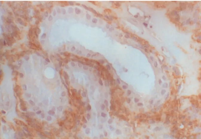
Fig. 5. Luminal cells of the tubules are positive for the anti-epithelial membrane antigen (EMA) antibody (EMA × 400).
Fig. 6. S-100 protein was detected only in the periph- eral layer of the tubular structures and stroma (S-100
× 400).
104 TW Lim, et al.
Annals of Dermatology Vol. 14, No. 2, April 2002 trunk, and axilla has also been reported 4,5.
In the Korean literature, Kang et al6 reported two cases of TAA. One patient was a male who had had a finger-tip sized cystic mass on the scalp, and the other patient was a female who had had a dark red hard nodule on the left forearm. And You et al7 reported a case of multiple TAA, which ap- peared as three separated nodules on the right deltoid area. In our patient, the lesions occurred on the nostril, which is a very uncommon site.
The occasional association of TAA with nevus se- baceous and syringocystadenoma papilliferum (SCAP) suggests the hamartomatous charac- ter1,4,8.
Fisher9criticized that this neoplasm simply repre- sented a variant of SCAP because of location on the scalp, concurrence with what was interpreted as basal cell carcinoma, two layers of epithelial cells lin- ing tubules, arrangement in tubules, plasma cell in the stroma and connection to the skin surface.
However, according to Umbert and Winkel- mann10, TAA was different histopathologically from SCAP in that neither invaginations nor pap- illary projections into lumina was seen in TAA, and they suggested that TAA is an independent clinical entity consisting of a benign appendage tumor of apocrine origin that is often associated with an organoid nevus.
PEA, first reported in 1977 by Rulon and Hel- wig11, has similar histological features with TAA, and some authors2,12propose that a single term, tubu- lopapillary hidradenoma be employed for both le- sions.
The histopathological feature is the presence of nu- merous irregularly shaped tubular structures that are lined by two layers of epithelial cells. The pe- ripheral layer consists of cuboidal or flattened cells, and the luminal layer is composed of columnar cells with characteristic decapitation secretion13.
Several immunohistochemical studies have been done which supported either apocrine or eccrine differentiation of the neoplasm2-5, 8-18. The findings in favor of apocrine differentiation include connec- tion of some tubular structures of the neoplasm to pre-existing inflammation, presence of apocrine secretion in cells along the luminal border that lined tubules, and immunohistochemical positivity of human milk fat globule and GCDFP-15, posi- tivity for acid phosphatase, lysozyme, α1- anti- chymotrypsin and negativity for phosphorylase5,18.
The findings in favor of eccrine differentiation seem to be positivity of some of the neoplastic cells for S-100 protein and ferritin 17. In our case, def- inite decapitation secretion was noted on the lumi- nal cells of tubules, and immunohistochemically, cells comprising tubules were positive for cytokeratin and EMA. S-100 was negative in luminal cells but weakly positive in cells of outer layer of the tubules, which might be exhibiting myoepithelial cells of the secretory portion of the apocrine gland.
TAA can be determined to be benign on the basis of symmetry, sharp circumscription, smooth bor- ders of tubules . Complete surgical excision is always curative and no metastasis has ever been reported.
Burket and Zelickson19 reported a case of TAA which showed tubules formed by neoplastic cells in perineural space and between thick bundles of col- lagen, and they proposed that TAA might not be be- nign but behave in a quietly aggressive fashion , so wide excision should be recommended.
Our case should be considered apocrine in na- ture on the basis of the presence of apocrine secre- tion in cells that line lumina and negativity for S- 100 protein of tumor cells except some cells of pe- ripheral layer. It is also interesting that the lesion in our patient occurred on nostril which is a very un- common site for this tumor.
REFERENCES
1. Landry M, Winkelmann RK. An unusual tubular apocrine adenoma. Arch Dermatol 105:869-879, 1972.
2. Falck VG, Jordaan HF. Papillary eccrine adenoma.
A tubulopapillary hidradenoma with eccrine differ- entiation. Am J Dermatopathol 8:64-72, 1986.
3. Ishiko A, Shimizu H, Inamoto N, Nakmura K. Is tubular apocrine adenoma a distinct clinical enti- ty?.Am J Dermatopathol 15:482-487, 1993.
4. Civatte J, Belaich S, Lauret P. Adenoma tubulare apocrine (quntre cas) Ann Dermatol Venereol 1979:106:665-669
5. Fox SB, Cotton DW . Tubular apocrine adenoma and papillary eccrine adenoma. Entities or unity?.
Am J Dermatopathol 14:149-154, 1992.
6. Kang MJ, Choi YW, Choi HY, Myung KB. Two cases of tubular apocrine adenoma. Kor J Dermatol 37:1782-1788 , 1999.
7. You MY, Yun SK, Ihm CW. A case of multiple
tubular apocrine adenoma. Kor J Dermatol 38:659- 663, 2000.
8. Epstein BA, Argenyi ZB, Goldstein G, Whitaker D. An unusual presentation of a congenital benign apocrine hamartoma. J Cutan Pathol 17:53-58, 1990.
9. Fisher TL. Tubular apocrine adenoma. Arch Der- matol 107:137, 1973.
10. Umbert P, Winkelmann RK. Tubular apocrine adenoma. J Cutan Pathol 3:75-87, 1976.
11. Rulon DB, Helwig EB. Papillary eccrine adenoma.
Arch Dermatol 113:596-598, 1977.
12. Oka K, Katsumata M. apoccrine tubular adenoma-a histopathological, histochemical, and ultrastructur- al study. J Dermatol 13:285-292, 1986.
13. Elder D, Elenitas R, Ragsdale BD: Tumors of the epidermal appendages, in Elder D, Elenitas R, Ja- worsky C, Johnson B Jr(eds):Levers histopathology of the skin, 8th ed, LippincottRaven, Philadelphia, 1997, pp747-803.
14. Hashimoto K, Kato I, Taniguchi Y, Eto H, Hori K,
Daneshvar S . Papillary eccrine adenoma. Immuno- histochemical and ultrastructural analyses. J Der- matol Sci 1:65-71, 1990.
15. Requena L, Pena M, Sanchez M, Sanchez Yus E.
Papillary eccrine adenoma-a light-microscopic and immunohistochemical study. Clin Exp Dermatol 15:425-428, 1990.
16. Megahed M, Holzle E. Papillary eccrine adenoma.
A case report with immunohistochemical examina- tion. Am J Dermatopathol 15:150-155, 1993.
17. Tellechea O, Reis JP, Marques C, Baptista AP.
Tubular apocrine adenoma with eccrine and apoc- rine immunophenotypes or papillary tubular adeno- ma?. Am J Dermatopathol. 17:499-505, 1995.
18. Wollina U, Rulke D, Schaarschmidt H. Dermal cylindroma. Expression of intermediate filaments, epithelial and neuroectodermal antigens. Histol Histopathol 2:575-582, 1992.
19. Burket JM, Zelickson AS. Tubular apocrine adeno- ma with perineural invasion. J Am Acad Dermatol 11:639-642, 1984.
A Case of Tubular Apocrine Adenoma 105