64 Ann Dermatol
Received December 28, 2009, Revised January 24, 2010, Accepted for publication January 28, 2010
Corresponding author: Deborah Lee, M.D., Department of Derma- tology, Busan Paik Hospital, College of Medicine, Inje University, 633-165 Gaegum-dong, Busanjin-gu, Busan 614-735, Korea. Tel:
82-51-890-6135, Fax: 82-51-897-6391, E-mail: [email protected]
Ann Dermatol Vol. 23, No. 1, 2011 DOI: 10.5021/ad.2011.23.1.64
CASE REPORT
Fig. 1. Skin-colored, 2.5×1.6×1.4 cm sized, solitary, round and protruded nodule on the lateral side of the left great toe.
Giant Acquired Digital Fibrokeratoma Occurring on the Left Great Toe
Joon Hee Choi, M.D., So Young Jung, M.D.
1, Ji Sung Chun, M.D.
1, Jong Keun Seo, M.D.
1, Deborah Lee, M.D.
1, Seon Wook Hwang, M.D.
1, Ho Suck Sung, M.D.
1Department of Dermatology, Maryknoll Hospital, 1Department of Dermatology, Busan Paik Hospital, College of Medicine, Inje University, Busan, Korea
Acquired digital fibrokeratoma is an uncommon, benign fibrous tumor which usually occurs in adults as a solitary lesion. The most frequent locations are fingers and toes and the size of the tumor is generally small, around 3∼5 mm. An 18-year-old female presented with a solitary, skin-colored, round and protruded nodule of the left great toe. The size of nodule was 2.5×1.6×1.4 cm. Histopathologic examination revealed typical findings of acquired digital fibrokeratoma.
Herein, we report a giant acquired digital fibrokeratoma.
(Ann Dermatol 23(1) 64∼66, 2011) -Keywords-
Acquired digital fibrokeratoma, Great toe
INTRODUCTION
Acquired digital fibrokeratoma is rare, benign fibrous tumor. It commonly occurs in adults as a solitary nodule on fingers and toes1,2. The size of the tumor is generally small, less than 1 cm. Even though an exact definition of the standard size of giant acquired digital fibrokeratoma has not been established, 3 cases of acquired digital fibrokeratoma larger than 1 cm were reported, as giant acquired digital fibrokeratomas, in the dermatologic literatures3-5.
Herein, we report the biggest case in Korean dermatologic literature of a giant acquired digital fibrokeratoma occurr- ing on the left great toe.
CASE REPORT
An 18-year-old woman presented with an asymptomatic, solitary nodule on her left great toe. Two years ago, she noticed a slow growing, flesh-colored nodule on her left great toe. There was no history of trauma. One year ago the lesion was totally excised. After several months, the lesion recurred and gradually enlarged. Physical examina- tion revealed a 2.5×1.6×1.4 cm sized, skin-colored, solitary, round and protruded nodule on the lateral side of her left great toe (Fig. 1).
Histopathologic examination showed marked hyperkera- tosis, acanthosis and elongated rete ridges in the epider- mis (Fig. 2). The dermis showed thick collagen bundles with dilated capillaries oriented in the direction of the longitudinal axis of the lesion. The histopathologic fea- tures were compatible with acquired digital fibrokera- toma. The lesion was completely excised without recur- rence for 1 year.
Giant Acquired Digital Fibrokeratoma Occurring on the Left Great Toe
Vol. 23, No. 1, 2011 65 Fig. 2. (A) Histopathologic examination demonstrated a dome-shaped tumor consisting of a thick core lesion and peripheral lesion (H&E, ×4). (B) The core of tumor showed thick dermal collagen bundles oriented along the longitudinal axis of the lesion (H&E,
×40). (C) The peripheral lesion of tumor showed irregular acanthosis, thickening of rete ridges and increased capillaries (H&E, ×40).
(D) The close-up view of the peripheral lesion showed thin walled dilated capillaries and thick collagen bundles in dermis (H&E,
×100).
DISCUSSION
Acquired digital fibrokeratoma is an uncommon tumor first reported by Bart et al.1 in 1968. It is a benign tumor, almost always solitary, can be seen in adults and does not show spontaneous regression. In most cases, acquired digital fibrokeratoma appears as a small solitary nodules mainly on the fingers and toes, occasionally occurring on the lower lip, nose, elbow, pre-patellar area, nail bed and heel3,6-8.
The size of acquired digital fibrokeratoma is generally small, less than 1 cm. Exceptionally, there have been reported cases of giant acquired digital fibrokeratoma.
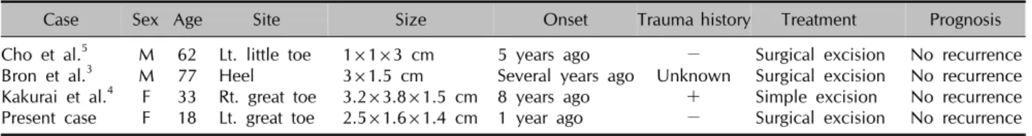
Kakurai et al.4 and Bron et al.3 reported 3.2×3.8×1.5 cm and 3.0×1.5 cm acquired digital fibrokeratoma on the toe and heel as giant one respectively. In Korean dermatologic
literatures, Cho et al.5 reported 1×1×3 cm giant acquired digital fibrokeratoma (Table 1). In our case, the size of the tumor was 2.5×1.6×1.4 cm, so we suggest that it is a proper case of giant acquired digital fibrokeratoma.
The pathophysiology of an acquired digital fibrokeratoma is unknown. In general, trauma is often thought to be a predisposing factor, especially on the digits9. In 1985, Kint et al.2 suggested that the acquired digital fibrokeratoma resulted from a neoformation of collagen by the fibro- blasts. Because when comparing with the surrounding dermis, the tumor was composed of denser fibers than normal skin and contained more capillaries, more fibro- blasts and coarser elastic fibers. Nemeth and Penneys10 reported that Factor XIIIa, which was found in fibrovas- cular tumors, was also found in acquired digital fibro- keratoma; therefore this factor might play an important
JH Choi, et al
66 Ann Dermatol
Table 1. Summary of previously reported cases and present case of giant acquired digital fibrokeratoma
Case Sex Age Site Size Onset Trauma history Treatment Prognosis
Cho et al.5 M 62 Lt. little toe 1×1×3 cm 5 years ago − Surgical excision No recurrence Bron et al.3 M 77 Heel 3×1.5 cm Several years ago Unknown Surgical excision No recurrence Kakurai et al.4 F 33 Rt. great toe 3.2×3.8×1.5 cm 8 years ago + Simple excision No recurrence Present case F 18 Lt. great toe 2.5×1.6×1.4 cm 1 year ago − Surgical excision No recurrence
role in the pathogenesis of these tumors. After then, Suh et al.11 observed the factor XIIIa positive dermal dendrocytes were increased in acquired digital fibrokeratoma by immunohistochemical stain and suggested that these cells were associated with collagen synthesis regulation.
Histopathologic examination shows hyperkeratosis and irregular acanthosis in epidermis and thick collagen bundles with dilated capillaries oriented in the direction of the longitudinal axis in dermis. Kint et al.2 described 3 types of acquired digital fibrokeratoma by clinical and histopathologic features. Type I acquired digital fibrokera- toma is a dome-shaped lesion which contains fibroblast between collagen bundles, fine elastic fibers and numer- ous capillaries in dermis. Type II is a mainly tall and hyperkeratotic lesion which contains many more fibro- blasts and reduced elastic fibers. Type III is a flat to dome-shaped lesion which is characterized by poorly cellular and edematous structure and no elastic fibers.
This case showed typical histopathologic findings of acquired digital fibrokeratoma and was compatible with type I.
The differential diagnosis for acquired digital fibrokera- toma includes supernumerary digit, cutaneous horn and neurofibroma. Supernumerary digit is a congenital digital anomaly, which contains neural bundles in the dermis.
Cutaneous horn is uncommon epidermal tumor which consists of a column of keratin arising from a wide range of benign, premalignant or malignant underlying pro- cesses. Neurofibroma is soft, polypoid skin colored tumor.
On histopathologic examination, the dermis is composed of loosely spaced spindle cells and wavy collagenous strands.
Simple excision is curative. Usually, there is no recur-
rence after complete surgical excision.
Herein, we report an acquired digital fibrokeratoma which occurred as a giant one on the left great toe.
REFERENCES
1. Bart RS, Andrade R, Kopf AW, Leider M. Acquired digital fibrokeratomas. Arch Dermatol 1968;97:120-129.
2. Kint A, Baran R, De Keyser H. Acquired (digital) fibro- keratoma. J Am Acad Dermatol 1985;12:816-821.
3. Bron C, Noël B, Panizzon RG. Giant fibrokeratoma of the heel. Dermatology 2004;208:271-272.
4. Kakurai M, Yamada T, Kiyosawa T, Ohtsuki M, Nakagawa H. Giant acquired digital fibrokeratoma. J Am Acad Der- matol 2003;48:S67-68.
5. Cho EA, Lee WS, Kim SY. A case of giant acquired digital fibrokeratoma resembling supernumerary digit. Korean J Dermatol 2007;45:319-320.
6. Kim HJ, Lee SH, Park EJ, Kim CW, Jo HJ, Kim KH, et al. A case of acquired fibrokeratoma of the lower lip. Korean J Dermatol 2006;44:1438-1440.
7. Hur J, Suh KS, Kim ST. Two cases of acquired fibrokeratoma.
Korean J Dermatol 2000;38:1116-1117.
8. Jee MS, Lee DP, Chang SE, Choi JH, Sung KJ, Moon KC, et al. Two cases of acquired fibrokeratoma occuring on the prepatellar area and the nail bed. Korean J Dermatol 2003;
41:403-405.
9. Altman DA, Griner JM, Faria DT. Acquired digital fibro- keratoma. Cutis 1994;54:93-94.
10. Nemeth AJ, Penneys NS. Factor XIIIa is expressed by fibroblasts in fibrovascular tumors. J Cutan Pathol 1989;16:
266-271.
11. Suh HS, Ryu BJ, Choi JH, Sung KJ, Koh JK. A case of acquired digital fibrokeratoma: immunohistochemical stain with anti-factor XIIIa antibody. Korean J Dermatol 1994;32:
1131-1135.