Received March 6, 2011, Revised March 14, 2011, Accepted for publication April 7, 2011
Corresponding author: Jin-Wou Kim, M.D., Department of Derma- tology, Uijeongbu St. Mary’s Hospital, The Catholic University of Korea College of Medicine, 271 Cheonbo-ro, Uijeongbu 480-717, Korea. Tel: 82-31-820-3031, Fax: 82-31-846-4799, E-mail: jwkim52@
catholic.ac.kr
This is an Open Access article distributed under the terms of the Creative Commons Attribution Non-Commercial License (http://
creativecommons.org/licenses/by-nc/3.0) which permits unrestricted non-commercial use, distribution, and reproduction in any medium, provided the original work is properly cited.
ORIGINAL ARTICLE
Effect of Spa Spring Water on Cytokine Expression in Human Keratinocyte HaCaT Cells and on
Differentiation of CD4 + T Cells
Ho-Pyo Lee, M.D., Yoon-Jung Choi, M.D., Kyung-Ah Cho1, So-Youn Woo, M.D.1, Seong-Taek Yun, Ph.D.2, Jong Tae Lee, M.D.3, Hong Jig Kim, M.D.4, Kyung-Ho Lee, M.D.5, Jin-Wou Kim, M.D.
Department of Dermatology, Uijeongbu St. Mary’s Hospital, The Catholic University of Korea College of Medicine, Uijeongbu,
1Department of Microbiology, Ewha Womans University School of Medicine, Seoul, 2Department of Earth and Environmental Sciences, Korea University, Seoul, 3The Korea Central Institute of Hot Spring, Seoul, 4The Korean Academy of Hot Spring, Seoul, 5Department of Dermatology, The Catholic University of Korea College of Medicine, Bucheon, Korea
Background: Skin acts as the first line of defense against any foreign materials outside of our body. In inflammatory skin disease, the pathogenesis is due to an immune reaction in the keratinocytes, immune cells and soluble mediators. Balneo- therapy is widely used for the treatment of inflammatory skin disease, but the mechanisms are only partly understood by immune regulation. Balneotherapy in dermatologic disease can affect the secretion of pro- inflammatory cytokines, IL-1 α and tumor necrosis factor from keratinocytes, and possibly affect the T cell differentiation. Objective: In this study, we evaluated the effect of spa spring water from Yong-gung oncheon on the cells, and investigated the skin immune reaction. Methods: We investigated the immuno- modulatory or anti-inflammatory effect of thermal spring water on the expression of pro-inflammatory cytokines in the HaCaT cells under Toll-like receptor (TLR) stimulation, as well as the effect on the differentiation of CD4+ T cells under spring water. Results: The treatment of spa spring water from Yong-gung oncheon decreased the expression of pro- inflammatory cytokines under TLR stimulation to the HaCaT
cells and antigen presenting cells. In addition, spa spring water attenuated the differentiation process of subsets of CD4+ T cells, i.e., Th1, Th2 and Th17 cells. All these immune parameters can be used to evaluate the efficacy of spa spring water in Korea, in terms of the immune modulatory effect. Conclusion: Spa spring water treatment suppressed the inflammatory cytokines production and also modulated the differentiation of CD4+ T cells into Th1, Th2, and Th17 cells, but not the Tregs cells. (Ann Dermatol 24(3) 324∼336, 2012)
-Keywords-
Balneotherapy, CD4+ T cells, Cytokines, HaCaT cells, Skin inflammation
INTRODUCTION
Skin contributes to the first line of defense against any foreign materials outside of our body, as a physical barrier. As immune sentinels, keratinocytes can recognize foreign or danger stimuli from the outside via pattern- recognition receptors, such as Toll-like receptors (TLRs), and release innate immune mediators, including cytokines and chemokines, under the stimulation of the keratino- cytes. Epidermal keratinocytes express several TLRs, including TLR1, TLR2, TLR3, TLR4, TLR5, TLR6, TLR7 and TLR91, and through the TLRs, keratinocytes initiate and promote immune responses in the skin.
For inflammatory skin disease, pathogenesis is due to the sum of the immune reactions of the keratinocytes, immune cells and soluble mediators. The three main types
of CD4+ T cells can be found in the skin during inflam- matory skin disease, i.e., Th1, Th2 and Th17. For example, Th1-dominant immune reactions were reported to be associated with the autoimmunity or psoriasis, and Th2-dominant responses were related to that of asthma or atopic dermatitis. The Th17 cells were reported to con- tribute to defend against various fungal or bacterial infec- tions, and possibly induced atopic dermatitis and epidermal changes in psoriasis2,3 via secretion of inter- leukin (IL)-17 and IL-22. IL-17 and IL-22 increased antimicrobial peptides, β-defensins and cathelicidins, from keratinocytes4. In addition to the antimicrobial peptides, keratinocytes secrete cytokines, including IL-1, IL-6, IL-10, IL-18 and tumor necrosis factor (TNF)5.
Spa therapy is widely used for the treatment of inflam- matory skin diseases, such as atopic dermatitis, psoriasis, pruritus, rosacea, seborrheic dermatitis and others6. The efficacy of spa therapy for inflammatory skin diseases and the mechanisms are only partly understood, and presumably incorporate chemical, thermal, mechanical and immuno- modulatory effects7,8. Among them, we investigated the immunomodulatory or anti-inflammatory effect of thermal spring water on the expression of pro-inflammatory cytokines in the HaCaT cells under TLR stimulation, as well as the effect on differentiation of CD4+ T cells under spring water.
MATERIALS AND METHODS
Cell culture
HaCaT (human keratinocyte cell line) was kindly provided by Dr. Tae-Yoon Kim (College of Medicine, The Catholic University of Korea) and was cultured in Dulbeco's Modified Eagle Medium (DMEM, Gibco-BRL, Grand Island, NY, USA) supplemented with 10% fetal bovine serum (FBS, Gibco- BRL) and 100 U/ml penicillin /streptomycin (Gibco- BRL), at 37oC in an incubator containing 5% CO2.
Preparation of spa spring water
Water was collected from the Yong-gung oncheon (Incheon-si, Gangwha-gun, Korea) spring and filtered through 0.44 um filters. Waters were stored at 4oC, until used for the experiments. For the measurement of water osmolarity, we used a Micro-Osmometer 210 (FISKE Associate, Norwood, MA, USA).
MTT assay
For the 3-(4,5-dimethylthizol-2-yl) 2,5-diphenyl tetrazolium bromide (MTT, Sigma-Aldrich, St. Louis, MO, USA) assay, cells were plated onto 96-well microtiter plates at a
density of 3×10⁴/200 μl in fresh medium, and then treated with hot spring water at a serially diluted concen- tration. Cells were cultured for 1, 4, 10, and 24 hours, respectively, to observe a time dependent effect. After the indicated time, 20 μl of MTT (5 mg/ml in phosphate- buffered saline) was added to each well, and the plates were returned to the incubator for an additional 4 hours.
At the end of the incubation period, the supernatants were discarded by a suction and 200 μl dimethyl sulfoxide was added to all the wells, in order to dissolve the dark blue formazan crystals. The plates were subsequently covered with aluminum foil, gently shaken for 15 minutes, and read at a wavelength of 570 nm.
TLR stimulation
TLR agonists were treated with the following final concentrations for 24 hours. Tripamitoyl-S-glyceryl-cysteine (Pam3Cys, 1 μl/ml), heat-killed Listeria monocytogenes (HKLM, 106 cells/ml), polyriboinosinic polyribocytidylic acid (poly (I : C), 10 μl/ml), lipopolysaccharide (LPS, 10 μl/ml), flagellin (10 μl/ml), and Pam2CGDPKHPKSF (FSL-1, 1 μl/m) were from InvivoGen (San Diego, CA, USA). Spa spring waters were added simultaneously or pre-treated 2 hours before the TLR agonist treatment. Cells were cultured for 1, 4, 10, or 24 hours in each treatment group.
ELISA
Levels of IL-6, IL-8, granulocyte-macrophage colony- stimulating factor (GM-CSF), and TNF-α (BD OptEIATM, BD Biosciences Pharmingen, San Diego, CA, USA), IL-1α (Biolegend, San Diego, CA, USA) in HaCaT supernatant treated with TLR agonist in the presence or absence of hot spring water were quantified according to the manufac- turer’s protocol. IL-6 and TNF-α in mouse antigen presenting cells (APC) supernatant were also measured. In brief, the wells were coated with 100 μl of capture antibody in coating buffer (0.1 M sodium carbonate, pH 9.5) and the plates were incubated overnight at 4oC. After washing the wells with washing buffer (phosphate buffered saline [PBS] with 0.05% Tween-20), the wells were blocked with assay diluents (10% FBS in PBS) for 1 hour at room temperature (RT), followed by the addition of 100 μl/well of cell supernatant and cytokine standard solutions for 2 hours at RT. After washing, 100 μl of detection antibody and streptavidin-conjugated horseradish peroxidase reagent were added to the wells, which was then incubated for 1 hour at RT. After extensive washing, 100 μl of substrate solution (tetramethylbenzidine and hydrogen peroxide, BD Biosciences Pharmingen) was added to each well and the plates were incubated for 30
Fig. 1. Cell culture with spa spring water led to detach HaCaT cells. The HaCaT cells cultured in Dulbeco's Modified Eagle Medium (DMEM, Gibco-BRL, Grand Island, NY, USA) with 10% fetal bovine serum (FBS, Gibco-BRL) (A) and DMEM prepared from undiluted spa spring water with 10% FBS (B). Original magnification ×200. Tx: Treatment.
minutes at RT, in darkness. Stop solutions (2 N H2SO4) were added, and the absorbance was read at 450 nm within 30 minutes.
Mouse spleen cell isolation
Naïve CD4+ T cells were purified from the mouse spleens, via magnetic isolation (Miltenyi Biotec GmbH, Gladbach, Germany). For the preparation of the spleen cell suspen- sions, spleens from 8 weeks-old female Balb/c mice were removed and minced with a Nylon mesh (70 μm pore).
After the cells were pelleted, erythrocytes were lysed using hypotonic buffer (0.15 M NH4Cl, 10 mM KHCO3, 0.1 mM Na2EDTA). The cells were washed in PBS and incubated with anti-CD4 antibody for 15 minutes, at 4oC.
Directly following, the cells were conducted onto a magnetic separator to isolate the CD4+ cells and collected with positive selection. CD4− cells were seeded onto a culture dish of 100 mm in diameter and incubated for 4 hours for the collection of the adherent cells (used as APC in these experiments) by gentle pipetting.
CD4+ T cell differentiation
CD4+ naïve cells were seeded at a density of 2×10⁵ per well in 96-well plates, and were cultured in DMEM, containing 10% FBS. For each of the helper T cell differentiation, the skewing conditions are as followed;
IL-12 (25 ng/ml, Biolegend) and anti-CD28 (1 μg/ml, Biolegend) for Th1, IL-4 (25 ng/ml, Biolegend) and anti-CD28 (1 μg/ml, Biolegend) for Th2, IL-6 (25 ng/ml, Biolegend), transforming growth factor (TGF)-β (2 ng/ml,
R&D system) and anti-CD28 (1 μg/ml, Biolegend) for Th17, IL-2 (25 ng/ml, Biolegend) and anti-CD28 (1 μg/ml, Biolegend) for T reg. All cells stimulated with anti-CD3 antibodies with serial dilutions, in which the initial concentration was 3 μg/ml. Cells were incubated for 3 days in the presence or absence of spa spring water.
Cell proliferation assay
Before seeding for the differentiation of each of the helper T cell differentiation, the CD4+ cells were labeled with carboxyfluorescein diacetate, succinimidyl ester (CFSE, CellTraceTM CFSE Cell Prolifetration kit, Invitrogen, Paisley, UK). First, the cells were resuspended in pre- warmed PBS/0.1% bovine serum albumin, at a concen- tration of 106 cells/ml, and 10 μM of CFSE were added.
After incubation at 37oC for 10 minutes, 5 volumes of ice-cold culture media was added to the cells to quench the staining, incubated for 5 minutes on ice, and pelleted by centrifugation. The cells were resuspended in fresh media for a total of three washes, and seeded under each condition for the differentiation of Th1, Th2, Th17, and Treg cells. After 3 days, CD4+ and CFSE+ cells were measured for the degree of proliferation, via a flow cytometry, and were analyzed using ModFit LT software (Verity Software House, Topsham, ME, USA), based on the reduction of CFSE positive cells.
Flow cytometry
APCs were stimulated with TLR3 agonist, poly (I : C) (10 μg/ml) for 24 hours with or without spa spring waters.
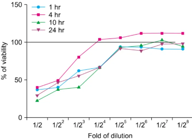
Fig. 2. Viability of HaCaT cells was not affected by spa spring water in the 1:32 fold of dilution. The HaCaT cells were plated at a density of 3×10⁴/200 μl in fresh medium and treated with spa spring water as indicated. After 1, 4, 10, and 24 hours of culture, 20 μl of MTT (5 mg/ml in PBS) was added to each of the wells. After 4 hours, dark blue formazan crystals were dissolved, and the plates were read at a wavelength of 570 nm.
100% viability was determined based on the result of MTT assay of normally cultured HaCaT cells. PBS: peripheral blood smear examination.
Fig. 3. Schematic diagram of experimental design. (A) The HaCaT cells were pre-incubated for 2 hours with spa spring water at a concentration of 1 : 30 fold dilution, and then stimulated with TLR1 to TLR6 agonist as indicated time. (B) The HaCaT cells were simultaneously exposed to spa spring water and TLR agonist and incubated as indicated time. TLR: toll-like receptor.
APCs stimulated with poly (I : C) with or without hot spring water were collected, washed and stained with anti-mouse I-A/I-E (Biolegend, clone M5/114.15.2, Rat IgG2b, κ) to analyze the expression of MHC II on the surface of APCs for 20 minutes, at RT. Samples were acquired on FACSCalibur system (BD Bioscience, San Jose, CA, USA) and were analyzed using CellQuest software (BD Bioscience).
Statistical analysis
Data are expressed as the means±standard error of the mean. Non-parametric Mann-Whitney tests were applied to analyze the results for significant differences (at p<
0.05), using GraphPad Prism software (GraphPad Software Inc., San Diego, CA, USA).
RESULTS
Determination of dilution concentration of spa spring water for HaCaT cell culture
Because the osmolarity of spa spring water from Yong-gung oncheon was 700 mOsm/kg, the HaCaT cells were not able to maintain the growth in undiluted culture medium (Fig. 1). Therefore, we first determined to set an optimal dilution concentration for HaCaT culture condi- tions by the MTT assay. For this, HaCaT cells were cultured with spa spring water at a concentration of serially 2-fold dilution for 1, 4, 10, and 24 hours and the
minimum concentration of spa spring water was determined. We found that each group of cells was up to 100% of viability under a 1 : 32 fold dilution (Fig. 2).
Cytokine production of HaCaT cells under the TLR agonist treatment with spa spring water
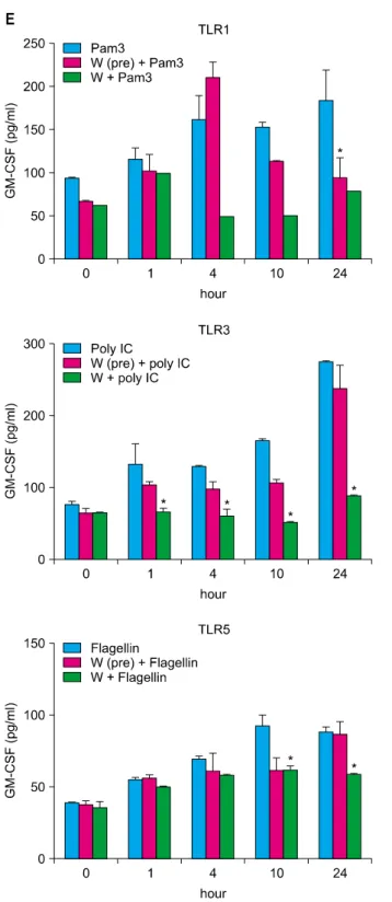
TLRs act as primary sensors that detect a wide variety of microbial components and elicit innate immune response, which guides the acquired immunity. Therefore, kera- tinocyte constitutively expressing TLRs may affect skin immune response, by regulating the inflammatory res- ponses, via TLR signaling. We attempted to determine whether the spa spring water affected the TLR stimulated production of pro-inflammatory cytokines, including IL-1 α, IL-6, IL-8, TNF-α, and GM-CSF, on the HaCaT cells.
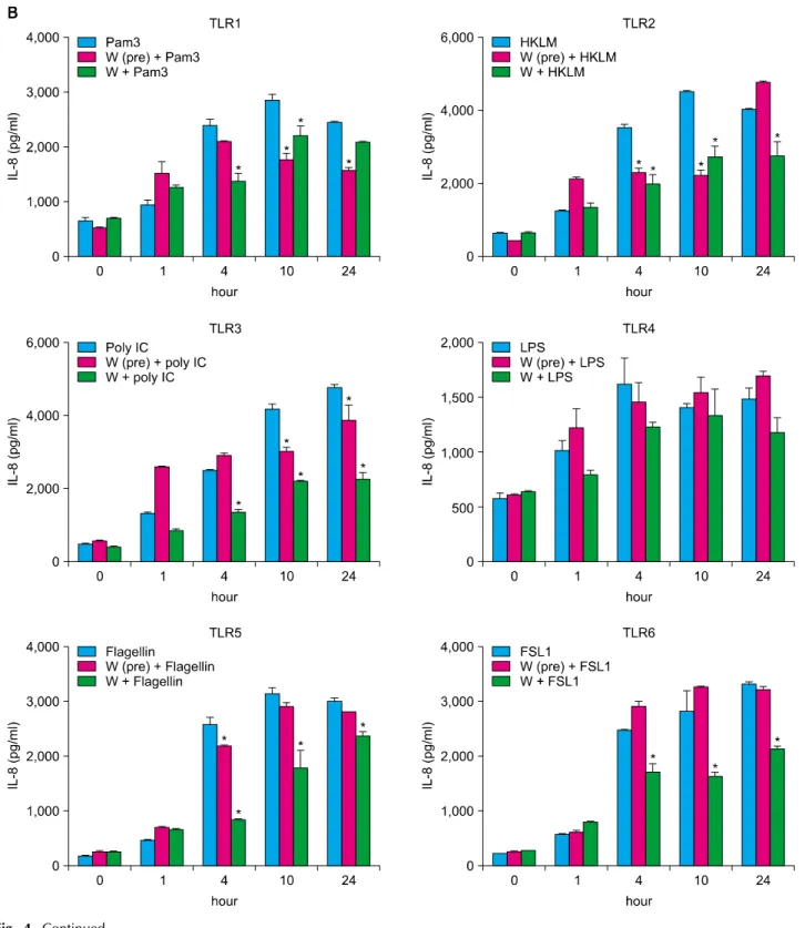
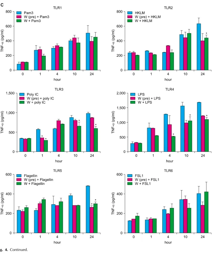
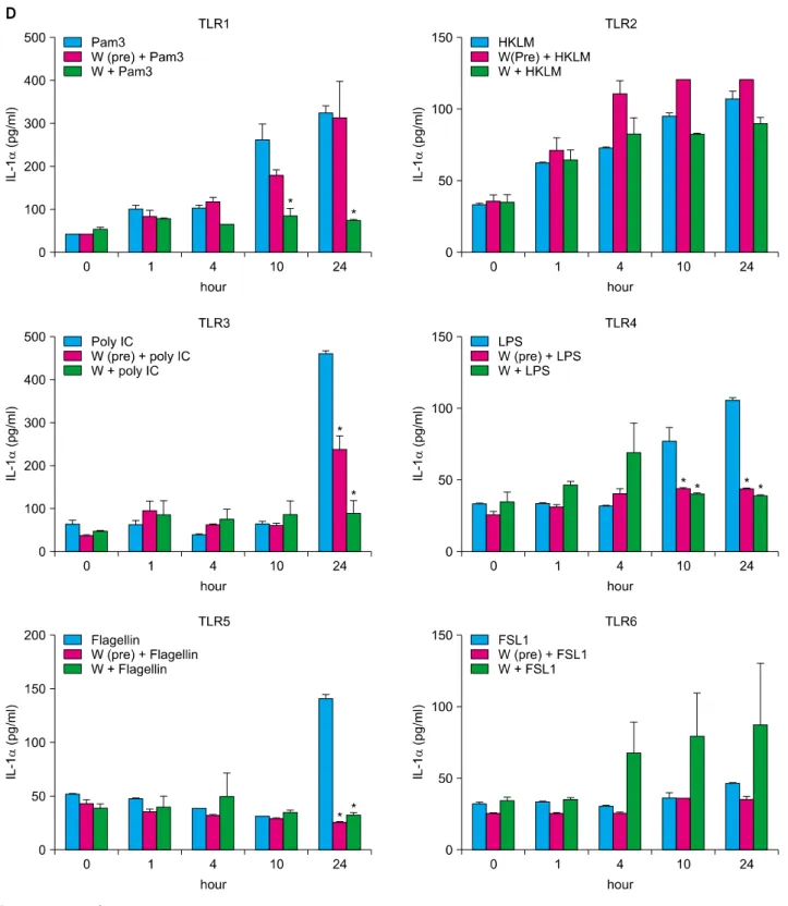
TLR1 agonist, through TLR6 agonist treatment, induced the attenuation of cytokine production in the exposure to spa spring in advance or simultaneously with TLR agonist treatment (Fig. 3, 4).
Proliferation characteristics of T cells under spa spring water
We subsequently attempted to ascertain the potential involvement of spa spring water in the differentiation of the helper T cells. Because the effector T cells, such as
Fig. 4. Spa spring water impairs pro-inflammatory cytokine production on HaCaT cells under TLR stimulation. The HaCaT cells were treated with TLR1 through TLR6 agonist for 1, 4, 10, and 24 hours, respectively. Spa spring water diluted 1 : 30 fold was added prior to adding TLR agonists or simultaneously. After the indicated time, the supernatants were collected to measure secreted level of IL-6 (A), IL-8 (B), TNF-α (C), IL-1α (D), and GM-CSF (E) via ELISA. The data are expressed as the means±SEM (*p<0.05) of three independent experiments. TLR: Toll-like receptor, IL: interleukin, HKLM: heat-killed Listeria monocytogenes, LPS:
lipopolysaccharide, FSL1: Pam2CGDPKHPKSF, TNF: tumor necrosis factor, GM-CSF: granulocyte macrophage-colony stimulating factor, SEM: standard error of the mean.
Fig. 4. Continued.
Th1, Th2, and Th17 cells act on various skin resident cells and potentiate the immune reaction, regulating T cell differentiation may modulate pro-inflammatory responses.
To evaluate the in vitro suppressive capacity of spa spring water toward the helper T cell differentiation, magnetic sorted CD4+ T cells, from the spleen, were prepared from
Balb/c mice. CFSE-labeled CD4+ T cells were cultured under various conditions, which polarized Th1, Th2, Th17, and Treg with or without spa spring water. After 3 days, the cells were harvested and stained with CD4 and interferon (IFN)-γ for Th1, IL-4 for Th2, IL-17 for Th17 and Foxp3 for Treg cells, respectively. Then, the flow
Fig. 4. Continued.
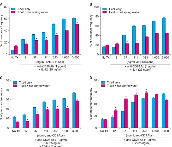
cytometry was employed to evaluate the precursor frequency in terms of the T cell differentiation. As shown in Fig. 5, CD4+ naïve T cells were profoundly proliferated and differentiated under each skewing condition, along with anti-CD3 stimulation. Spa spring water suppressed the proliferation of Th1, Th2 and Th17 cells. In contrast to
the suppressive effect on Th1, Th2 and Th17 cells, proliferation and differentiation to Treg cells were pro- moted under spa spring water treatment. These results indicate that spa spring water may affect the distribution of the helper T cells in the immune response, by suppressing the polarization of the Th1, Th2 or Th17 cells.
Fig. 4. Continued.
Cytokine production and major histocompatibility complex (MHC) II molecule expression of APCs under TLR3 agonist treatment were affected by spa spring water
Skin immunity is achieved by an interaction of different
kinds of immune cells. In particular, APCs, such as Langerhans cells found in the epidermis, are the best- characterized dendritic cell population. They have the ability to process antigens in the periphery, transport it to the draining lymph nodes, where they are able to cluster with, and activate the antigen-specific naive T cells. APCs
Fig. 4. Continued.
in the dermis may also provide alternative routes of antigen presentation, which can be important in the regulation of skin immune responses. Therefore, APCs are vital for the induction of immune responses to antigens encountered via the skin. To determine whether the expression of MHC II on the surface of APC is modulated
by spa spring water, we isolated APC from BALB/C mouse spleen, and stimulated it with 20 μg/ml of poly (I : C) for 24 hours. TLR3 stimulation via poly (I : C) strongly induced MHC II expression on the APCs. By contrasts, APC treated spa spring water down-regulated the surface level of class II MHC expression, under TLR 3 stimulation
Fig. 5. Spa spring water inhibited the differentiation and proliferation of effector T cells and promoted Treg proliferation. The CD4+ naïve T cells were isolated, labeled with carboxyfluorescein diacetate, succinimidyl ester (CFSE, CellTraceTM CFSE Cell Prolifetration kit, Invitrogen, Paisley, UK), and cultured under skewing conditions for Th1, Th2, Th17, and Treg cells, respectively, in the presence or absence of spa spring water. Serially diluted (1 : 3) anti-CD3 antibodies were treated at a concentration starting from 3 μg/ml to monitor dose-dependent proliferation. On day 3, cells were harvested and proliferation was measured by flow cytometry. CD4+ IFN-γ+ Th1 cells (A), CD4+ IL-4+ Th2 cells (B), and CD4+ IL-17+ Th17 cells (C) were proliferated along with the increase of anti-CD3 stimulation, whereas they were inhibited under spa spring water. In contrast, spa spring water promoted proliferation of CD4+Foxp3+ Tregs (D) relative to those of the HaCaT cells cultured alone. Percentages of proliferative cells were represented as precursor frequency via analysis using ModFit LT software (Verity Software House, Topsham, ME, USA) based on the reduction of CFSE positive cells.
The data are expressed as the means±SEM (*p<0.05) from three independent experiments. Tx: treatment, IFN: interferon, IL: interleukin, TGF: transforming growth factor, SEM: standard error of the mean.
(Fig. 6A). In addition, we observed that TLR3 agonist, poly (I : C) is a very potent inducer of inflammatory responses in the HaCaT cells9. Therefore, we measured and com- pared poly (I : C) mediated production of inflammatory cytokines from the APCs with or without spa spring water (Fig. 6B, C). APC induced TNF-α and IL-6 under poly (I : C) stimulation and cytokine production was reduced in the presence of spa spring water. These results showed that TLR-triggered inflammatory responses in APCs might
also be modulated under spa spring water treatment.
DISCUSSION
In this study, we evaluated the effect of spa spring water from Yong-gung oncheon on cells related to the skin immune reaction. The treatment of spa spring water from Yong-gung oncheon decreased the expression of pro- inflammatory cytokines under TLR stimulation to the HaCaT
Fig. 6. Spa spring water decreased class II MHC expression and impaired pro-inflammatory cytokine production on APCs under TLR3 stimulation. APCs were isolated and stimulated with 20 μg/ml of poly (I : C) for 24 hours. (A) Class II MHC expression was increased under poly (I : C) treatment and significantly reduced by adding spa spring water. Expressed levels were represented by histogram (left) and quantified as MFI (right). Poly (I : C) induced IL-6 (B) and TNF-α (C) were attenuated under spa spring water treatment.
The data are expressed as the means±SEM (*p<0.05) from three independent experiments. APC: antigen presenting cell, TLR: Toll-like receptor, Tx: treatment, MFI: mean fluorescence intensity, IL: interleukin, IFN: interferon, SEM: standard error of the mean.
cells and APCs. In addition, spa spring water attenuated the differentiation process of subsets of T helper cells, i.e., Th1, Th2 and Th17 cells.
The therapeutic mechanism of spa spring water in derma- tologic diseases can be divided into three categories:
active ingredient, thermal effects and mechanical effects.
Minerals in spa spring water, such as sulfur, magnesium, calcium or selenium induce anti-inflammatory, keratolytic, antibacterial or antifungal effects6. For anti-inflammatory effects, the inhibition of Th1 differentiation, inhibition of cytokine production from keratinocytes, and modulatory effects on epidermal Langerhans cells have been reported10. In this study, we observed the significant inhibitory effect on the secretion of pro-inflammatory cytokines, including
IL-6, IL-8, IL-1α, TNFα and GM-CSF from keratinocytes (Fig. 4). In addition, we observed the differentiation of Th1, Th2, or Th17, but not of Foxp3+ Treg cells under the treatment with water from Yong-gung oncheon (Fig. 5).
Th17 cells are related to the autoimmunity or inflam- matory skin disease, such as psoriasis or atopic dermatitis via IL-17 and IL-22 secretion. IL-17 acts directly on keratinocytes and induces the production of MIP-3α, IL-8 and β-defensin, whereas, IL-22 regulates the keratinocyte differentiation11,12. In addition, IL-17 promotes the neu- trophil recruitment by inducing the neutrophil-attracting chemokines (CXCL1, CXCL2, CXCL5, and CXCL8) and stimulates neutrophil production by inducing granulo- poiesis factors (G-CSF or GM-CSF)13. In case of Staphy-
lococcus aureus infection, the role of Th17 was reported to be critical: in mice bacterial clearance was impaired in IL-17R-deficient mice, and in human hyper immuno- globulin E syndrome, atopic dermatitis, human immuno- deficiency virus/acquired immune deficiency syndrome or mucocutaneous candidiasis, in which there is a common deficiency of Th17 cells. These IL-17-mediated defenses against S. aureus infection are involved with promotion of neutrophil recruitment via cytokines, chemokines or adhesion molecules and production of antimicrobial peptides. In this context, reduced Th17 cell differentiation, by spa spring water, implies that spa spring water may reduce the immune reaction in the epidermal layer, partly by affecting the antimicrobial peptide production and epidermal differentiation irrespective of antibacterial effect of IL-17.
Regulatory T cells (Treg cells) mediate immunosuppression and tolerogenic responses through contact-dependent or -independent mechanisms14-16. Foxp3+ Tregs produce IL-10 or TGF-β as effector molecules, and the balance between Treg cells and effector T cells is crucial for the maintenance of homeostasis and self-tolerance17. In this study, spa spring water from Yong-gung oncheon treatment induced Foxp3+ Treg cell differentiation in vitro, implying that the immune modulatory effect of spa spring water also includes Treg cell-induced immune suppressive effects.
TLRs are a type of pattern recognition receptor, which recognize microbial products known as pathogen-associated molecular patterns, i.e., bacterial lipoproteins, zymosan, LPS, flagellin, ssRNA, dsRNA, and unmethylated CpG DNA. TLRs are transmembrane receptors and present on the cell surface or on the surface of endosomal compartments. It was reported that TLR3 ligand poly (I : C) was the most potent stimulator of IL-8, IL-6 and TNFα secretion18 in the primary keratinocyte and HaCaT cell lines. In this experiment, we used TLR1 to TLR6 agonists to activate the HaCaT cells inducing pro-inflammatory cytokines. We observed that spa spring water treatment led to reduced IL-6, IL-8, TNF-α, IL-1α, and GM-CSF from the HaCaT cells, following TLR ligand treatments, and spa spring water from Yong-gung oncheon suppressed the expression of IL-6, TNF-α and class II MHC expression of APC in this experiment (Fig. 6). In case of atopic asthma, IL-5, IL-13, IL-1β, IFN-γ, IL-12, GM-CSF, IL-4, and IL-10 were elevated, compared with those of the non-atopic patients19. Further studies are required to compare the cytokine profile in non-atopic and atopic dermatitis patients after a spa treatment.
These results showed that spa spring water treatment suppressed the inflammatory cytokines production, and also modulated differentiation of CD4+ T cells into Th1,
Th2, and Th17 cells, but not Tregs cells. With these experimental protocols, we can evaluate and compare the efficacy of spa spring waters in Korea, immunologically.
Furthermore, we would define immune-active ingredient from this spa spring water to reveal immune modulatory mechanism of spa spring waters in Korea.
ACKNOWLEDGMENT
This work was partly supported by the Korean Academy of Hot Spring and the Basic Science Research Program though the National Research Foundation of Korea (NRF) funded by the Ministry of Education, Science, and Technology [NRF-2010-0003185] to S-Y. Woo.
This manuscript has been presented at the Korean Dermatological Association, the 63th Autumn Meeting.
REFERENCES
1. Lebre MC, van der Aar AM, van Baarsen L, van Capel TM, Schuitemaker JH, Kapsenberg ML, et al. Human keratino- cytes express functional Toll-like receptor 3, 4, 5, and 9. J Invest Dermatol 2007;127:331-341.
2. Di Cesare A, Di Meglio P, Nestle FO. The IL-23/Th17 axis in the immunopathogenesis of psoriasis. J Invest Dermatol 2009;129:1339-1350.
3. Di Cesare A, Di Meglio P, Nestle FO. A role for Th17 cells in the immunopathogenesis of atopic dermatitis? J Invest Dermatol 2008;128:2569-2571.
4. Liang SC, Tan XY, Luxenberg DP, Karim R, Dunussi- Joannopoulos K, Collins M, et al. Interleukin (IL)-22 and IL- 17 are coexpressed by Th17 cells and cooperatively enhance expression of antimicrobial peptides. J Exp Med 2006;203:2271-2279.
5. Albanesi C, Scarponi C, Giustizieri ML, Girolomoni G. Kera- tinocytes in inflammatory skin diseases. Curr Drug Targets Inflamm Allergy 2005;4:329-334.
6. Matz H, Orion E, Wolf R. Balneotherapy in dermatology.
Dermatol Ther 2003;16:132-140.
7. Valitutti S, Castellino F, Musiani P. Effect of sulfurous (thermal) water on T lymphocyte proliferative response. Ann Allergy 1990;65:463-468.
8. Celerier P, Richard A, Litoux P, Dreno B. Modulatory effects of selenium and strontium salts on keratinocyte-derived inflammatory cytokines. Arch Dermatol Res 1995;287:680- 682.
9. Lee KH, Cho KA, Kim JY, Kim JY, Baek JH, Woo SY, et al.
Filaggrin knockdown and Toll-like receptor 3 (TLR3) stimulation enhanced the production of thymic stromal lymphopoietin (TSLP) from epidermal layers. Exp Dermatol 2011;20:149-151.
10. Wiedow O, Streit V, Christophers E, Ständer M. Liberation of human leukocyte elastase by hypertonic saline baths in psoriasis. Hautarzt 1989;40:518-522.
11. Nograles KE, Zaba LC, Guttman-Yassky E, Fuentes-Duculan
J, Suárez-Fariñas M, Cardinale I, et al. Th17 cytokines interleukin (IL)-17 and IL-22 modulate distinct inflammatory and keratinocyte-response pathways. Br J Dermatol 2008;
159:1092-1102.
12. Boniface K, Bernard FX, Garcia M, Gurney AL, Lecron JC, Morel F. IL-22 inhibits epidermal differentiation and induces proinflammatory gene expression and migration of human keratinocytes. J Immunol 2005;174:3695-3702.
13. Cua DJ, Tato CM. Innate IL-17-producing cells: the sentinels of the immune system. Nat Rev Immunol 2010;10:479-489.
14. Shevach EM. Mechanisms of foxp3+ T regulatory cell- mediated suppression. Immunity 2009;30:636-645.
15. Vignali DA, Collison LW, Workman CJ. How regulatory T cells work. Nat Rev Immunol 2008;8:523-532.
16. Tang Q, Bluestone JA. The Foxp3+ regulatory T cell: a jack of all trades, master of regulation. Nat Immunol 2008;9:
239-244.
17. Wing K, Sakaguchi S. Regulatory T cells exert checks and balances on self tolerance and autoimmunity. Nat Immunol 2010;11:7-13.
18. Lai Y, Di Nardo A, Nakatsuji T, Leichtle A, Yang Y, Cogen AL, et al. Commensal bacteria regulate Toll-like receptor 3-dependent inflammation after skin injury. Nat Med 2009;
15:1377-1382.
19. Kim CK, Choi J, Callaway Z, Iijima K, Volcheck G, Kita H.
Increases in airway eosinophilia and a th1 cytokine during the chronic asymptomatic phase of asthma. Respir Med 2010;104:1436-1443.