ISSN 0378-6471 (Print)⋅ISSN 2092-9374 (Online)
https://doi.org/10.3341/jkos.2019.60.10.994
Case Report
성형수술 중 각막기질 내 리도카인 국소마취제 주입으로 인해 생긴 각막부종
Corneal Stromal Edema during Lidocaine Injection for Blepharoplasty
전성연⋅최연정⋅조영주
Sung Yeon Jun, MD, Yeon Jung Choi, MD, Young Joo Cho, MD
한길안과병원
Hangil Eye Hospital, Incheon, Korea
Purpose: To report a case of corneal edema caused by an iatrogenic lidocaine injection into the corneal stroma created while performing a local anesthetic (lidocaine) injection into the eyelid for a blepharoplasty procedure.
Case summary: A 15-year-old female visited our clinic after the onset of severe pain and decreased visual acuity while receiving a local anesthetic injection in the upper blepharon for a blepharoplasty procedure. At the first clinical visit, visual acuity was hand motion and an accurate anterior chamber examination was difficult because of corneal edema. The Seidel test was negative. On corneal optical coherence tomography, the corneal thickness was 1,580 µm without any sign of Descemet’s membrane detachment. We prescribed 5% NaCl four times a day and prednisolone acetate eight times a day. On the next day after injury, the corneal edema was significantly improved (central corneal thickness: 660 µm), and Descemet’s membrane detachment was still not observed. One week after injury, the naïve visual acuity was 20/20, the central corneal thickness was 560 µm, and the endothelial cell count was 3,260 cells/cm2. Three weeks after injury, the corneal edema was fully resolved and only slight tempo- ral corneal haziness remained. After 2 months, the cornea was clear without any subjective discomfort.
Conclusions: Corneal edema without Descemet’s membrane detachment can be resolved spontaneously without aggressive treatment such as gas or air injection. However, endothelial cell count and corneal opacity need to be monitored on follow up exam. This clinical experience suggests that severe corneal edema in anterior stromal layer could be spontaneously resolved without severe complication.
J Korean Ophthalmol Soc 2019;60(10):994-998
Keywords: Blepharoplasty, Corneal edema, Local anesthesia
■Received: 2019. 4. 4. ■ Revised: 2019. 5. 14.
■Accepted: 2019. 9. 24.
■Address reprint requests to YoungJoo Cho, MD
Hangil Eye Hospital, #35 Bupyeong-daero, Bupyeong-gu, Incheon 21388, Korea
Tel: 82-32-503-3322, Fax: 82-32-504-3322 E-mail: [email protected]
* This study was presented as an e-poster at the 121th Annual Meeting of the Korean Ophthalmology Society 2019.
*Conflicts of Interest: The authors have no conflicts to disclose.
ⓒ2019 The Korean Ophthalmological Society
This is an Open Access article distributed under the terms of the Creative Commons Attribution Non-Commercial License (http://creativecommons.org/licenses/by-nc/3.0/) which permits unrestricted non-commercial use, distribution, and reproduction in any medium, provided the original work is properly cited.
안검 국소마취는 흔히 시행되는 술기로 다래끼를 포함한 안검수술 시 시행하게 된다. 본 증례와 같이 소아 환자에서 도 흔히 시행하게 되며, 소아 환자에서 다래끼 시술 중 각 막 내 리도카인 주입으로 생긴 데스메막 분리를 동반한 각 막부종에 대해 반복 전방내 가스 주사 후 호전된 국내 증례 보고가 있다.1
그러나 시술 빈도에 비해 안검 국소마취 시 마취제의 각 막주입으로 발생한 각막부종은 흔하지 않은 경우로 이에 대한 다양한 임상양상이 나타날 수 있음을 보고하는 바이 다. 본 증례에서는 소아 환자에서 성형외과 수술 중 리도카 인 국소마취제가 각막기질 내에 주입되어 생긴 각막부종이
A
B
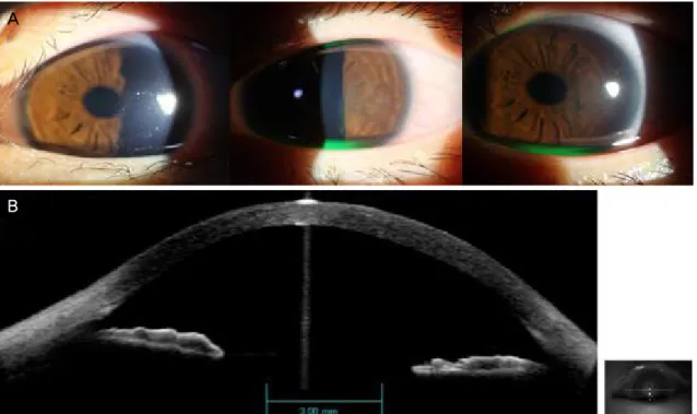
Figure 1. The images were taken on the injury day. On slit examination, corneal edema could be visualized (A). Anterior segment optical coherence tomography revealing significant corneal edema (thickness: 1,580 μm), no evidence of Descemet’s membrane de- tachment was observed (B).
해 보고하고자 한다. 본 증례는 한길안과병원 임상시험심 사위원회와 윤리위원회의 심사를 통과하였다.
증례보고
15세 여자 환자가 타 병원에서 상안검성형술을 위해 좌 안 상안검에 1% 리도카인과 1:10만 에피네프린 국소마취 제를 주입하는 시도 중에 눈을 뜬 상태로 바늘로 각막을 찌 른 뒤 갑자기 발생한 통증과 급격한 시력저하를 호소하여 술기를 중단하고 본원으로 전원되었다. 내과적 과거력이 없는 환아로 초진 당시 교정시력 우안 1.0, 좌안 안전수지 로 측정되었다. 세극등현미경검사상 각막부종이 관찰되었 으며, 전반적인 각막부종이 매우 심하여 전방 내 세부 구조 는 제대로 관찰되지 않았으나 다른 이상 소견은 발견되지 않았고, Seidel test는 음성이었다. 내원 당일 시행한 각막단 층촬영(Visante Anterior Segment OCT, Carl Zeiss Meditec, Dublin, CA, USA)검사상 각막중심두께는 1,580 μm였고 데스메막 분리 소견은 관찰되지 않았다(Fig. 1). 검사 결과 만으로 데스메막 분리 소견을 완전히 배제할 수는 없었으 나 일단 각막부종의 호전을 위해 5% NaCl 점안액 4회, 1%
프레드니솔론아세테이트 점안액(Predforte, Allergan, Inc.,
기로 하였다.
다음날 각막부종은 매우 호전되었으며 각막단층촬영검 사상 각막중심두께는 660 μm로 줄었고 역시 데스메막 분 리 소견은 관찰되지 않았다(Fig. 2). 그러나 각막상피 결손 이 관찰되어 증상 호전을 위해 각막보호렌즈를 착용하고 항생제 점안액을 추가하였으며, 5% NaCl 점안액은 4회로 유지하고 프레드니솔론아세테이트를 4회로 줄였다.
수상 후 6일째 세극등현미경검사상 각막부종은 호전양상 을 보였고 각막상피 결손은 모두 호전되었다. 각막단층촬 영검사상 각막중심두께는 560 μm로 줄었고 나안시력은 1.0으로 측정되었으며, 중심부 각막내피세포 수는 3,206개 로 측정되었다. 수상 후 10일째 나안시력은 1.0으로 측정되 었으며 이측 부분의 경미한 각막부종 외에 모두 호전 소견 을 보였다(Fig. 3A).
수상 후 3주째 각막부종은 없었고 이측 주변부에 아주 경미한 혼탁이 관찰되었으나 나안시력은 1.0으로 측정되었 고 다른 주관적 불편감은 없었다. 중심부 각막내피세포 수 는 3,098개로 측정되었으며 인공눈물 외에 모든 안약은 중 단하였다(Fig. 3B). 수상 후 2달째 나안시력은 1.0으로 측정 되었으며, 중심부 각막내피세포 수는 2,984개로 측정되었 다(Fig. 3C).
A
B
Figure 2. The images were taken on the next day after injury. On slit lamp examination, corneal edema was significantly improved and small corneal abrasion was visualized on temporal side of it (A). Anterior segment optical coherence tomography showing sig- nificantly improved corneal edema (thickness: 660 μm) and still no evidence of Descemet’s membrane detachment (B).
A
B
C
Figure 3. The images were taken at 1 week (A), 3 weeks (B) and 2 months (C) after the injury. Corneal edema was resolved (A) and slight corneal haziness was observed at injury site (B, C).
고 찰
교과서적으로 성형안과 시술 시에는 각막보호를 위해 shield를 시술 전에 착용하도록 하고 있으나 성형외과에서 는 일반적으로 시행하지 않고 있으며, 국내 문헌상에는 다 래끼 시술 중 발생한 각막기질 내 리도카인 주사로 인한 데 스메막 분리를 동반한 각막부종에 대한 증례 보고가 있었 다.1 해당 증례에서는 수상 후 25일째에 전방 내 가스주입 술을 시행하였고, 수상 후 4달째에 나안시력 1.0, 각막부종 이 모두 호전된 것이 관찰되었다. 다만 각막내피세포에 대 한 언급은 없었다.
다행히 본 증례에서는 데스메막 분리가 관찰되지 않았으 며, 이로 인해 부종이 빠른 시일 내로 호전된 것으로 추정 된다. 초기 각막부종이 심할 경우 세극등현미경뿐 아니라 각막단층촬영에서도 데스메막 분리 여부를 정확히 알기 어 려울 수 있으며, 심한 경우 파열까지도 생각해보아야 한다.
본 증례에서는 첫 내원 당시 심한 각막부종으로 데스메막 분리 여부를 완전히 배제할 수 없었으나 추후 검사상 데스 메막 분리가 없었고 각막부종이 다음날부터 급격히 호전되 는 것을 관찰하였다. 따라서 본 증례를 통해 심한 각막부종 이 있을 경우 초기에 경과 관찰기간을 줄여 각막부종이 빠 르게 호전되는지 여부를 확인하는 것이 좋을 것으로 생각 된다.
는 것을 유발하며, 동물 실험을 통해 전방 내 2% 리도카인 주입은 각막내피세포에 독성이 있고, 염화벤잘코늄의 경우 비가역적인 각막부종을 야기한다고 알려져 있다.2-4 한 보고 에서는 72세 환자에서 안검성형술을 위해 시행한 2% 리도 카인과 0.5% bupivacaine 국소마취 주사가 전방 천공 없이 각막기질 내에 유입된 경우, 수상 후 6주째 시력 0.7, 각막 부종두께 550 μm로 임상경과는 호전되었으나 각막내피세 포가 반대 안에 비해 mm2당 450개 정도 적어져 있는 소견 이 관찰되었다.5
그러나 1% 리도카인의 경우 높은 농도의 리도카인에 노 출된 것과는 달리 각막내피세포 변화는 1% 리도카인과 대 조군의 큰 차이가 없었다는 동물 실험과 백내장수술 시 1%
리도카인을 전방 내에 주입한 경우 각막내피세포 수와 모 양은 대조군과 큰 차이가 없으며 안전한 마취 방법으로 소 개되는 연구들이 다수 있다.6-9 본 증례에서는 1% 리도카인 과 1:10만 에피네프린이 각막기질 내에 주입되었을 때 수 상 후 2달째까지 수상 안과 비수상 안의 각막내피세포 수 와 내피세포 모양(hexagonality)에는 차이가 없어 이로 인 한 내피세포 영향은 없는 것으로 관찰되었다.
이는 수상 당시 각막 내 정확한 주사 깊이를 알기는 어려 우나 다음날 각막상피 결손이 관찰되고 데스메막 분리가 관찰되지 않는 것으로 보아 상대적으로 전부 기질쪽에 주 사가 유입되었을 것으로 생각이 되며, 이전 증례 보고에 비 하여 상대적으로 낮은 농도인 1% 리도카인의 각막기질 내 주입 때문으로도 추정해 볼 수 있다. 또한 환자가 15세라는 젊은 나이로 인해 회복 속도가 더 빨랐을 가능성도 있다.
상안검성형술은 성형안과뿐 아니라 성형외과에서도 보 편적으로 시행하는 시술로서 본 증례와 같이 국소마취 합 병증으로 인해 생긴 각막부종의 경과에 대해서는 잘 알려
있어서도 주의를 기울여 시술을 하여야 하며, 소아 환자나 협조가 어려운 경우는 각막 보호를 위한 shield를 시술 전 착용하도록 하는 것이 안전할 것으로 사료된다.
REFERENCES
1) Kim BR, Park SY, Lee HK, et al. A case of Descemet’s membrane detachment during lidocaine injection for hordeolum incision and drainage. J Korean Ophthalmol Soc 2016;57:1790-4.
2) Schellini SA, Creppe MC, Gregório EA, Padovani CR. Lidocaine effects on corneal endothelial cell ultrastructure. Vet Ophthalmol 2007;10:239-44.
3) Rosenwasser GO. Complications of topical ocular anesthetics. Int Ophthalmol Clin 1989;29:153-8.
4) Britton B, Hervey R, Kasten K, et al. Intraocular irritation evalua- tion of benzalkonium chloride in rabbits. Ophthalmic Surg 1976;7:46-55.
5) Ghosh S, Mukhopadhyay S, Mukhopadhyay S, et al. Inadvertent intracorneal injection of local anesthetic during lid surgery. Cornea 2010;29:701-2.
6) Eggeling P, Pleyer U, Hartmann C, Rieck PW. Corneal endothelial toxicity of different lidocaine concentrations. J Cataract Refract Surg 2000;26:1403-8.
7) Heuermann T, Hartmann C, Anders N. Long-term endothelial cell loss after phacoemulsification: peribulbar anesthesia versus intra- cameral lidocaine 1%: prospective randomized clinical trial. J Cataract Refract Surg 2002;28:639-43.
8) Chuang LH, Yeung L, Ku WC, et al. Safety and efficacy of topical anesthesia combined with a lower concentration of intracameral li- docaine in phacoemulsification: paired human eye study. J Cataract Refract Surg 2007;33:293-6.
9) Ezra DG, Allan BD. Topical anaesthesia alone versus topical an- aesthesia with intracameral lidocaine for phacoemulsification.
Cochrane Database Syst Rev 2007;18:CD005276.
= 국문초록 =
성형수술 중 각막기질 내 리도카인 국소마취제 주입으로 인해 생긴 각막부종
목적: 성형외과 수술 중 국소마취제의 각막기질 주입으로 발생한 각막부종 1예의 보고하고자 한다.
증례요약: 15세 여자 환자가 타원에서 상안검성형술을 위하여 좌안 상안검 리도카인 국소마취제 주입 시도 시 발생한 시력저하와 통증 을 주소로 내원하였다. 초진 당시 시력은 안전수동이었으며, 세극등현미경검사에서 각막부종이 심하여 전방 내 세부 구조가 제대로 관찰되지 않았으나, Seidel test 음성이었다. 같은 날 시행한 각막단층촬영검사에서 각막중심두께는 1,580 μm였고, 데스메막 분리는 관찰되지 않아 5% NaCl, 프레드니솔론아세테이트를 점안하였다. 다음날 각막부종은 매우 호전되어 각막중심두께는 660 μm로 줄었 으며, 여전히 데스메막 분리는 관찰되지 않았다. 수상 후 6일째에는 나안시력 1.0, 각막중심두께 560 μm, 각막내피세포 수는 3,206개 로 측정되었으며, 수상 후 2달째 주변부 경미한 각막혼탁도 호전되었다.
결론: 데스메막 분리가 없는 각막부종은 전방내 가스주입술 등 시술 없이 호전될 수 있고, 추후 각막내피세포와 혼탁 여부에 대한 경과 관찰이 필요하나 상대적으로 앞쪽 기질층 부종으로 추정되는 경우에는 특별한 합병증 없이 회복되었음을 보고하는 바이다.
<대한안과학회지 2019;60(10):994-998>
전성연 / Sung Yeon Jun
한길안과병원 Hangil Eye Hospital