An assessment of template-guided implant
surgery in terms of accuracy and related factors
Jee-Ho Lee1, BS, DDS, MSD, PhD, Ji-Man Park2, DDS, MSD, PhD, Soung-Min Kim3, DDS, MSD, PhD, Myung-Joo Kim4, DDS, MSD, PhD, Jong-Ho Lee3, DDS, MSD, PhD, Myung-Jin Kim3*, DDS, MSD, PhD
1Department of Oral and Maxillofacial Surgery, Seoul Asan Medical Center, Seoul, Republic of Korea
2Department of Prosthodontics, Ewha Womans University, School of Medicine, Seoul, Republic of Korea
3Department of Oral and Maxillofacial Surgery, Seoul National University Dental Hospital, Seoul, Republic of Korea
4Department of Prosthodontics, Seoul National University Dental Hospital, Seoul, Republic of Korea
PURPOSE. Template-guided implant therapy has developed hand-in-hand with computed tomography (CT) to improve the accuracy of implant surgery and future prosthodontic treatment. In our present study, the accuracy and causative factors for computer-assisted implant surgery were assessed to further validate the stable clinical application of this technique. MATERIALS AND METHODS. A total of 102 implants in 48 patients were included in this study. Implant surgery was performed with a stereolithographic template. Pre- and post-operative CTs were used to compare the planned and placed implants. Accuracy and related factors were statistically analyzed with the Spearman correlation method and the linear mixed model. Differences were considered to be
statistically significant at P≤.05. RESULTS. The mean errors of computer-assisted implant surgery were 1.09 mm at the coronal center, 1.56 mm at the apical center, and the axis deviation was 3.80°. The coronal and apical errors of the implants were found to be strongly correlated. The errors developed at the coronal center were magnified at the apical center by the fixture length. The case of anterior edentulous area and longer fixtures affected the accuracy of the implant template. CONCLUSION. The control of errors at the coronal center and stabilization of the anterior part of the template are needed for safe implant surgery and future prosthodontic treatment. [J Adv Prosthodont 2013;5:440-7]
KEY WORDS: Computer-assisted surgery; Dental implant; Accuracy
INTRODUCTION
Dental implant therapy has improved for the biomechanical
restoration of function and the esthetic appearance of edentulous patients.1,2 For successful implant therapy, prosthodontic and anatomical considerations should be included at the time of treatment planning. Panorama imag- es have been widely used for many years to aid treatment planning. However, some limitations of panorama images are evident when assessing anatomical images, such as those of the inferior alveolar nerve and maxillary sinus floor, as well as when determining the path of implant fix- tures for subsequent prosthodontic treatment. Computed tomography (CT) can overcome these faults and enables a stereoscopic approach to prosthodontic treatment.3 Three- dimensional (3D) reformatted CT images have the potential to aid the spatial analysis of anatomical objects. For implant dentistry, CT imaging began to be actively applied in the 2000s through the use of 3D image-based guidance sys- tems. Fortin et al.4 suggested the following classification of
Corresponding author:
Myung-Jin Kim
Department of Oral and Maxillofacial Surgery, School of Dentistry, Seoul National University, 275-1, Yeongeon-dong, Jongno-gu, Seoul, 110-768 Republic of Korea
Tel. 82220722631: e-mail, omsljh@gmail.com
Received May 7, 2013 / Last Revision October 18, 2013 / Accepted October 21, 2013
© 2013 The Korean Academy of Prosthodontics
This is an Open Access article distributed under the terms of the Creative Commons Attribution Non-Commercial License (http://creativecommons.
org/licenses/by-nc/3.0) which permits unrestricted non-commercial use, distribution, and reproduction in any medium, provided the original work is properly cited.
This research was supported by Basic Science Research Program through the
guide template; and active systems, which use robotic con- sole that is operated by surgeons.
Surgical templates greatly assist implant surgery, provid- ing prosthodontic considerations before surgery, guiding the drilling procedure and preventing unexpected surgical complications. Conventional surgical templates used to be made on a dental study model with resin, but anatomical information was not directly transmitted to surgical tem- plates.5 Stereolithographic templates, recently developed, have begun to be used in implant therapy. Treatment plan- ning, established via computer software, can be transmitted to the fabricating machine that creates the stereolithograph- ic templates.6 These surgical templates can also provide additional information on the accurate location of anatomi- cal structures and the bone quality under the oral mucosa, something that conventional templates cannot currently do.
However, stereolithographic template errors between the planned and the placed implants need to be minimized for the correct clinical application. The accuracy of the sur- gical template at surgery should be considered when per- forming dental implant therapy. Some studies that examine general template errors to evaluate surgical implant tem- plates have been carried out, with several studies suggesting the causative factors that affect implant template errors to be the positioning of the template in the oral cavity, the type of guide fixation, the rotational allowance of the drill in the tube, and limited mouth opening.7-13 Although the accuracy of the template has been standardized through previous studies, few evaluated the correlation between the errors and the suggested causative factors.5,14,15 The present template accuracy should be improved upon for a future wider use in implant therapy. Therefore, the suggested causative factors need to be evaluated and minimized.
The purpose of the present study was to evaluate the accuracy of computer-assisted template surgery for implant therapy and to study the related factors that affect accuracy so as to support the further clinical application of the tech- nique.
MATERIALS AND METHODS
Forty-eight patients who had received implant therapy with template-guided surgery from 2009 to 2012 in three insti- tutes (two dental hospitals and a department of dentistry in one general hospital) were included in this study. The tem- plate supporting type (mucosa supported or tooth support- ed), maxillary or mandibular arch, anterior-posterior (AP) location (anterior, premolar, molar), the length of the implants, and the institute where the procedure was per- formed were considered as error-related causative factors (Table 1).
A total of 102 fixtures were implanted. These were the Osstem US II (Osstem Implant, Seoul, Korea), Superline (Dentium, Seoul, Korea), and Branemark MKIII Groovy (Nobel Biocare, Kloten, Switzerland). Fixtures were select- ed according to prosthodontic options and patient choice.
Preoperative 3D CT was taken for the fabrication of the stereolithographic template with the computer software (OnDemand3D; Cybermed Co., Seoul, Korea). A postoper- ative 3D CT was taken within 2 months after the computer- assisted implant surgery for comparison. Patients who had surgical complications, such as infection or early implant failure, were excluded from the study. The study protocol was reviewed by the Institutional Review Boards of the participating institutes. All patients were given the protocol information and informed consent was obtained.
The dental study models were made after taking the arch impressions of the patient. According to the prosth- odontic plan, diagnostic wax-up was performed and the preliminary position of the implant fixture was determined.
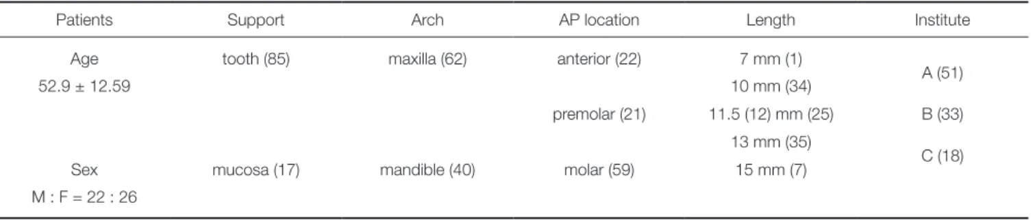
The dental study models were scanned and matched with the preoperative CT images for simulation surgery by the computer software (OnDemand3D) and the drill sequence, size of fixture, and the location were determined according to the prospective prosthodontic consideration (Fig. 1).
Three-dimensional stereolithographic models were printed out through surface registration and model scan-
Table 1. Patients and causative factors
Patients Support Arch AP location Length Institute
Age tooth (85) maxilla (62) anterior (22) 7 mm (1)
A (51)
52.9 ± 12.59 10 mm (34)
premolar (21) 11.5 (12) mm (25) B (33) 13 mm (35)
C (18)
Sex mucosa (17) mandible (40) molar (59) 15 mm (7)
M : F = 22 : 26
AP location: anterior-posterior location of implants (anterior, premolar, molar).
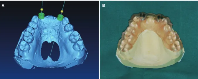
ning. Information from the simulation surgery permitted the creation of the stereolithographic template for the guided implant surgery with a three dimensional printer (Connex350®3D printing system; Object Geometries Inc., Billerica, MA, USA). Metal sleeves for drill guidance were attached to the template (Fig. 2).
Routine preparation of the surgical field and adminis-
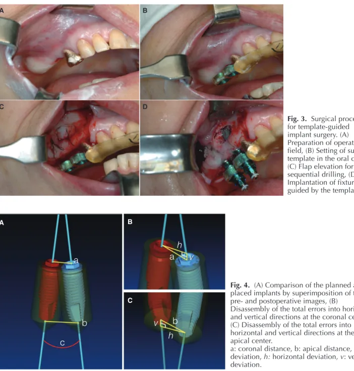
mucoperiosteal flap was elevated. The surgical template was adapted in the patient’s oral cavity for the planned sequen- tial drilling. Fixtures were inserted through metal sleeves, after being locked at connecting implant mounters (Fig. 3).
Cover screws were engaged and suturing was performed without tension. The tooth-supported (partially edentulous) type of template acquired retention from the residual teeth.
Fig. 1. The location and angulation of the implant were determined through the use of planning software and virtual surgery by considering the critical anatomical structures and the future dental prostheses.
Fig. 2. The information from the planned fixture was used to create the stereolithographic implant template.
(A) Completion of planning of implant surgery on 3D software, (B) Stereolithographic template for computer-assisted implant surgery.
A B
Antibiotics and analgesics were prescribed after surgery.
Patients were periodically checked for wound care, followed by prosthodontic treatment.
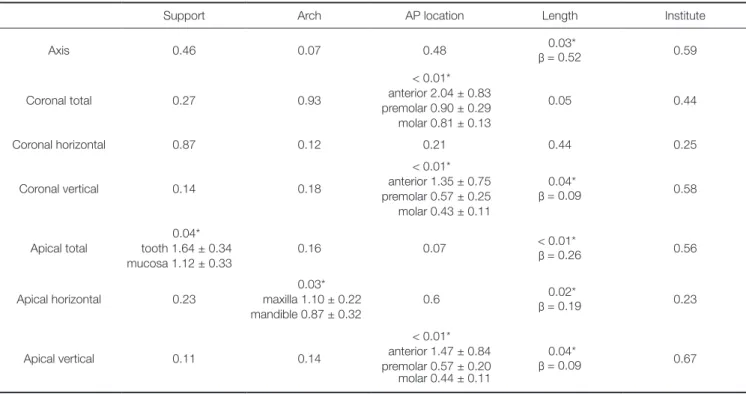
Pre- and postoperative CT images were loaded and the two serial images were fused through the use of a maxi- mum mutual information (MMI) algorithm on the comput- er program (OnDemand3D).16 The planned and placed implants from the same patients were superimposed and the errors were calculated. The coronal distances, apical dis- tances, and axis deviations between the two implants were compared, and the linear distances (total errors) of the cor- onal and apical centers were disassembled into horizontal and vertical deviations (Fig. 4).
The mean values of each measure were calculated and correlations were assessed with each causative factor: tem- plate supporting type, maxillary or mandibular arch, AP location (anterior, premolar, molar), length of implants, and the institutes involved.
The mean errors of the coronal and apical distances and the axis deviations were calculated and their correla- tions were analyzed with the Spearman correlation method.
Correlations of errors with causative factors were analyzed with the linear mixed model and each factor variable was compared. The statistical analyses were performed with SPSS (version 20.0 for Windows; SPSS, Chicago, IL, USA).
Differences were considered to be statistically significant at P≤.05.
Fig. 3. Surgical procedure for template-guided implant surgery. (A) Preparation of operation field, (B) Setting of surgical template in the oral cavity, (C) Flap elevation for sequential drilling, (D) Implantation of fixture guided by the template.
A B
C D
Fig. 4. (A) Comparison of the planned and placed implants by superimposition of the pre- and postoperative images, (B)
Disassembly of the total errors into horizontal and vertical directions at the coronal center, (C) Disassembly of the total errors into horizontal and vertical directions at the apical center.
a: coronal distance, b: apical distance, c: axis deviation, h: horizontal deviation, v: vertical deviation.
A B
C
a a
b c
h v
v b
h
RESULTS
The mean errors between the planned and placed implants were 1.09 ± 1.10 mm at the coronal center, 1.56 ± 1.48 mm at the apical center, and 3.80 ± 3.24° in the axis. The hori- zontal deviation was 0.72 ± 0.75 mm at the coronal center and 1.23 ± 1.25 mm at the apical center. The vertical devia- tion was 0.66 ± 0.95 mm at the coronal center and 0.69 ± 1.03 mm at the apical center (Table 2). The total errors at the coronal and apical centers were strongly correlated with their horizontal deviations (r = 0.83 and r = 0.89, respec- tively). Vertical deviations were also strongly correlated between the coronal and apical centers (r = 0.92) (Table 3).
The difference in the template supporting type was sig- nificant for the total error at the apical center of the implants. Only the horizontal deviation of the implant apex of the mandibular arch was less than that of the maxillary arch. Significant errors were found according to the AP location of the implants. These errors were larger in the anterior area than in the premolar and molar areas in the coronal total and vertical, and the apical vertical dimen- sions. The errors were all increased in proportion to the length of the implants, except in the coronal total and hori- zontal dimension. The institutes where the procedures were performed did not affect the accuracy of the template (Table 4).
Table 2. Mean errors between the planned and placed implants (Mean ± SD)
Coronal (mm) Apical (mm) Axis (°) Total 1.09 ± 1.10 1.56 ± 1.48
3.80 ± 3.24 Horizontal 0.72 ± 0.75 1.23 ± 1.25
Vertical 0.66 ± 0.95 0.69 ± 1.03 SD means standard deviation.
Table 3. Correlations between errors
Correlation of variables r P value
Coronal total Apical total 0.65 < .01
Coronal horizontal Apical horizontal 0.52 < .01 Coronal vertical Apical vertical 0.92* < .01 Coronal total Coronal horizontal 0.83* < .01 Coronal total Coronal vertical 0.66 < .01 Apical total Apical horizontal 0.89* < .01 Apical total Apical vertical 0.42 < .01
*When the correlation coefficient (r) was between 0.7 and 1.0, the items were considered to be strongly correlated.
Table 4. Significance (P value) of each error that correlated with causative factors for implant fixtures
Support Arch AP location Length Institute
Axis 0.46 0.07 0.48 0.03*
β = 0.52 0.59
Coronal total 0.27 0.93
< 0.01*
anterior 2.04 ± 0.83 premolar 0.90 ± 0.29 molar 0.81 ± 0.13
0.05 0.44
Coronal horizontal 0.87 0.12 0.21 0.44 0.25
Coronal vertical 0.14 0.18
< 0.01*
anterior 1.35 ± 0.75 premolar 0.57 ± 0.25
molar 0.43 ± 0.11
0.04*
β = 0.09 0.58
Apical total
0.04*
tooth 1.64 ± 0.34 mucosa 1.12 ± 0.33
0.16 0.07 < 0.01*
β = 0.26 0.56
Apical horizontal 0.23
0.03*
maxilla 1.10 ± 0.22 mandible 0.87 ± 0.32
0.6 0.02*
β = 0.19 0.23
Apical vertical 0.11 0.14
< 0.01*
anterior 1.47 ± 0.84 premolar 0.57 ± 0.20 molar 0.44 ± 0.11
0.04*
β = 0.09 0.67
*Considered significant at P<.05. β means increasing amount of error according to one millimeter increase of length of fixture.
When P value was less than .05, mean errors in each causative factor were presented as mean (mm) ± C.I (95% confidence interval).
Support: the type of template supporting (tooth-supported and mucosa-supported), Arch: the arch on which implant template was positioned (maxilla and mandibular
DISCUSSION
CT imaging is a very helpful tool for implant surgery in the case of an unfavorable alveolar bone condition.5 Conven- tional surgical templates used to be made with resin on dental study models to guide the planned positions of implant fixtures during the surgical procedure. Due to the development of the stereolithographic modeling technique, surgical templates can be designed with accuracy and repro- ducibility through 3D imaging of a patient’s maxilla and mandible. The templates also contain information about the planned location and direction of implant fixtures when planning the prosthodontic treatment. Clinicians can there- fore establish and carry out a safer treatment plan in the actual treatment of the patients.17
Template-guided implant surgery can improve accuracy and reduce the need for additional bone grafts in atrophied alveolar ridge. However, errors between the planned and placed implant appeared in all systems of template-guided implant surgery, which caused various deviations.8,13,18 Studies that evaluated template-guided implants have been performed for decades but few studies have dealt with the errors of template-guided implant surgery in terms of causative factors that should be resolved for future wide- spread clinical use of the technique.19
Sarment et al.20 and Kramer et al.21 found that computer- guided surgical templates were more accurate than conven- tional surgical templates, but these studies were performed in vitro, with the in vivo studies showing poorer accuracy.14,15 Cassetta et al.8 suggested that the potential mechanical (intrinsic) error is a more significant factor than the other variables that could affect the accuracy of template guided systems. The intrinsic errors affecting the accuracy were caused by the diameter and length of the guide sleeve of the template and the distance between the undersurface of the template and the targeted alveolar crest.22 The potential intrinsic error developing from the sleeve unit was pointed out by Vrielinck et al.15 to be caused by the fact that the diameter of the sleeve was slightly larger than that of the drill. However, mechanical errors were found to be relative- ly smaller than the errors that occurred in the clinical pro- cedure,14,15 and can be controllable in the fabrication phase.
Most studies that have investigated template-guided implant surgery showed similar levels of accuracy.
Schneider et al.23 conducted a meta-regression analysis of the systematic review of studies in the accuracy of various surgical templates. In that study, the mean error was 1.07 mm at the coronal center and 1.63 mm at the apical center and the axis deviation was 5.26°. In another systematic review performed by Van Assche et al.,24 the mean error was 0.99 mm at the coronal center, 1.24 mm at the apical center, and the axis deviation was 3.81°. In our present study, the errors at the coronal and apical centers and direc- tion were also analyzed; the mean values were 1.09 mm at the coronal center, 1.56 mm at the apical center, and 3.80°
in the axis deviation. Thus, our present data are similar to those of the previously presented systematic reviews.
However, some outliers appeared due to severe errors between the planned and placed fixtures. These errors were enough to cause complications in the case of complex ana- tomical structures, such as the maxillary sinus and inferior alveolar nerve. Such cases should be considered by clini- cians so that they use CT to become well informed about critical anatomical structures rather than depend on com- puter-guided surgical templates alone. Further effort should be exerted to decrease the deviation of errors among the cases as well as the errors between the planned and the placed fixtures.
Strong correlations were shown between the coronal vertical and apical vertical, coronal total and coronal hori- zontal, and apical total and apical horizontal dimensions (Table 3). The vertical errors changed simultaneously in both the coronal and apical centers. The implant deviation between the planned and placed implants was more affect- ed by horizontal deviations than by vertical deviations.
Errors in horizontal deviation can limit prospective prosth- odontic placement, whereas errors in vertical depth can cause damage to anatomical structures such as the inferior alveolar nerve and floor of the maxillary sinus. The control of errors in the surgical procedure should be focused on securing the prospective placement of dental prostheses by decreasing horizontal deviation as well as protecting ana- tomical structures. The errors increased in proportion to the length of the implant fixtures except in the coronal total and horizontal dimensions, which implies that errors developed at the coronal center would be magnified at the apical center by the fixture length.9 The fundamenal errors occurred in the coronal portion and all efforts to decrease errors during surgery should be exerted in the coronal point.
Ozan et al.5 compared the accuracy of three different types of computer-guided implant templates, which were the tooth-supported, bone-supported, and mucosa-sup- ported types, and suggested that the tooth-supported type were more accurate. In the present study, although mucosa- supported templates had less error than the tooth-support- ed type (1.12 mm vs. 1.64 mm, respectively) in the apical total dimension, significant differences were not found in the other dimensions. When comparing the arch of the maxilla and mandible, a difference was only found in the horizontal deviation of the apical center (maxilla = 1.10 mm, mandible = 0.87 mm). The template supporting type and a maxillary or mandibular arch did not largely affect the accuracy-at least in this study-but a relative magnification of the horizontal deviation was found at the apical center.
It may be therefore conceded that the template-guided implant surgery can be performed stably regardless of sup- porting type and arch but, the control of errors at the coro- nal level is needed to decrease the error at the apical center for the protection of anatomical structures in the surgical procedure.
With respect to the anterior-posterior locations of implants, the coronal total, coronal vertical, and apical ver- tical dimensions showed significant differences, and the
anterior area (incisor and canine) was more deviated than the premolar and molar areas. The importance of accurate positioning of implant fixtures in anterior teeth cannot be too highly emphasized for esthetic considerations. Vertical errors in the anterior area are thought to be due, in particu- lar, to the metal sleeve housing parts, which, in the case of anterior edentulous ridge, should be stabilized to control the vertical movement.
Template-guided implant surgery was intended to improve both the accuracy of prospective prosthodontic treatment and the safety of implant surgery. In our present study, accuracy did not differ between the different insti- tutes. Template-guided implant surgery was thus thought to be performed with stable errors, regardless of different environments and surgeons.
The causative factors related to accuracy of surgical template during surgery were suggested by several studies.
In the present study, the related factor; template supporting type, maxillary or mandibular arch, anterior-posterior loca- tion, length of implants and clinicians (different institutes) were considered for clinical implication. Template-guided surgery system was stable related to suggested causative factors in this study. But, in the case of anterior edentulous ridge and drilling control at the coronal part, special pre- caution was needed for clinicians.
This retrospective study had some limitations, such as too few and unevenly distributed cases to evaluate the caus- ative factors. For a more reliable evaluation of template- guided surgery, further studies, based on a more suitable prospective study design, are needed.
CONCLUSION
The stereolithographic template-guided implant surgery in the present study had errors of 1.09 mm at the coronal center, 1.56 mm at the apical center, and 3.80° in axis devia- tion. Controlling the accuracy in horizontal deviation at the coronal center and ensuring template stabilization in the case of anterior edentulous areas should be considered for safe implant surgery and prospective prosthodontic treat- ment.
ACKNOWLEDGEMENTS
The authors would like to thank Min-Ju Kim, Biostatistician in Asan Biomedical Research Center, for her support in sta- tistical data analysis.
REFERENCES
1. Besimo C, Lambrecht JT, Nidecker A. Dental implant treat- ment planning with reformatted computed tomography.
Dentomaxillofac Radiol 1995;24:264-7.
2. Fortin T, Coudert JL, Champleboux G, Sautot P, Lavallée S.
Computer-assisted dental implant surgery using computed
Van Steenberghe D. Predictability of reformatted computed tomography for pre-operative planning of endosseous im- plants. Dentomaxillofac Radiol 1999;28:37-41.
4. Fortin T, Champleboux G, Bianchi S, Buatois H, Coudert JL.
Precision of transfer of preoperative planning for oral im- plants based on cone-beam CT-scan images through a robot- ic drilling machine. Clin Oral Implants Res 2002;13:651-6.
5. Ozan O, Turkyilmaz I, Ersoy AE, McGlumphy EA, Rosenstiel SF. Clinical accuracy of 3 different types of com- puted tomography-derived stereolithographic surgical guides in implant placement. J Oral Maxillofac Surg 2009;67:394- 401.
6. Jacobs R, Adriansens A, Verstreken K, Suetens P, van Steen- berghe D. Predictability of a three-dimensional planning sys- tem for oral implant surgery. Dentomaxillofac Radiol 1999;
28:105-11.
7. Arisan V, Karabuda ZC, Ozdemir T. Accuracy of two stereo- lithographic guide systems for computer-aided implant place- ment: a computed tomography-based clinical comparative study. J Periodontol 2010;81:43-51.
8. Cassetta M, Di Mambro A, Giansanti M, Stefanelli LV, Cavallini C. The intrinsic error of a stereolithographic surgi- cal template in implant guided surgery. Int J Oral Maxillofac Surg 2013;42:264-75.
9. D’haese J, Van De Velde T, Elaut L, De Bruyn H. A prospec- tive study on the accuracy of mucosally supported stereo- lithographic surgical guides in fully edentulous maxillae. Clin Implant Dent Relat Res 2012;14:293-303.
10. Park C, Raigrodski AJ, Rosen J, Spiekerman C, London RM.
Accuracy of implant placement using precision surgical guides with varying occlusogingival heights: an in vitro study.
J Prosthet Dent 2009;101:372-81.
11. Stumpel LJ. Deformation of stereolithographically produced surgical guides: an observational case series report. Clin Implant Dent Relat Res 2012;14:442-53.
12. Valente F, Schiroli G, Sbrenna A. Accuracy of computer-aid- ed oral implant surgery: a clinical and radiographic study. Int J Oral Maxillofac Implants 2009;24:234-42.
13. Vercruyssen M, Jacobs R, Van Assche N, van Steenberghe D.
The use of CT scan based planning for oral rehabilitation by means of implants and its transfer to the surgical field: a crit- ical review on accuracy. J Oral Rehabil 2008;35:454-74.
14. Di Giacomo GA, Cury PR, de Araujo NS, Sendyk WR, Sendyk CL. Clinical application of stereolithographic surgical guides for implant placement: preliminary results. J Periodontol 2005;76:503-7.
15. Vrielinck L, Politis C, Schepers S, Pauwels M, Naert I. Image- based planning and clinical validation of zygoma and ptery- goid implant placement in patients with severe bone atrophy using customized drill guides. Preliminary results from a pro- spective clinical follow-up study. Int J Oral Maxillofac Surg 2003;32:7-14.
16. Lee JH, Kim MJ, Kim SM, Kwon OH, Kim YK. The 3D CT superimposition method using image fusion based on the maximum mutual information algorithm for the assessment
17. Verstreken K, Van Cleynenbreugel J, Marchal G, Naert I, Suetens P, van Steenberghe D. Computer-assisted planning of oral implant surgery: a three-dimensional approach. Int J Oral Maxillofac Implants 1996;11:806-10.
18. Fortin T, Camby E, Alik M, Isidori M, Bouchet H. Panoramic images versus three-dimensional planning software for oral implant planning in atrophied posterior maxillary: a clinical radiological study. Clin Implant Dent Relat Res 2013;15:198- 204.
19. Cassetta M, Stefanelli LV, Giansanti M, Calasso S. Accuracy of implant placement with a stereolithographic surgical tem- plate. Int J Oral Maxillofac Implants 2012;27:655-63.
20. Sarment DP, Sukovic P, Clinthorne N. Accuracy of implant placement with a stereolithographic surgical guide. Int J Oral Maxillofac Implants 2003;18:571-7.
21. Kramer FJ, Baethge C, Swennen G, Rosahl S. Navigated vs.
conventional implant insertion for maxillary single tooth re- placement. Clin Oral Implants Res 2005;16:60-8.
22. Choi M, Romberg E, Driscoll CF. Effects of varied dimen- sions of surgical guides on implant angulations. J Prosthet Dent 2004;92:463-9.
23. Schneider D, Marquardt P, Zwahlen M, Jung RE. A systemat- ic review on the accuracy and the clinical outcome of com- puter-guided template-based implant dentistry. Clin Oral Implants Res 2009;20:73-86.
24. Van Assche N, Vercruyssen M, Coucke W, Teughels W, Jacobs R, Quirynen M. Accuracy of computer-aided implant placement. Clin Oral Implants Res 2012;23:112-23.