Journal of Bacteriology and Virology 2006. Vol. 36, No. 3 p.151 – 157
상피세포에 대한 Weissella cibaria 유산균의 부착력에 영향을 미치는 인자
전남대학교 의과대학 미생물학교실
강미선·MeiShan Piao·신부안·이현철·오종석
*Adhesion of Weissella cibaria to the Epithelial Cells and Factors Affecting its Adhesion
Mi-Sun Kang, MeiShan Piao, Boo-Ahn Shin, Hyun-Chul Lee and Jong-Suk Oh*
Department of Microbiology and Immunology, School of Medicine, Chonnam National University, Gwangju 501-746, Korea
Received : June 13, 2006 Accepted : July 25, 2006
We evaluated the ability of lactic acid bacteria, Weissella cibaria, isolated from the oral cavity to adhere to epithelial cells. W. cibaria efficiently adhered to KB cells and HeLa cells. In addition, W. cibaria efficiently adhered to Fusobacterium nucleatum. But the adhesiveness of W. cibaria disappeared upon exposure to LiCl or pronase, suggesting that the S-layer proteins of W. cibaria mediated the adhesiveness. The molecular mass of the S-layer proteins extracted from W. cibaria was approximately 50 kDa. When W. cibaria strains were washed with 0.45% saline, the bacteria were efficiently adhered to the epithelial cells. In conclusion, W. cibaria has the ability to adhere to epithelial cells through the S-layer proteins.
Key Words: Adhesion, Weissella cibaria, KB cell, HeLa cell, Fusobacterium nucleatum
서 론
유산균은 인간의 입, 장내, 여성의 질내 등에서 정상세균 총을 이루고 있으며 (2,21), 대부분의 유산균은 유익한 작용 을 하는 것으로 알려져 있다. 특히, 장내에 정착한 유산균은 병원성 세균이 소화관 상피에 부착하는 것을 방해하여 질병 발생을 막아주고, 유산균에 의해 생성된 항생물질이 설사를 일으키는 병원성 미생물이나 장내유해균을 죽이거나 증식을 억제하며, Lactobacillus acidophilus는 직접 콜레스테롤을 분 해한다고 보고된 바 있다 (10,11). 따라서, 최근에는 이와 같 은 프로바이오틱 유산균에 대해 활발히 연구가 진행되고 있 다 (8,9).
Weissella는 최근 DNA 기술의 발달에 따라 Lactobacillus
속에서 새로 분리된 유산균으로서 (26), Weissella cibaria는 그람양성, 비포자 형성, 비운동성, hetero 유산발효, 카탈라 아제 음성 간균이고, 자당으로부터 덱스트란을 생성할 수 있으며, 발효식품과 사람과 동물의 임상시료에서 정상적으 로 분리되는 세균이다 (3). 강산을 만드는 Lactobacillus와 같 은 유산균이 구강에서 충치의 진행을 도우나, 최근 Kang 등 (12,13)은 구강에서 분리한 W. cibaria 분리균주가 강산을 만 들지 않고 구취 및 충치를 억제하여 구강에 이로운 역할을 한다고 보고하였다.
구강에는 수백 여종의 세균이 서로 상호작용하며 살고 있으며, 이들 균들이 구강에 상주하기 위해서는 상피표면 에 부착할 수 있어야 한다. 구취유발 원인균 중의 하나인 Fusobacterium nucleatum은 구강에 다수 존재할 뿐만 아니라, 부착과 상호결합에 의해서 다른 균들이 쉽게 구강에 정착할 수 있게 하는 bridge-organism으로서 역할을 하고 있다 (16).
이렇게 상피표면에 부착할 수 있는 능력을 가진 세균들은 부착능력이 없는 세균에 비해 타액에 의해 쉽게 제거되지
151
*교신저자: 오종석. 501-746, 광주광역시 동구 학동 5번지, 전남대학교 의과대학 미생물학교실
Phone: 062-220-4134, Fax: 062-228-7294, e-mail: [email protected]
다 (4,22). 최근에는 W. cibaria가 구취원인균 중의 하나인 F.
nucleatum과 상호결합하고, 상피세포에 부착능력이 있음이 보고된 바 있다 (12). Schneitz 등 (25)은 Lactobacillus의 sur- face layer (S-layer) 단백질이 부착에 관여한다고 제안하였 으며, 이들 단백질은 세포벽의 펩티도글리칸층에 결합되어 있다고 보고되었다 (20). 단백질 단량체로 구성된 S-layer는 Lactobacillus 세포벽에 결정 구조로 배열되어 있으며, 수소 결합을 파괴하는 물질인 urea, guanidine hydrochloride, 세제인 SDS, 양이온 치환물질인 Lithium chloride (LiCl)에 의해서 추출할 수 있다고 보고되었다 (20).
따라서 본 연구에서는 W. cibaria가 상피세포 및 F. nucle- atum에 대한 부착력이 있음을 현미경을 이용하여 확인하고, 그 부착력에 영향을 미치는 인자들을 살펴보고자 하였다.
재료 및 방법
1. 공시세균 및 배양
구강이 건강한 4~5세의 유아의 타액에서 분리한 W. cibaria CMU, CMS2, CMS3 균주와 F. nucleatum ATCC 10953을 실 험에 이용하였다. W. cibaria 분리균주는 De Man, Rogosa, Sharpe broth (MRS broth, Difco, Detroit, MI, USA)에 접종하여 37℃ 호기조건에서 16시간 배양하였다. F. nucleatum은 0.5%
yeast extract (Difco), 5 µg/ml haemin (Sigma, St Louis, MO, USA), 1 µg/ml menadione (Sigma)이 포함된 brain heart infu- sion broth (BHI broth, Difco)에 접종하여 37℃ 혐기조건 (85%
N2, 10% H2, 5% CO2)에서 1일간 배양하였다. 실험에 이용하 기 전에 본 배지에서 2회 계대배양한 후 실험에 이용하였다.
2. 상피세포에 대한 부착실험
실험에 이용한 세포주는 인간의 구강상피세포인 KB 세 포 (KCLB 10017)와 자궁경부암세포인 HeLa 세포 (ATCC CCL-2)로서, 2 mM L-glutamine과 10% fetal bovine serum (FBS)이 첨가된 minimal essential medium with Earle's salts (MEM, Hyclone, Logan, Utah, USA)를 사용하여 37℃, 5%
CO2 배양기에서 배양하였다.
W. cibaria의 부착실험은 Scaletsky 등 (24)의 방법에 따라 수행하였다. 4-well Lab-Tek II chamber slide system (Nalge Nunc International, Naperville, USA)에 well당 105 세포를 18 시간 배양한 후, PBS를 이용하여 세포를 2회 세척하고 5 M LiCl를 처리한 것과 처리하지 않은 W. cibaria CMS2 (109
(BX51, Olympus, Tokyo, Japan)을 이용하여 관찰하였으며, 100개의 상피세포에 부착한 세균의 평균 개수를 측정하였다.
통계적 처리는 SPSS 통계분석 프로그램 (SPSS version 12.0) 을 사용하였으며, Mann-Whitney test로 분석하였다.
3. SDS-PAGE를 이용한 S-layer 단백질 분석
5 M LiCl 추출방법은 일반적으로 Lactobacillus의 S-layer 단백질을 선택적으로 분리하기 위한 방법으로 이용되어져 왔으며 (19), W. cibaria의 S-layer 단백질을 조사하기 위해서 이 방법을 이용하였다. W. cibaria를 회수한 후 5 M LiCl로 현탁시켜 얼음에 15분 동안 방치하였다. 원심분리 (14000 × g, 15 min, 4℃)하여 얻은 상청액을 Laemmli buffer와 혼합한 후, 10% polyacrylamide gel과 Coomassie blue를 이용하여 SDS-PAGE로 분석하였다 (17).
4. W. cibaria와 F. nucleatum간의 부착력
W. cibaria와 F. nucleatum간의 부착력은 광학현미경과 trans- mission electron microscope (TEM, H-7600, Hitachi, Nissei, Japan)를 이용하여 관찰하였다. 광학현미경은 Gram 염색하 여 관찰하였으며, TEM은 다음과 같은 방법을 수행하여 관 찰하였다. 세균의 부착력이 표면 단백질임을 확인하기 위하 여 표면 단백질을 제거하는 pronase를 처리한 것과 처리하지 않은 W. cibaria CMS2와 F. nucleatum를 각각 혼합시켜 0.1 M sodium cacodylate buffer (pH 7.2)에 녹인 2.5% glutaraldehyde 로 4℃에서 6시간 고정시킨 다음, 본 buffer로 3회 세척하였 다. Sodium cacodylate buffer (0.1 M, pH 7.2)에 녹인 2% (w/v) osmium tetroxide에 4℃에서 16시간 침지시킨 후, ethanol 용 액으로 탈수시키고 Epon에 박은 다음, ultramicrotome MT- 7000 (RMC-EM, Tucson, AZ, USA)으로 절단하였다. Uranyl acetate와 lead citrate로 염색하여 80 kV에서 TEM으로 관찰 하였다.
5. W. cibaria의 부착력에 영향을 미치는 인자
세균의 배양온도와 세균을 세척하는 식염수 농도와 같은 인자들이 W. cibaria CMS2의 세포에 대한 부착력에 미치는 영향을 알아 보았다. 4-well Lab-Tek II chamber slide system에 well당 105 KB 세포와 HeLa 세포를 각각 37℃, 5% CO2 배 양기에서 18시간 배양하였다. 세포를 2회 세척하고 30℃와 37℃에서 각각 배양한 W. cibaria CMS2 (109 bacteria/ml)를 0.9% 식염수, 0.45% 식염수, 멸균수로 각각 세척하여 접종
한 후 37℃, 5% CO2 배양기에서 10분 동안 배양한 다음, 세 포를 고정, 염색한 후, 광학현미경을 이용하여 세포에 대한 부착력을 관찰하였으며, 100개의 상피세포에 부착한 세균의 평균 개수를 측정 비교하였다. 통계적 처리는 SPSS 통계분 석 프로그램의 ANOVA로 분석하였으며 사후검정은 Duncan 을 사용하였다.
결 과
1. W. cibaria의 표면 단백질이 상피세포에 대한 부착력에 미치는 영향
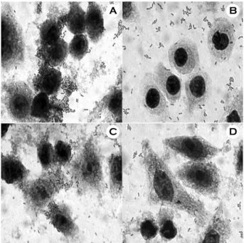
W. cibaria의 상피세포에 대한 부착실험한 결과, LiCl을 처 리하지 않은 정상균주 W. cibaria CMS2는 KB 세포와 HeLa 세포에 많이 부착하였으나, LiCl을 처리한 W. cibaria는 세 포에 부착하지 못하였다 (Fig. 1). 1개의 KB 세포에 대한 W. cibaria의 평균 부착수는 LiCl을 처리하지 않은 것이 약 101.7±12.6개인데 반하여 LiCl을 처리한 균의 부착수는 약 4.8±1.3개였으며, HeLa 세포에 대해서는 LiCl을 처리하지 않 은 W. cibaria의 부착수는 약 74.0±10.1개 인데 반하여 LiCl 을 처리한 균의 부착수는 8.2±1.8개였다 (Fig. 2). 이와 같은 결과는 부착과정에 W. cibaria의 표면 단백질이 연관이 있음 을 시사하여 주었다. 또한, LiCl를 이용하여 W. cibaria CMU,
CMS2, CMS3의 S-layer 단백질을 각각 SDS-PAGE로 분석 한 결과, 단백질의 분자량이 약 50 kDa 이었다 (Fig. 3).
2. W. cibaria의 표면 단백질이 F. nucleatum에 대한 부 착력에 미치는 영향
W. cibaria CMS2와 F. nucleatum간의 부착력을 광학현미경 및 TEM으로 검경한 결과, W. cibaria는 F. nucleatum과 결합 되어 있는 것을 확인할 수 있었다 (Fig. 4A). 또한, TEM을 이용하여 W. cibaria의 구조 및 F. nucleatum간의 부착을 관찰 한 결과, pronase를 처리하지 않은 W. cibaria는 바깥층에 3 층 구조를 이루고 있는 것을 볼 수 있었다 (Fig. 4B). Pronase Figure 2. Numbers of adhered W. cibaria CMS2 to epithelial cells. Treatment of W. cibaria CMS2 was as follows: □, control;
, 5 M LiCl. Values are expressed as means ± SD. **Signifi- cances of differences were defined as p<0.05.
Figure 1. Effect of 5 M LiCl treatment on the adherence of W.
cibaria CMS2 to epithelial cells. A, untreated W. cibaria CMS2 vs. KB cells; B, treated W. cibaria CMS2 vs. KB cells; C, untrea- ted W. cibaria CMS2 vs. HeLa cells; D, treated W. cibaria CMS2 vs. HeLa cells. Magnification, × 1000. Shown in the figures are the representative data of three separate experiments.
Figure 3. SDS-PAGE of S-layer proteins of W. cibaria strains extracted by LiCl (5 mol l-1) treatment. Lane 1, W. cibaria CMU;
lane 2, W. cibaria CMS2; lane 3, W. cibaria CMS3. Shown in the figures are the representative data of three separate experiments.
A B
C D
를 처리하지 않은 W. cibaria는 F. nucleatum과 결합하는 것 을 볼 수 있는 반면 (Fig. 4C), pronase를 처리한 W. cibaria는 부착능력이 없어짐을 확인할 수 있었다 (Fig. 4D).
3. 상피세포에 대한 W. cibaria의 부착력에 영향을 미치 는 인자
세균의 배양온도와 세균을 세척하는 식염수 농도가 KB 세포 및 HeLa 세포에 대한 부착력에 미치는 영향을 알아 본 결과, KB 세포에 대해서는 W. cibaria CMS2를 37℃에서 배 양한 후, 0.45% 식염수로 세척하였을 때 가장 부착력이 좋았 다 (Fig. 5). 1개의 KB 세포당 부착된 W. cibaria 평균 개수는 W. cibaria를 30℃에서 배양한 후, 0.9% 식염수, 0.45% 식염 수, 멸균수로 세척하였을 때 각각 16.4±5.1, 10.3±2.5, 7.7±
1.5개였으며, 37℃에서 배양한 후, 0.9% 식염수, 0.45% 식염 수, 멸균수로 세척하였을 때 각각 20.6±2.2, 125.3±9.0, 8.5±
3.1개로서, 37℃에서 배양한 후 0.45% 식염수로 세척하였을 때 W. cibaria의 부착수가 가장 많았다 (Fig. 7). 또한, HeLa 세포에 대해서는 W. cibaria CMS2를 30℃에서 배양한 후, 0.45% 식염수로 세척하였을 때 가장 부착력이 좋았으며 (Fig.
6), 1개의 HeLa 세포당 부착된 W. cibaria 평균 개수는 W.
cibaria를 30℃에서 배양한 후, 0.9% 식염수, 0.45% 식염수, 멸균수로 세척하였을 때 각각 24.3±6.0, 95.7±6.0, 12.3±3.1 개였으며, 37℃에서 배양한 후, 0.9% 식염수, 0.45% 식염수, 멸균수로 세척하였을 때 각각 34.3±6.0, 8.0±2.6, 6.4±1.5개 로서, 30℃에서 배양한 후 0.45% 식염수로 세척하였을 때 W. cibaria의 부착수가 가장 많았다 (Fig. 7). 따라서, 두 상피 세포 모두에 대해 0.45% 식염수로 세척한 W. cibaria가 부착 력이 가장 좋았으며, 배양온도에 대해서는 각각 차이를 보 임을 알 수 있었다.
고 찰
W. cibaria는 Bjorkroth 등 (3)에 의해 최근에 새로 명명되 었으며, 그리스 살라미, 스페인 소시지, 한국 김치와 같은 발효식품에서부터 동물과 인간의 분변까지 다양하게 분리되 Figure 5. Effect of incubation temperature and saline solution on the adherence of W. cibaria CMS2 to KB cells. A, bacterial cells were incubated at 30℃ and washed with 0.9% saline; B, bacterial cells were incubated at 30℃ and washed with 0.45%
saline; C, bacterial cells were incubated at 30℃ and washed with distilled water; D, bacterial cells were incubated at 37℃ and washed with 0.9% saline; E, bacterial cells were incubated at 37℃
and washed with 0.45% saline; F, bacterial cells were incubated at 37℃ and washed with distilled water. Magnification, × 1000.
Shown in the figures are the representative data of three separate experiments.
C D
A
B
C
D
E
F Figure 4. Microscopic observations of adhesion between W.
cibaria strain and F. nucleatum ATCC 10953. A, bright-field micro- graphs of W. cibaria CMS2 vs. F. nucleatum; Magnification,
× 1000; B, TEM micrographs of untreated W. cibaria CMS2 whole cell only; C, and D, TEM micrographs of adhesion between W.
cibaria CMS2 and F. nucleatum before and after pronase treat- ment, respectively. Wc represents W. cibaria; Fn represents F.
nucleatum. Shown in the figures are the representative data of three separate experiments.
었고 (3,5,6,23), 충치 유발균인 Streptococcus mutans의 치태 형성을 억제하는 W. cibaria가 사람의 타액에서 분리되었다 (12). W. cibaria가 생성하는 수용성 중합체는 S. mutans의 glu- cosyltransferase (GTF) 작용을 전환시켜 비수용성 글루캔 대 신 수용성 글루캔을 합성하도록 하여 S. mutans의 치태형성 을 억제하는 것으로 보고되었다 (12).
그동안 다양한 항생제를 비롯하여 chlorhexidine, triclosan, sanguinarine, fluoride와 같은 약제를 사용하여 구강의 치태형 성을 억제하기 위한 많은 노력들이 있어 왔다. 그러나 이들 약제들은 일시적인 효과일 뿐, 오랜 기간 사용하면 오히려 부작용을 일으킬 수 있었다. 그러므로, 프로바이오틱 (9)은 구강의 충치뿐만 아니라, 치태로 유발되는 질병에 대한 방 어책으로서 약제에 대한 매력적인 대체품으로 여겨져 왔다.
Ahola 등 (1)은 짧은 기간 동안 프로바이오틱이 포함된 치 이즈를 섭취하게 하여 충치 위험 인자를 관찰하였을 때, 치 이즈를 섭취하는 동안과 그 후에도 대체로 충치 위험 인자 가 감소되었음을 보고하였다.
Weissella속은 아직까지 구강 건강에 대한 프로바이오틱으 로서 개발되지 않았으며, 또한, Weissella의 그 어떤 종도 아 직까지 병원성을 가지고 있다고 보고되어진 바 없다. 본 실 험실에서는 구강에서 분리한 W. cibaria 분리균주가 Lacto- bacillus보다는 더 적은 양의 유산을 생성하므로 프로바이오 틱으로서 구강에 적용가능할 것이라 제안하였다 (12,13). 구 강에서 프로바이오틱으로 정착하기 위해서는 구강상피세포 에 부착할 수 있는 능력이 있어야 한다 (18). 많은 연구자들 은 결합능력이 세포 부착능력과 상호 연관이 있다고 보고하 였다 (7,14,22). 또한 Lactobacillus의 S-layer 단백질이 세포 에 대한 부착력과 관련이 있다고 알려져 있으며 (25), 많은 Lactobacillus에서 S-layer가 발견되어졌다 (27). 대체로, 이들 균의 S-layer 단백질은 당이 결합되지 않은 단백질로 이루어 져 있으며, 분자량은 약 40~50 kDa이다. 이 단백질들은 세 포벽의 펩티도글리칸층에 결합되어 있으며 LiCl를 처리하여 선택적으로 얻을 수 있다 (20). 본 연구에서는 W. cibaria의 Figure 7. Numbers of adhered W. cibaria CMS2 to epithelial cells at various conditions. A, adherence of W. cibaria CMS2 to KB cell; B, adherence of W. cibaria CMS2 to HeLa cell. W. cibaria CMS2 were incubated at 30℃ or 37℃ and washed with saline solutions as follows: , 0.9% saline; □, 0.45% saline; , di- stilled water. Means ± SD followed by distinct letters differ stati- stically (p<0.05).
B A
B
A D
C
E
F Figure 6. Effect of incubation temperature and saline solution on the adherence of W. cibaria CMS2 to HeLa cells. A, bacterial cells were incubated at 30℃ and washed with 0.9% saline; B, bacterial cells were incubated at 30℃ and washed with 0.45%
saline; C, bacterial cells were incubated at 30℃ and washed with distilled water; D, bacterial cells were incubated at 37℃ and washed with 0.9% saline; E, bacterial cells were incubated at 37℃
and washed with 0.45% saline; F, bacterial cells were incubated at 37℃ and washed with distilled water. Magnification, × 1000.
Shown in the figures are the representative data of three separate experiments.
에 중요한 역할을 하고 있음을 알게 되었다.
상호결합은 단백질과 탄수화물들을 가지고 있는 adhesin 과 complementary receptor로서 작용을 하는 세포표면물질간 의 상호작용과 관련된 고도로 특이성을 갖는 과정이다 (15).
Kang 등 (14)은 구강에서 분리한 W. cibaria가 F. nucleatum 과 적절하게 상호결합하며 상피세포에 잘 부착한다고 보고 하였다. 본 연구에서는 W. cibaria와 F. nucleatum간의 부착력 을 광학현미경과 TEM을 이용하여 좀 더 명확히 관찰하였 으며, 세균의 배양온도와 식염수 농도가 세포에 대한 부착력 에 영향을 미치는지에 대해 알아 보고자 하였다. 프로바이오 틱으로서 W. cibaria를 구강에 적용할 때 인체에 거의 영향을 미치지 않는 생리 식염수 농도 내에서 NaCl의 농도 변화를 주었을 때 세정액이 세균의 부착력에 미치는 영향을 알아보 고자 실험하였으며, 실험 결과, KB 세포와 HeLa 세포에 대 한 적절한 부착온도는 W. cibaria를 각각 37℃와 30℃에서 배양하였을 때였으며, 0.45% 식염수를 사용하여 W. cibaria 를 세척하여 부착실험을 하였을 때 두 상피세포 모두에서 가장 효과적으로 부착력을 보였다.
이상의 결과를 요약하면 W. cibaria 분리균주는 상피세포 및 F. nucleatum에 대해 부착을 잘 하였으며, 부착력은 S- layer 단백질과 같은 세균표면의 다양한 단백질들에 의해 매 개 되어진다고 생각된다. 따라서, 이와 같은 W. cibaria 분리 균주의 특성은 구강에 적용할 수 있는 프로바이오틱으로서 개발 가능성이 있음을 시사하여 주었다.
Acknowledgements
이 논문은 전남대학교병원 학술연구비 (CNUHRICM-U- 2006-22)에 의하여 연구되었음.
참 고 문 헌
1) Ahola AJ, Yli-Knuuttila H, Suomalainen T, Poussa T, Ahlström A, Meurman JH, Korpela R: Short-term con- sumption of probiotic-containing cheese and its effect on dental caries risk factors. Arch Oral Biol 47: 799-804, 2002.
2) Ahrne S, Nobaek S, Jeppsson B, Adlerberth I, Wold AE, Molin G: The normal Lactobacillus flora of healthy human rectal and oral mucosa. J Appl Microbiol 85: 88-94, 1998.
3) Bjorkroth KJ, Schillinger U, Geisen R, Weiss N, Hoste B, Holzapfel WH, Korkeala HJ, Vandamme P: Taxonomic
gation promoting factor from Lactobacillus gasseri, a vaginal isolate. J Appl Microbiol 83: 413-420, 1997.
5) Choi HJ, Cheigh CI, Kim SB, Lee JC, Lee DW, Choi SW, Park JM, Pyun YR: Weissella kimchii sp. nov., a novel lactic acid bacterium from kimchi. Int J Syst Evol Microbiol 52:
507-511, 2002.
6) De Vuyst L, Schrijvers V, Paramithiotis S, Hoste B, Van- canneyt M, Swings J, Kalantzopoulos G, Tsakalidou E, Messens W: The biodiversity of lactic acid bacteria in Greek traditional wheat sourdoughs is reflected in both composition and metabolite formation. Appl Environ Microbiol 68: 6059 -6069, 2002.
7) Del Re B, Sgorbati B, Miglioli M, Palenzona D: Adhesion, autoaggregation and hydrophobicity of 13 strains of Bifido- bacterium longum. Lett Appl Microbiol 31: 438-442, 2000.
8) Drago L, Gismondo MR, Lombardi A, de Haen C, Gozzini L: Inhibition of in vitro growth of enteropathogens by new Lactobacillus isolates of human intestinal origin. FEMS Mi- crobiol Lett 153: 455-463, 1997.
9) Fuller R: Probiotics in human medicine. Gut 32: 439-442, 1991.
10) Gill HS, Shu Q, Lin H, Rutherfurd KJ, Cross ML: Protec- tion against translocating Salmonella typhimurium infection in mice by feeding the immuno-enhancing probiotic Lacto- bacillus rhamnosus strain HN001. Med Microbiol Immunol 190: 97-104, 2001.
11) Gilliland SE, Nelson CR, Maxwell C: Assimilation of cho- lesterol by Lactobacillus acidophilus. Appl Environ Microbiol 49: 377-381, 1985.
12) Kang MS, Chung J, Kim SM, Yang KH, Oh JS: Effect of Weissella cibaria isolates on the formation of Streptococcus mutans biofilm. Caries Res 2006 (In press).
13) Kang MS, Kim BG, Chung J, Lee HC, Oh JS: Inhibitory effect of Weissella cibaria isolates on the production of vola- tile sulfur compounds. J Clin Periodon 33: 226-232, 2006.
14) Kang MS, Na HS, Oh JS: Coaggregation ability of Weissella cibaria isolates with Fusobacterium nucleatum and their adhesiveness to epithelial cells. FEMS Microbiol Lett 253:
323-329, 2005.
15) Kinder SA, Holt SC: Coaggregation between bacterial species.
Methods Enzymol 236: 254-270, 1994.
16) Kolenbrander PE: Oral microbial communities: biofilms, in- teractions, and genetic systems. Annu Rev Microbiol 54: 413 -437, 2000.
17) Laemmli UK: Cleavage of structural proteins during the assembly of the head of bacteriophage T4. Nature 227: 680 -685, 1970.
18) Lamont RJ, Jenkinson HF: Adhesion as an ecological determinant in the oral cavity. pp131-168, In Oral bacterial ecology: the molecular basis, Kuramitsu HK and Ellen RP (Ed), Horizon Scientific Press, Wymondham. 2000.
19) Lortal S, van Heijenoort J, Gruber K, Sleytr UB: S-layer of Lactobacillus helveticus ATCC 12046: isolation, chemical characterization and reformation after extraction with lithium chloride. J Gen Microbiol 138: 611-618, 1992.
20) Messner P, Allmaier G, Schäffer C, Wugeditsch T, Lortal S, König H, Niemetz R, Dorner M: Biochemistry of S-layers.
FEMS Microbiol Rev 20: 25-46, 1997.
21) Redondo-López V, Cook RL, Sobel JD: Emerging role of lactobacilli in the control and maintenance of the vaginal bacterial microflora. Rev Infect Dis 12: 856-872, 1990.
22) Reid G, McGroarty JA, Angotti R, Cook RL: Lactobacillus inhibitor production against Escherichia coli and coaggrega- tion ability with uropathogens. Can J Microbiol 34: 344-351, 1988.
23) Santos EM, Jaime I, Rovira J, Lyhs U, Korkeala H, Bjor- kroth J: Characterization and identification of lactic acid bacteria in "morcilla de Burgos". Int J Food Microbiol 97:
285-296, 2005.
24) Scaletsky IC, Silva ML, Trabulsi LR: Distinctive patterns of adherence of enteropathogenic Escherichia coli to HeLa cells.
Infect Immun 45: 534-536, 1984.
25) Schneitz C, Nuotio L, Lounatma K: Adhesion of Lacto- bacillus acidophilus to avian intestinal epithelial cells media- ted by the crystalline bacterial cell surface layer (S-layer). J Appl Bacteriol 74: 290-294, 1993.
26) Stiles ME, Holzapfel WH: Lactic acid bacteria of foods and their current taxonomy. Int J Food Microbiol 36: 1-29, 1997.
27) Yasui T, Yoda K, Kamiya T: Analysis of S-layer proteins of Lactobacillus brevis. FEMS Microbiol Lett 133: 181-186, 1995.