Heterogeneous Characteristics of Korean Patients with Dysferlinopathy
Dysferlinopathy is caused by mutations in the DYSF gene. To characterize the clinical spectrum, we investigated the characteristics of 31 Korean dysferlinopathy patients confirmed by immunohistochemistry. The mean age of symptom onset was 22.23 ± 7.34 yr. The serum creatine kinase (CK) was highly increased (4- to 101-fold above normal). The pathological findings of muscle specimens showed nonspecific dystrophic features and frequent inflammatory cell infiltration. Muscle imaging studies showed fatty atrophic changes dominantly in the posterolateral muscles of the lower limb. The patients with dysferlinopathy were classified by initial muscle weakness: fifteen patients with Miyoshi myopathy phenotype (MM), thirteen patients with limb girdle muscular dystrophy 2B phenotype (LGMD2B), two patients with proximodistal phenotype, and one asymptomatic patient. There were no differences between LGMD2B and MM groups in terms of onset age, serum CK levels and pathological findings. Dysferlinopathy patients usually have young adult onset and high serum CK levels. However, heterogeneity of clinical
presentations and pathologic findings upon routine staining makes it difficult to diagnose dysferlinopathy. These limitations make immunohistochemistry currently the most important method for the diagnosis of dysferlinopathy.
Key Words: Dysferlin; Immunohistochemistry; Limb-Girdle Muscular Dystrophy Type 2B;
Miyoshi Myopathy Hyung Jun Park1, Ji-Man Hong1,
Gyoung Im Suh2, Ha Young Shin1, Seung Min Kim1, Il Nam Sunwoo1, Bum Chun Suh3, and Young-Chul Choi1
1Department of Neurology, Brain Korea 21 Project for Medical Science, Yonsei University College of Medicine, Seoul; 2Department of Neurology, The Catholic University of Korea College of Medicine, Seoul; 3Department of Neurology, Kangbuk Samsung Hospital, Sungkyunkwan University School of Medicine, Seoul, Korea
Received: 26 August 2011 Accepted: 20 January 2012 Address for Correspondence:
Young-Chul Choi, MD
Department of Neurology, Gangnam Severance Hospital, Yonsei University College of Medicine, 211 Eonju-ro, Gangnam-gu, Seoul 135-720, Korea
Tel: +82.2-2019-3323, Fax: +82.2-3462-5904 E-mail: [email protected]
This study was not sponsored by any industries, governments or institutions.
http://dx.doi.org/10.3346/jkms.2012.27.4.423 • J Korean Med Sci 2012; 27: 423-429
INTRODUCTION
Dysferlinopathy is an autosomal recessive disease caused by a DYSF (MIM*603009) gene mutation, located on chromosome 2p13 (1). Dysferlin encoded by the DYSF gene is a 230 kDa pro- tein with seven C2 domains and a single transmembrane do- main at the C terminus. Dysferlin is homologous to the Cae- norhabditis elegans spermatogenesis factor fer-1 protein that mediates fusion of intracellular vesicles to the sperm plasma membrane. It was suggested that dysferlin might be a vesicle- associated membrane protein involved in docking and fusion of vesicles in muscle cells (2). Therefore, dysferlin has multiple roles in the process of membrane repair, myoblast differentia- tion, T tubulogenesis and muscle regeneration (3-5). Although dysferlinopathy is caused by a single DYSF gene, it is well-known that dysferlinopathy has various clinical presentations such as distal Miyoshi myopathy (MM), limb girdle muscular dystrophy 2B (LGMD2B), mixed proximodistal (PD), distal anterior com- partment myopathy (DACM) and asymptomatic groups (1, 6, 7).
This heterogeneity was observed even in the same family with the same mutation (8, 9). It is well-known that LGMD2B is one of the most common forms of limb girdle muscular dystrophy (10, 11). However, there are few reports of patients with dysfer-
linopathy in Korea (12-14). To characterize the clinical spectrum of Korean dysferlinopathy, we investigated clinical, pathologi- cal, laboratory, and radiological features of dysferlinopathy ex- hibited by Korean patients.
MATERIALS AND METHODS Subjects
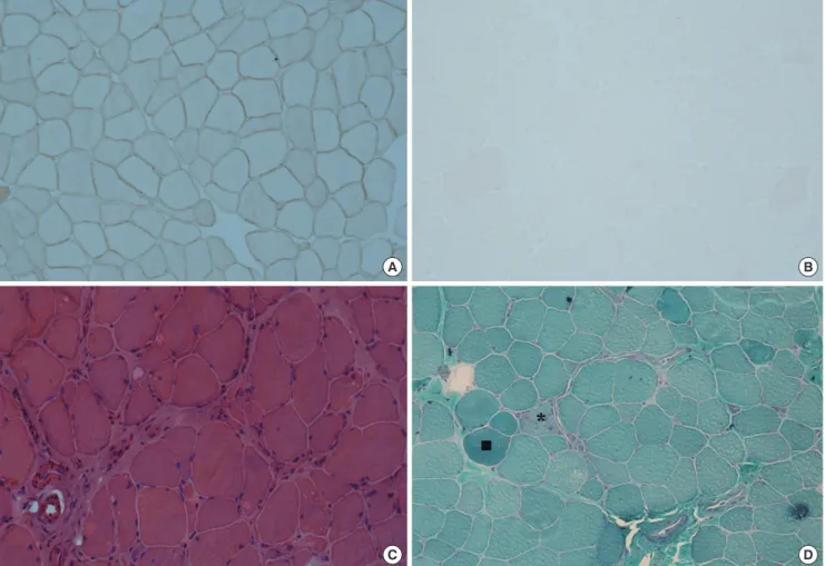
For this study, we reviewed muscle specimens referred to Gang- nam Severance Hospital from March 2004 to June 2011. Each muscle specimen was obtained from the patients with suspected myopathy. We found 33 cases with complete or nearly complete dysferlin protein loss in the muscle specimens by immunohis- tochemistry (Fig. 1A, B). Two patients were withheld due to in- complete medical records. Finally, 31 patients were enrolled for this study. We studied clinical, laboratory, pathologic and radio- logic features of each patient. Clinical data included sex, age of symptom onset, disease duration, initial pattern of muscle in- volvement, family history, and distribution of weakness at diag- nosis. Muscle involvement was evaluated clinically using medi- cal research council scales. Laboratory findings included cre- atine kinase (CK) levels, needle electromyography, and electro- cardiography. The serum CK levels were expressed as ×-fold the
upper limit of normal values according to the local reference range. Radiological findings were included muscle CT or MRI scans. Muscle specimens were taken from the following muscles:
the vastus lateralis (n = 12), biceps brachii (n = 9), deltoid (n = 1), and unspecified muscles (n = 9). The frozen muscle sections (5 μm thickness) from all muscle specimens were stained with routine histopathological stains such as hematoxylin and eosin (H&E), modified Gomori trichrome (modified GT), and nico- tinamide adenine dinucleotide-tetrazolium reductase (NADH- TR) stains.
Immunohistochemistry of muscle specimens
The tissues were processed for immunohistochemistry as fol- lows: 5-μm transverse serial sections were obtained from all mus- cle specimens. Sections were fixed in acetone at 4°C for 10 min, rinsed in 50 mM Tris-buffered saline (pH 7.5) for 20 min, and incubated for 30 min with a blocking solution containing 2%
bovine serum albumin and 5% normal goat serum as described (15). The sections were then incubated overnight in a humid chamber at 4°C with one of the following antibodies: C-termi- nal of dystrophin (Leica Microsystems, Newcastle upon Tyne,
UK), rod domain of dystrophin (Leica Microsystems), N-termi- nal of dystrophin (Leica Microsystems), dysferlin (Leica Micro- systems), α-sarcoglycan (Leica Microsystems), β-sarcoglycan (Leica Microsystems), γ-sarcoglycan (Leica Microsystems), and δ-sarcoglycan (Leica Microsystems), α-dystroglycan (Millipore, Billerica, MA, USA), and caveolin (BD Biosciences, San Diego, CA, USA). After washing for 20 min in Tris-buffered saline, the sections were incubated with biotinylated goat anti-mouse IgG (Vector Laboratories Inc., Burlingame, CA, USA) for 30 min at room temperature followed by detection with streptavidin-bio- tin complex immunoperoxidase (VECTASTAIN Elite ABC kit;
Vector Laboratories Inc.). Then the slides were developed with 3, 3´-diaminobenzidine (DAB substrate kit for peroxidase; Vec- tor Laboratories Inc.) substrate for 1-5 min. After being washed, the slides were mounted in mounting medium (VectaMount;
Vector Laboratories Inc.).
Statistical analysis
The chi-square test and the Fischer’s exact tests were used to compare discrete variables, and the independent t-test was used to compare the means of two samples for continuous variables.
Fig. 1. Pathological features of dysferlinopathy. (A) Dysferlin in a normal muscle specimen by immunohistochemistry (× 200). (B) Loss of dysferlin in a nearly total muscle speci- men by immunohistochemistry (× 200). (C) Prominent inflammatory cell infiltration and increased endomysial fibrosis on H&E stain (× 200). (D) A few necrotic (*) and regener- ating muscle fibers (■) on modified Gomori trichrome stain (× 200).
A B
C D
The relationship between serum CK levels and age at diagnosis or disease duration was assessed using Pearson correlation co- efficients. Differences were considered statistically significant at P < 0.05. Statistical analyses were performed with SPSS (ver- sion 17.0).
Ethics statement
This study protocol was approved by the institutional review board of Gangnam Severance hospital (IRB Number: 3-2011-0190).
Written informed consent was exempted by the board because this was a retrospective study.
RESULTS
Demographic and clinical characteristics
In the study, 12 (38.9%) men and 19 (61.3%) women were in- cluded. The mean age of symptom onset was 22.23 ± 7.34 yr
(range, 12-36 yr), and 22 patients of 30 symptomatic patients (73.3%) experienced their first symptoms between 15 and 30 yr of age. The mean disease duration was 7.97 ± 6.94 yr (range, 1- 30 yr). Table 1 summarizes the clinical and laboratory findings.
Three patients had family history of myopathy. Twenty nine pa- tients showed slowly progressive muscle weakness. However, one patient had become symptomatic rapidly during military training. One patient had only extremely high serum CK levels without weakness. Seven patients (23%) described very active and sporty life before symptom onset. Seven patients (two LG- MD2B, four MM and one PD patients) had initially asymmetric muscle weakness. Muscle assessment at diagnosis revealed rel- atively preserved muscle function in the upper limbs.
Laboratory findings
The mean serum creatine kinase (CK) level was elevated 42-fold (min; 4, max; 131) above the upper limit of normal values. There Table 1. Clinical and laboratory characteristics of 31 patients with dysferlinopathy
No.
Sex/
Age (yr)
Age (yr) of onset
Clinical diagnosis before
immunohistochemistry F/H
Distribution of muscle weakness
EKG Serum CK (fold)
U/Ex L/Ex
P D P AD PD
Group 1: LGMD2B phenotype 1
2 3 4 5 6 7 8 9 10 11 12 13
F/15 F/43 F/45 F/18 F/36 F/37 F/30 M/31 F/51 M/20 F/34 F/40 F/37
12 36 25 12 20 32 18 15 30 14 26 35 33
LGMD LGMD LGMD LGMD LGMD LGMD Inflammatory myopathy
LGMD LGMD LGMD LMGD LGMD Inflammatory myopathy
0 0 0 0 0 0 0 0 0 0 0 0 0
4+
3 5 4 4+
3 4+
4- 4- 4 5 5 3
5 4+
5 5 5 4 4+
4 4+
5 5 5 4
3 4- 4 4 4- 3 4+
4- 3+
4 4- 4+
2
4 4 5 5 4+
4+
4+
4 4 4+
4- 5 4
4 4 5 5 4+
4+
4+
4 4 4+
4- 5 4
NR NR WNL
NR NR NR WNL WNL NR NR NR WNL
NR
111 13 27 128 17 20 65 40 NR 4 25 31 10 Group 2: Miyoshi distal myopathy phenotype
14 15 16 17 18 19 20 21 22 23 24 25 26 27 28
F/38 M/25 F/26 M/21 M/20 M/22 M/33 F/23 F/33 F/36 F/33 M/50 M/22 M/30 F/26
36 23 16 16 17 21 18 16 19 30 29 20 21 29 19
Miyoshi myopathy Miyoshi myopathy Miyoshi myopathy Miyoshi myopathy Miyoshi myopathy Inflammatory myopathy
Miyoshi myopathy Miyoshi myopathy Miyoshi myopathy Miyoshi myopathy Miyoshi myopathy Inflammatory myopathy
Miyoshi myopathy Miyoshi myopathy Miyoshi myopathy
0 0 0 0 0 0 1 0 1 0 0 0 0 0 0
5 5 4+
5 5 5 5 4+
4+
5 5 4 5 5 5
5 5 5 5 5 5 5 4+
5 5 5 4 5 5 5
5 5 4- 5 5 4+
5 4+
4+
5 4+
4 5 4+
4+
5 5 4 4+
4+
4- 4+
4 4- 5 5 2 5 5 4+
4+
4 4 4+
4+
4 4 4- 4 4+
4+
2 4+
4 4+
NR NR NR NR NR NR NR NR WNL WNL NR NR WNL WNL WNL
14 25 40 48 50 41 25 100 12 24 33 8 48 34 30 Group3: proximodistal phenotype
29 30
F/19 M/21
17 16
LGMD LGMD
0 0
4 5
5 5
4 4-
5 4+
4 4
NR NR
131 60 Group 4: asymptomatic phenotype
31 M/21 18 HyperCKemia 1 5 5 5 5 5 NR 43
U/Ex, upper extremities; L/Ex, lower extremities; P, proximal; D, distal; AD, anterior distal; PD, posterior distal; LGMD, limb girdle muscular dystrophy; NR, no record; WNL, within normal limit.
was significant negative correlation between the serum CK and the age of patients (R = -0.652, P < 0.001). However, there was no significant correlation between the serum CK level and the disease duration (P = 0.123). Nine patients recalled that they had had persistent elevation of liver function tests before the on- set of weakness and one patient (patient 29) had even received a liver biopsy. Needle electromyography in all patients showed short duration, low amplitude motor unit potentials with full interference patterns. Nine patients had undergone electrocar-
Table 2. Histopathological findings of muscle biopsy in 31 patients with dysferlinopathy
Findings No. (%)
Muscle size variation Mild
Moderate Marked
7 (23) 8 (26) 13 (42)
Small angulated fiber 0 (0)
Internal nuclei 17 (55)
Necrotic and regenerative fiber 28 (90)
Cellular response
Inflammatory cell infiltration Endomysial fibrosis
22 (71) 20 (65) Architectural change
Lobulated fiber Rimmed vacuoles Target fiber Targetoid fiber Moth-eaten fiber Ring fiber
1 (3) 0 (0) 0 (0) 0 (0) 0 (0) 0 (0)
Fig. 2. Radiological features of dysferlinopathy. Muscle CT scans revealed that muscles of the upper limb (A) are relatively spared and lateral posterior compartments of thigh (B) and calf (C) muscles had dominantly fatty atrophic changes. Transverse T1 weighted images through thigh (D) and calf (E) muscles revealed that lateral posterior muscles had dominantly fatty atrophic changes.
A B C
D E
diograms and all showed normal findings.
Pathological findings
Table 2 shows pathological findings of muscle specimens. The muscle specimens on routine histopathological stains (H&E, modified GT and NADH-TR stains) showed nonspecific dystro- phic features of varying degrees with regard to fiber size variabil- ity, increased endomysial fibrosis, and necrotic and regenerat- ing muscle fibers. One muscle specimen presented lobulated fibers. However, routine histopathological staining of three mus- cle specimens did not reveal any pathological changes. Inflam- matory cells were observed in the majority of the muscle biopsies and were located especially in the perivascular area (Fig. 1C, D).
In the present study, four patients had been misdiagnosed with inflammatory myopathy after muscle biopsy and were treated by steroids and immunomodulating drugs. They could have been properly diagnosed with immunohistochemistry for dys- ferlin. By immunohistochemistry, all muscle specimens revealed normal staining pattern for other sarcolemmal proteins such as dystrophin, sarcoglycans, dystroglycan and caveolin.
Radiological findings
Muscle CT scans were performed in four patients (two LGMD2B and two MM patients). CT scans identified the extent of replace- ment of skeletal muscle by fat or fibrotic tissue. The upper limb muscles are relatively spared and the lateral posterior compart-
ments of the lower limb muscles had dominantly fatty atrophic changes (Fig. 2A). Muscle MRI scans of the lower limbs were performed in four patients (one LGMD2B and three MM pa- tients). MRI scans revealed that the gastrocnemius, soleus, ad- ductor magnus, hamstrings and vastus lateralis muscles had dominantly fatty atrophic changes and diffuse patchy edema (Fig. 2B).
Comparison between MM and LGMD2B types
We classified 30 symptomatic patients according to their initial patterns of muscle weakness. Fifteen patients had initial clinical presentation of calf weakness in one or both legs (MM group).
Thirteen patients had proximal leg weakness (LGMD2B group), and two patients showed both proximal and distal weakness of the leg simultaneously (PD group). There was no difference be- tween the LGMD2B and MM groups in the age of onset (LGM- D2B group: 23.69 ± 8.99 yr; MM group: 22.00 ± 6.16 yr; P = 0.562) and the mean disease duration (LGMD2B group: 10.23 ± 6.02 yr; MM group: 7.20 ± 7.74 yr; P = 0.264). There was a tendency for a higher proportion of women in the LGMD2B group (11/13 patients, 85%) than in the MM group (7/16 patients, 47%; P = 0.055). Table 3 shows the difference of clinical and laboratory characteristics between the LGMD2B and MM groups. The weak- ness of proximal arm and leg is more frequently present in the LGMD2B group than the MM group. Anterior compartment mus- cles of the lower leg were frequently affected in both LGMD2B and MM groups. In pathological findings and serum CK levels, there were no differences between LGMD2B and MM groups.
DISCUSSION
Our study reveals that clinical and pathological features of Korean dysferlinopathy patients vary greatly. Even though the majority experienced their first symptoms between the ages of 15 and 30, the patients had a broad range of age of onset from 12 to 36 yr.
Although classical MM and LGMD2B phenotypes are most com-
mon, 31 patients evaluated in this study had a heterogeneous clinical spectrum from isolated hyperCKemia to proximodistal phenotype as shown in previous studies (7, 16-18). The factors responsible for distinct clinical features are yet unknown in our study. Muscle assessment and radiological studies demonstrat- ed that the upper limb muscles were relatively preserved, but posterolateral compartments of the lower limb muscles were dominantly impaired. In the comparison between the MM and LGMD2B groups in our study, there were no differences of on- set age, serum CK levels, and pathological findings. The dysfer- linopathy patients had markedly elevated serum CK levels. In addition, the serum CK level correlated with the patient’s age, but did not correlate with disease duration in contrast with pre- vious studies (7, 19). This is probably associated with a poor re- lationship with disease progression and the serum CK or histo- pathological changes of each patient (20). The pathological find- ings of dysferlinopathy also revealed various features from nor- mal findings to severe dystrophic changes in routine histopath- ologic stains as were shown in previous studies (21). In our study, four patients had been misdiagnosed with an inflammatory my- opathy similar to other reports (22-24). Dysferlinopathy is poten- tially misdiagnosed as inflammatory myopathy due to several reasons. First, inflammatory cell infiltration is frequently observed in muscle specimens of dysferlinopathy as well as inflammato- ry myopathy. Inflammatory cell infiltration may be associated with impaired secretion of cytokines and prolonged release of endogenous molecules such as heat shock proteins, high mo- bility group box 1, and adenosine triphosphates due to compro- mised membrane repair (25-27). Second, dyferlinopathy and inflammatory myopathy have common features such as very high serum CK levels and young adult onset. Third, many pa- tients with dysferlinopathy had good muscle strength prior to the onset of symptoms (18), which can be suggestive of an acquired myopathy. Recent study revealed the analyzing inflammatory cells, MHC class I expressions and MAC deposits in a muscle specimen may contribute to differentiating dysferlinopathy from inflammatory myopathy (28). However, the best way for differ- ential diagnosis between dysferlinopathy and inflammatory myopathy is to confirm the loss of dysferlin protein or gene.
Dysferlinopathy usually has young adult onset and shows high serum CK levels. However, heterogeneity of clinical presenta- tions and pathological findings on routine histopathological stains makes it difficult to diagnose dysferlinopathy. Currently the most important and easy method for differential diagnosis of dysferlinopathy is immunohistochemical staining. Even though western blotting can quantitatively measure the amount of pro- tein present, it is time-consuming and not compulsory for all proteins on a routine basis. Western blotting can detect the pres- ence of a protein, not the protein function (11). In addition, im- munohistochemistry and western blotting both require assess- ment by invasive muscle biopsy. Even if genetic testing is a less Table 3. Comparison of clinical and laboratory features for MM and LGMD2B groups
Findings LGMD2B
(n = 13) MM
(n = 15) P value Clinical finding
Proximal arm weakness Distal arm weakness Proximal leg weakness Anterior distal leg weakness Posterior distal leg weakness
10 (77%) 6 (46%) 13 (100%)
10 (77%) 10 (77%)
4 (27%) 2 (13%) 7 (47%) 9 (60%) 15 (100%)
0.008 0.096 0.002 0.435 0.087 Laboratory finding
Serum CK (fold) 40.92 ± 40.15 35.47 ± 22.19 0.658 Pathological finding
Internal nuclei
Necrotic and regenerative fiber Inflammatory cell infiltration Endomysial fibrosis
8 (62%) 13 (100%)
9 (69%) 7 (54%)
6 (40%) 12 (80%) 10 (67%) 10 (67%)
0.256 0.226 1.000 0.488
invasive and more accurate method, it is currently limited for diagnosing dysferlinopathy in clinical use due to the large size of dysferlin gene and the absence of a mutational hot spot (29, 30).
In conclusion, limitations in diagnosis by clinical and patho- logical means currently make immunohistochemistry the most important method for patients suspected to have dysferlinopa- thy. However, with advancing technology, genetic testing will be the most useful method for diagnosis of dysferlinopathy in the near future.
REFERENCES
1. Liu J, Aoki M, Illa I, Wu C, Fardeau M, Angelini C, Serrano C, Urtizberea JA, Hentati F, Hamida MB, Bohlega S, Culper EJ, Amato AA, Bossie K, Oeltjen J, Bejaoui K, McKenna-Yasek D, Hosler BA, Schurr E, Arahata K, de Jong PJ, Brown RH Jr. Dysferlin, a novel skeletal muscle gene, is mu- tated in Miyoshi myopathy and limb girdle muscular dystrophy. Nat Genet 1998; 20: 31-6.
2. Bansal D, Campbell KP. Dysferlin and the plasma membrane repair in muscular dystrophy. Trends Cell Biol 2004; 14: 206-13.
3. Bansal D, Miyake K, Vogel SS, Groh S, Chen CC, Williamson R, McNeil PL, Campbell KP. Defective membrane repair in dysferlin-deficient mus- cular dystrophy. Nature 2003; 423: 168-72.
4. Han R. Muscle membrane repair and inflammatory attack in dysferlinop- athy. Skeletal Muscle 2011; 1: 10.
5. Klinge L, Laval S, Keers S, Haldane F, Straub V, Barresi R, Bushby K. From T-tubule to sarcolemma: damage-induced dysferlin translocation in ear- ly myogenesis. FASEB J 2007; 21: 1768-76.
6. Illa I, Serrano-Munuera C, Gallardo E, Lasa A, Rojas-Garcia R, Palmer J, Gallano P, Baiget M, Matsuda C, Brown RH. Distal anterior compartment myopathy: a dysferlin mutation causing a new muscular dystrophy phe- notype. Ann Neurol 2001; 49: 130-4.
7. Nguyen K, Bassez G, Krahn M, Bernard R, Laforêt P, Labelle V, Urtiz- berea JA, Figarella-Branger D, Romero N, Attarian S, Leturcq F, Pouget J, Lévy N, Eymard B. Phenotypic study in 40 patients with dysferlin gene mutations: high frequency of atypical phenotypes. Arch Neurol 2007; 64:
1176-82.
8. Weiler T, Bashir R, Anderson LV, Davison K, Moss JA, Britton S, Nylen E, Keers S, Vafiadaki E, Greenberg CR, Bushby CR, Wrogemann K. Identi- cal mutation in patients with limb girdle muscular dystrophy type 2B or Miyoshi myopathy suggests a role for modifier gene(s). Hum Mol Genet 1999; 8: 871-7.
9. Weiler T, Greenberg CR, Nylen E, Halliday W, Morgan K, Eggertson D, Wrogemann K. Limb-girdle muscular dystrophy and Miyoshi myopathy in an aboriginal Canadian kindred map to LGMD2B and segregate with the same haplotype. Am J Hum Genet 1996; 59: 872-8.
10. Angelini C. Limb-girdle muscular dystrophies: heterogeneity of clinical phenotypes and pathogenetic mechanisms. Acta Myol 2004; 23: 130-6.
11. Moore SA, Shilling CJ, Westra S, Wall C, Wicklund MP, Stolle C, Brown CA, Michele DE, Piccolo F, Winder TL, Stence A, Barresi R, King N, King W, Florence J, Campbell KP, Fenichel GM, Stedman HH, Kissel JT, Griggs RC, Pandya S, Mathews KD, Pestronk A, Serrano C, Darvish D, Mendell JR. Limb-girdle muscular dystrophy in the United States. J Neuropathol Exp Neurol 2006; 65: 995-1003.
12. Cho HJ, Sung DH, Kim EJ, Yoon CH, Ki CS, Kim JW. Clinical and genetic analysis of Korean patients with Miyoshi myopathy: identification of three novel mutations in the DYSF gene. J Korean Med Sci 2006; 21: 724-7.
13. Oh SH, Kim TS, Choi YC. Identification of a dysferlin gene mutation in a Korean case with Miyoshi myopathy. Yonsei Med J 2004; 45: 927-30.
14. Oh SH, Kang SW, Lee JG, Na SJ, Kim TS, Choi YC. Clinical and patho- logical characteristics of four Korean patients with limb-girdle muscular dystrophy type 2B. J Korean Med Sci 2004; 19: 447-52.
15. Choi YC, Park GT, Kim TS, Sunwoo IN, Steinert PM, Kim SY. Sporadic inclusion body myositis correlates with increased expression and cross- linking by transglutaminases 1 and 2. J Biol Chem 2000; 275: 8703-10.
16. Albrecht DE, Garg N, Rufibach LE, Williams BA, Monnier N, Hwang E, Mittal P. 4th Annual Dysferlin Conference 11-14 September 2010, Wash- ington, USA. Neuromuscul Disord 2011; 21: 304-10.
17. Rosales XQ, Gastier-Foster JM, Lewis S, Vinod M, Thrush DL, Astbury C, Pyatt R, Reshmi S, Sahenk Z, Mendell JR. Novel diagnostic features of dysferlinopathies. Muscle Nerve 2010; 42: 14-21.
18. Klinge L, Aboumousa A, Eagle M, Hudson J, Sarkozy A, Vita G, Charl- ton R, Roberts M, Straub V, Barresi R, Lochmuller H, Bushby K. New as- pects on patients affected by dysferlin deficient muscular dystrophy. J Neurol Neurosurg Psychiatry 2010; 81: 946-53.
19. Mahjneh I, Marconi G, Bushby K, Anderson LV, Tolvanen-Mahjneh H, Somer H. Dysferlinopathy (LGMD2B): a 23-year follow-up study of 10 patients homozygous for the same frameshifting dysferlin mutations.
Neuromuscul Disord 2001; 11: 20-6.
20. Mahjneh I, Passos-Bueno MR, Zatz M, Vainzof M, Marconi G, Nashef L, Bashir R, Bushby K. The phenotype of chromosome 2p-linked limb-gir- dle muscular dystrophy. Neuromuscul Disord 1996; 6: 483-90.
21. Ueyama H, Kumamoto T, Horinouchi H, Fujimoto S, Aono H, Tsuda T.
Clinical heterogeneity in dysferlinopathy. Intern Med 2002; 41: 532-6.
22. McNally EM, Ly CT, Rosenmann H, Mitrani Rosenbaum S, Jiang W, Anderson LV, Soffer D, Argov Z. Splicing mutation in dysferlin produces limb-girdle muscular dystrophy with inflammation. Am J Med Genet 2000; 91: 305-12.
23. Nakagawa M, Matsuzaki T, Suehara M, Kanzato N, Takashima H, Higu- chi I, Matsumura T, Goto K, Arahata K, Osame M. Phenotypic variation in a large Japanese family with Miyoshi myopathy with nonsense muta- tion in exon 19 of dysferlin gene. J Neurol Sci 2001; 184: 15-9.
24. Rowin J, Meriggioli MN, Cochran EJ, Sanders DB. Prominent inflam- matory changes on muscle biopsy in patients with Miyoshi myopathy.
Neuromuscul Disord 1999; 9: 417-20.
25. Han R, Frett EM, Levy JR, Rader EP, Lueck JD, Bansal D, Moore SA, Ng R, Beltrán-Valero de Bernabé D, Faulkner JA, Campbell KP. Genetic abla- tion of complement C3 attenuates muscle pathology in dysferlin-deficient mice. J Clin Invest 2010; 120: 4366-74.
26. Nagaraju K, Rawat R, Veszelovszky E, Thapliyal R, Kesari A, Sparks S, Raben N, Plotz P, Hoffman EP. Dysferlin deficiency enhances monocyte phagocytosis: a model for the inflammatory onset of limb-girdle muscu- lar dystrophy 2B. Am J Pathol 2008; 172: 774-85.
27. Rawat R, Cohen TV, Ampong B, Francia D, Henriques-Pons A, Hoff- man EP, Nagaraju K. Inflammasome up-regulation and activation in dysferlin-deficient skeletal muscle. Am J Pathol 2010; 176: 2891-900.
28. Choi JH, Park YE, Kim SI, Kim JI, Lee CH, Park KH, Kim DS. Differenrial immunohistological feautres of inflammatory myopathies and dysfer- linopathy. J Korean Med Sci 2009; 24: 1015-23.
29. Aoki M, Liu J, Richard I, Bashir R, Britton S, Keers SM, Oeltjen J, Brown HE, Marchand S, Bourg N, Beley C, McKenna-Yasek D, Arahata K, Bohle- ga S, Cupler E, Illa I, Majneh I, Barohn RJ, Urtizberea JA, Fardeau M, Amato A, Angelini C, Bushby K, Beckmann JS, Brown RH Jr. Genomic organization of the dysferlin gene and novel mutations in Miyoshi my- opathy. Neurology 2001; 57: 271-8.
30. Takahashi T, Aoki M, Tateyama M, Kondo E, Mizuno T, Onodera Y,
Takano R, Kawai H, Kamakura K, Mochizuki H, Shizuka-Ikeda M, Nak- agawa M, Yoshida Y, Akanuma J, Hoshino K, Saito H, Nishizawa M, Kato S, Saito K, Miyachi T, Yamashita H, Kawai M, Matsumura T, Kuzuhara S, Ibi T, Sahashi K, Nakai H, Kohnosu T, Nonaka I, Arahata K, Brown RH Jr, Itoyama Y. Dysferlin mutations in Japanese Miyoshi myopathy: rela- tionship to phenotype. Neurology 2003; 60: 1799-804.