First Imported Case of Zika Virus Infection into Korea
Since Zika virus has been spreading rapidly in the Americas from 2015, the outbreak of Zika virus infection becomes a global health emergency because it can cause neurological complications and adverse fetal outcome including microcephaly. Here, we report clinical manifestations and virus isolation findings from a case of Zika virus infection imported from Brazil. The patient, 43-year-old Korean man, developed fever, myalgia, eyeball pain, and maculopapular rash, but not neurological manifestations. Zika virus was isolated from his semen, and reverse-transcriptase PCR was positive for the virus in the blood, urine, and saliva on the 7th day of the illness but was negative on the 21st day. He recovered spontaneously without any neurological complications. He is the first case of Zika virus infection in Korea imported from Brazil.
Keywords: Zika Virus; Travel; Virus Shedding; Brazil; Korea Hee-Chang Jang,1* Wan Beom Park,2*
Uh Jin Kim,1 June Young Chun,2 Su-Jin Choi,2 Pyoeng Gyun Choe,2 Sook-In Jung,1 Youngmee Jee,3 Nam-Joong Kim,2 Eun Hwa Choi,4 and Myoung-don Oh2
1Department of Infectious Diseases, Chonnam National University Medical School, Gwangju, Korea; 2Department of Internal Medicine, Seoul National University College of Medicine, Seoul, Korea; 3Center for Pathology and Immunology, National Institute of Health, Korea Centers for Disease Control and Prevention, Osong, Korea;
4Department of Pediatrics, Seoul National University College of Medicine, Seoul, Korea
* Hee-Chang Jang and Wan Beom Park have equally contributed to the work.
Received: 24 May 2016 Accepted: 25 May 2016 Address for Correspondence:
Myoung-don Oh, MD
Department of Internal Medicine, Seoul National University College of Medicine, 103 Daehak-ro, Jongno-gu, Seoul 03080, Korea
E-mail: [email protected]
Funding: Eun Hwa Choi and Nam-Joong Kim are participating investigators of the research program funded by the Korea Centers for Disease Control and Prevention (2016P2400100).
http://dx.doi.org/10.3346/jkms.2016.31.7.1173 • J Korean Med Sci 2016; 31: 1173-1177
INTRODUCTION
Zika virus is a mosquito-borne flavivirus related to dengue vi- rus, yellow fever virus, and West Nile virus. The virus was first isolated from a sentinel rhesus monkey stationed in the Ugan- da’s Zika Forest in 1947, during the epidemiological research of yellow fever (1). Human infection by Zika virus was first recog- nized in 3 patients in Nigeria in 1953 (2). Subsequently, only 14 sporadic cases have been reported until the first outbreak of Zika virus in Yap Islands of Micronesia in 2007 (3). Most cases during the Yap Islands outbreak were mild cases. Six years later, however, another outbreak occurred in French Polynesia, and neurological and autoimmune complications were reported for the first time (4,5).
The Zika virus outbreak in the Americas was first recognized in March 2015, when an epidemic of an illness characterized by fever, rash, arthralgia, myalgia, and conjunctivitis occurred in Bahia, Brazil (6). By September 2015, reports of an increase in the number of congenital microcephaly in Zika virus-affected
areas began to emerge (7). Because of the cluster of microceph- aly cases and other neurological disorders reported in Brazil and French Polynesia, the World Health Organization declared a Public Health Emergency of International Concern on Febru- ary 1, 2016. As of May 18, 2016, 46 countries are experiencing a first outbreak of Zika virus transmitted by mosquitos, and 10 countries have reported evidence of person-to-person trans- mission of Zika virus, probably via a sexual route (8).
In order to prevent an outbreak of Zika virus in Korea, early detection and isolation of returning travelers with Zika virus in- fection from countries with ongoing outbreak is of paramount importance. Here, we report the first imported case of Zika vi- rus infection into Korea.
CASE DESCRIPTION
A 43-year-old Korean man visited Chonnam National University Hospital due to fever and rash. The patient had history of stay- ing and mosquito bites at Cumbuco, Ceara, Brazil for 3 weeks
from 17 Feb 2016 to 9 Mar 2016, and returned to Republic of Korea on 11 Mar 2016. He had fever, chill, myalgia, and eyeball pain on 6 days after return from Brazil. Three days later, rash also developed. The patient visited nearby clinic and the blood was sampled for Zika virus reverse-transcriptase polymerase chain reaction (RT-PCR) on the 6th day of illness. The RT-PCR result was reported to be positive by the Korea Center for Dis- ease Control and Prevention. The patient was admitted to Chon- nam National University Hospital for further evaluation and ma nagement on the 7th day of illness.
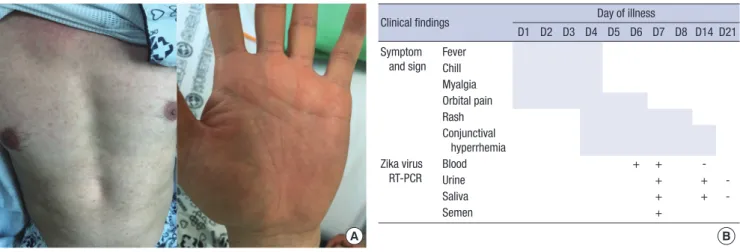
Upon admission, he had a blood pressure of 110/70 mmHg, pulse rate of 80 beats/min, respiratory rate of 20/min, and a body temperature of 36.3°C. Painless multiple erythematous maculopapular rash with itching was observed in trunk and both upper and lower extremity (Fig. 1A). Hyperemia was pres- ent in both eyes and the patient complained of feeling of dry- ness in both eyes. Enlarged lymph node was not observed. Neu- rologic symptoms or signs including headache, vomiting, de- crease in motor power, or abnormal sensation were absent. Ini- tial laboratory findings performed on the day of admission were as follows: white blood cell count 4,900/µL (neutrophils 55%, lymphocytes 29%, monocytes 15%), hemoglobin level 16.5 g/
dL, platelet count 221,000/µL, erythrocyte sedimentation rate 2 mm/hr, serum C-reactive protein 0.47 mg/dL, procalcitonin 0.05 ng/mL, serum neutrophil gelatinase-associated lipocalin 89.4 ng/mL, blood urea nitrogen 12.8 mg/dL, total protein 7.4 g/dL, albumin 4.6 g/dL. Serum aspartate aminotransferase, al- anine transaminase, and lactate dehydrogenase were 61 U/L, 92 U/L, and 459 U/L, respectively. Serum level of ferritin was 417 ng/mL and serum adenosine deaminase was 37 IU/L.
He was only medicated with cetirizine 10 mg/day per oral.
Intravenous fluid or other medication was not prescribed dur- ing the hospital stay. The patient no longer felt myalgia or fe- brile sense during the hospital stay. Conjunctival hyperemia continued but maculopapular rashes were fading out. He was
discharged from the hospital on 8th day of illness, and then was followed up weekly for 6 weeks. During the 6 weeks of follow- up, he did not have symptoms or signs of neurologic abnormal- ity. Zika virus RT-PCR was positive from saliva and urine for 2 weeks after the symptom onset but became negative after 3 weeks (Fig. 1B).
Culture isolate of Zika virus was obtained by inoculating mo- nolayers of Vero cells with his semen sample at the 7th day of the illness and culturing the cells at 37°C in a 5% carbon dioxide atmosphere. In the culture of first passage, the serial change of cycle threshold value by RT-PCR using RealStar Zika virus RT- PCR kit 1.0 (Altona Diagnostics, Hamburg, Germany) was shown in Fig. 2A.
For electron microscopic observation, Vero cell monolayer inoculated with culture supernatant of second passage was fixed as previously described (9). It was cut on ultramicrotome (RMC MT-XL) at 65 nm. Ultrathin sections were stained with saturat- ed 4% uranyl acetate and 4% lead citrate before examination with a transmission electron microscope (JEM-1400; JEOL Inc., Tokyo, Japan) at 80 kV. Virus particles were observed within the cytoplasm (Fig. 2B).
Immunofluorescence staining was performed by previously described method (10,11). Briefly, Zika virus-infected and mock- infected cells were fixed with 4% paraformaldehyde in PBS for 1h at room temperature. Slides were blocked and then incubat- ed with the 3 weeks convalescent serum of patient (1:40 dilu- tion) and serum of healthy control (1:40 dilution) at -4°C over- night. Cells were washed and incubated for 1 hour at room tem- perature with fluorescein isothiocyanate-conjugated anti-hu- man IgG. 4’,6-Diamidino-2-phenylindole dihydrochloride was used to stain the nucleus. Preparations were examined with a confocal microscope (Leica, Buffalo Grove, IL, USA). Immuno- fluorescence was observed in Zika virus-infected Vero cells ap- plied with convalescent serum rather than control serum (Fig.
2C and D).
Fig. 1. Clinical manifestation and virus shedding. (A) Maculopapular rash on the trunk and palm. (B) Time course of symptom, sign and the results of Zika virus RT-PCR.
+, positive; -, negative.
Clinical findings Day of illness
D1 D2 D3 D4 D5 D6 D7 D8 D14 D21 Symptom
and sign Fever Chill Myalgia Orbital pain Rash Conjunctival
hyperrhemia Zika virus
RT-PCR
Blood + + -
Urine + + -
Saliva + + -
Semen +
A B
DISCUSSION
In the present report, we describe clinical findings of the first imported patient (43 years old man) of Zika virus infection in Korea, from whom the virus was isolated. The incubation peri- od between the mosquito bite and the onset of clinical mani- festations of the current case was estimated to be 1 to 4 weeks, as his symptoms began 6 days after returning from Brazil, where he had stayed for 3 weeks. Clinical spectrum of Zika virus infec- tion includes acute febrile illness, neurologic complications, and adverse fetal outcomes (12). Most cases with Zika virus in- fections are mild or asymptomatic; only 19% of cases had symp- toms, and no patients were hospitalized during the Yap outbreak (3). Most common symptoms were macular or papular rash, fever, arthralgia, non-purulent conjunctivitis, myalgia, head- ache, and retro-orbital pain (3,13). A Brazilian study suggested
that clinical features of pruritic rash, conjunctival injection, and lymphadenopathy should raise the suspicion of Zika virus in- fection (13). In our case, conjunctival injection was present and it lasted longer than rash. Neurologic complications of Zika vi- rus infection include Guillain-Barré syndrome (14), myelitis, and meningoencephalitis (15). None of these neurologic com- plications were observed in our case. There is no antiviral treat- ment for Zika virus yet. Management consists of symptomatic treatment. The patient recovered spontaneously.
The diagnosis of Zika virus infection is made by detection of Zika viral RNA or serology. Zika viral RNA can be detected in blood, saliva, urine, and semen specimens by RT-PCR. Urine is the preferred sample for RT-PCR (16), as it yields higher detec- tion rates than serum, even 5 days after symptom onset, when all serum specimens were negative by RT-PCR (17). For patients presenting > 7 days after symptom onset, serologic tests should Fig. 2. Isolation of Zika virus from semen sample. (A) Temporal change of cycle threshold (Ct) value for Zika virus RT-PCR in the culture of first passage. (B) Transmission elec- tron microscopy image of Vero cells infected with Zika virus. White arrows denote virus particles. Black scale bar indicates 200 nm. (C, D) Immunofluorescence assay shows that Zika virus-infected Vero cells reacted with human convalescent anti-Zika virus IgG-positive serum (C) and did not with control serum (D). White scale bar denotes 100 μm.
1/Ct value
Day
0 2 4 6 0.055
0.050 0.045 0.040 0.035
0.030
A B
C D
also be included. However, serologic test results should be in- terpreted with caution, because Zika virus antibody can react extensively with other flavivirus antibodies (18). In our case, initial diagnosis was made by RT-PCR using blood sample. After 2 weeks, Zika virus RNA was detected only in saliva and urine but not in blood samples. Still, findings this case emphasize the sensitivity of urine and saliva than serum in the diagnosis of Zika virus infection, especially, in patients with time interval more than one week between symptom onset and specimen collection.
As for semen sample, which was obtains on day 7 of illness was positive by Zika virus RT-PCR and by virus culture. One of the possible modes of transmission of Zika virus infection is sexual transmission. Reports of the virus detected in semen as long as 62 days after onset of the illness (19), and the high viral load in semen compared to that of blood or urine (20) has brou- ght great concern, as prolonged presence of virus in semen may increase the possibility of sexual transmission to pregnant wo- man. Zika virus infection during pregnancy can cause micro- cephaly and ocular abnormalities of the fetus (12,14). These findings emphasize the importance of early detection of Zika vi- rus infected patients and the application of preventive measures including abstain from unprotected sex. Further study is need- ed on the duration of detection of viable Zika virus in semen, to determine the period of sexual precaution for infected cases. In this case, the duration of viral excretion in semen is still under investigation. Besides sexual contact, there are two more possi- ble route of transmission of Zika virus from this patient to other individuals in Korea, i.e. mosquito-borne and blood transfusion.
However, we think the possibilities are negligible because he had no mosquito bite after returning home and during the fe- brile period. Fortunately, the period was in March when active mosquitoes are absent in Korea. He also did not donate blood.
In conclusions, in order to prevent an outbreak of Zika virus in Korea, early detection and isolation of returning travelers with Zika virus infection is important. Zika virus infection should be suspected in patients with acute febrile illness who visit endem- ic area. Saliva and urine samples are useful for diagnosis of Zika virus using RT-PCR.
ACKNOWLEDGMENT
Patient care, follow-up, and specimen collection were perform- ed in collaboration with the Korea Center for Disease Control and Prevention. RT-PCR for Zika virus was performed at the Ko- rea Centers for Disease Control and Prevention using the fund for standard laboratory for infectious diseases (4837-301-210-13).
DISCLOSURE
The authors have no potential conflicts of interest to disclose.
AUTHOR CONTRIBUTION
Patient care: Jang HC, Kim UJ, Jung SI. Isolation of virus: Jang HC, Park WB, Kim UJ, Chun JY, Choi SJ, Choe PG, Oh MD. RT- PCR for virus: Jee Y. Study design and concept: Jang HC, Choi EH, Oh MD. Reference review: Jang HC, Park WB, Kim UJ, Oh MD. Writing: Jang HC, Park WB, Kim UJ, Oh MD. Critical review and revision: Jang HC, Park WB, Jung SI, Kim NJ, Oh MD.
ORCID
Hee-Chang Jang http://orcid.org/0000-0002-3407-8493 Wan Beom Park http://orcid.org/0000-0003-0022-9625 Uh Jin Kim http://orcid.org/0000-0002-8463-6297 June Young Chun http://orcid.org/0000-0001-9345-6645 Su-Jin Choi http://orcid.org/0000-0001-8732-3950 Pyoeng Gyun Choe http://orcid.org/0000-0001-6794-7918 Sook-In Jung http://orcid.org/0000-0002-1577-678X Nam-Joong Kim http://orcid.org/0000-0001-6793-9467 Eun Hwa Choi http://orcid.org/0000-0002-5857-0749 Myoung-don Oh http://orcid.org/0000-0002-2344-7695
REFERENCES
1. Dick GW, Kitchen SF, Haddow AJ. Zika virus. I. Isolations and serological specificity. Trans R Soc Trop Med Hyg 1952; 46: 509-20.
2. MacNamara FN. Zika virus: a report on three cases of human infection during an epidemic of jaundice in Nigeria. Trans R Soc Trop Med Hyg 1954; 48: 139-45.
3. Duffy MR, Chen TH, Hancock WT, Powers AM, Kool JL, Lanciotti RS, Pre- trick M, Marfel M, Holzbauer S, Dubray C, et al. Zika virus outbreak on Yap Island, Federated States of Micronesia. N Engl J Med 2009; 360: 2536- 43.
4. Oehler E, Watrin L, Larre P, Leparc-Goffart I, Lastere S, Valour F, Baudou- in L, Mallet H, Musso D, Ghawche F. Zika virus infection complicated by Guillain-Barre syndrome--case report, French Polynesia, December 2013.
Euro Surveill 2014; 19: 20720.
5. European Centre for Disease Prevention and Control. Zika virus infection outbreak, French Polynesia: rapid risk assessment [Internet]. Available at http://ecdc.europa.eu/en/publications/Publications/Zika-virus-French- Polynesia-rapid-risk-assessment.pdf [accessed on 23 May 2016].
6. Campos GS, Bandeira AC, Sardi SI. Zika virus outbreak, Bahia, Brazil. Emerg Infect Dis 2015; 21: 1885-6.
7. Schuler-Faccini L, Ribeiro EM, Feitosa IM, Horovitz DD, Cavalcanti DP, Pessoa A, Doriqui MJ, Neri JI, Neto JM, Wanderley HY, et al. Possible as- sociation between Zika virus infection and microcephaly - Brazil, 2015.
MMWR Morb Mortal Wkly Rep 2016; 65: 59-62.
8. World Health Organization. Zika situation report [Internet]. Available at http://who.int/emergencies/zika-virus/situation-report/19-may-2016/
en/ [accessed on 23 May 2016].
9. Graham L, Orenstein JM. Processing tissue and cells for transmission electron microscopy in diagnostic pathology and research. Nat Protoc 2007; 2: 2439-50.
10. Hamel R, Dejarnac O, Wichit S, Ekchariyawat P, Neyret A, Luplertlop N, Perera-Lecoin M, Surasombatpattana P, Talignani L, Thomas F, et al. Biol- ogy of Zika virus infection in human skin cells. J Virol 2015; 89: 8880-96.
11. Driggers RW, Ho CY, Korhonen EM, Kuivanen S, Jääskeläinen AJ, Smura T, Rosenberg A, Hill DA, DeBiasi RL, Vezina G, et al. Zika virus infection with prolonged maternal viremia and fetal brain abnormalities. N Engl J Med. Forthcoming 2016.
12. Petersen LR, Jamieson DJ, Powers AM, Honein MA. Zika virus. N Engl J Med 2016; 374: 1552-63.
13. Brasil P, Pereira JP Jr, Raja Gabaglia C, Damasceno L, Wakimoto M, Ribeiro Nogueira RM, Carvalho de Sequeira P, Machado Siqueira A, Abreu de Carvalho LM, Cotrim da Cunha D, et al. Zika virus infection in pregnant women in Rio de Janeiro - preliminary report. N Engl J Med. Forthcom- ing 2016.
14. Broutet N, Krauer F, Riesen M, Khalakdina A, Almiron M, Aldighieri S, Es- pinal M, Low N, Dye C. Zika virus as a cause of neurologic disorders. N Engl J Med 2016; 374: 1506-9.
15. Carteaux G, Maquart M, Bedet A, Contou D, Brugières P, Fourati S, Cleret de Langavant L, de Broucker T, Brun-Buisson C, Leparc-Goffart I, et al.
Zika virus associated with Meningoencephalitis. N Engl J Med 2016; 374:
1595-6.
16. Interim guidance for Zika virus testing of urine - United States, 2016. MMWR Morb Mortal Wkly Rep 2016; 65: 474.
17. Bingham AM, Cone M, Mock V, Heberlein-Larson L, Stanek D, Blackmore C, Likos A. Comparison of test results for Zika virus RNA in urine, serum, and saliva specimens from persons with travel-associated Zika virus dis- ease - Florida, 2016. MMWR Morb Mortal Wkly Rep 2016; 65: 475-8.
18. Lanciotti RS, Kosoy OL, Laven JJ, Velez JO, Lambert AJ, Johnson AJ, Stan- field SM, Duffy MR. Genetic and serologic properties of Zika virus associ- ated with an epidemic, Yap State, Micronesia, 2007. Emerg Infect Dis 2008;
14: 1232-9.
19. Atkinson B, Hearn P, Afrough B, Lumley S, Carter D, Aarons EJ, Simpson AJ, Brooks TJ, Hewson R. Detection of Zika virus in semen. Emerg Infect Dis 2016; 22: 940.
20. Mansuy JM, Dutertre M, Mengelle C, Fourcade C, Marchou B, Delobel P, Izopet J, Martin-Blondel G. Zika virus: high infectious viral load in semen, a new sexually transmitted pathogen? Lancet Infect Dis 2016; 16: 405.