Brief Report
Vol. 30, No. 6, 2018 755
Received August 2, 2017, Revised December 19, 2017, Accepted for publication January 15, 2018
Corresponding author: Kyung Chan Park, Department of Dermatology, Seoul National University Bundang Hospital, 82 Gumi-ro 173beon-gil, Bundang-gu, Seongnam 13620, Korea. Tel: 82-31-787-7311, Fax: 82-31-787-4058, E-mail:
ORCID: https://orcid.org/0000-0002-8994-8038
This is an Open Access article distributed under the terms of the Creative Commons Attribution Non-Commercial License (http://creativecommons.
org/licenses/by-nc/4.0) which permits unrestricted non-commercial use, distribution, and reproduction in any medium, provided the original work is properly cited.
Copyright © The Korean Dermatological Association and The Korean Society for Investigative Dermatology
Fig. 1. Effects of the stem cells in the living skin equivalents (LSE) reconstruction. Sections of LSE were stained for H&E (A, B, and C), activin A (D, E, and F), integrin β1 (G, H and I;
white arrows indicating basement membrane), PCNA (J, K, and L; yellow arrows indicating basal keratinocytes), and p63 (M, N and O; yellow arrows indicating basal keratinocytes). Original magnification: ×400 in (A∼C) and ×200 in (D∼O). Fb: fibro- blast, ADSC: adipose-derived stem cell, BMSC: bone marrow mesenchymal stem, PCNA: proliferating cell nuclear antigen.
https://doi.org/10.5021/ad.2018.30.6.755
Interactive Roles of Activin A in Epidermal Regeneration
Jee Woong Choi, Kyung Mi Nam1, Hye Ryung Choi1, Chang Hun Huh1, Kyung Chan Park1
Department of Dermatology, Ajou University School of Medicine, Suwon, 1Department of Dermatology, Seoul National University Bundang Hospital, Seoul National University College of Medicine, Seongnam, Korea
Dear Editor:
Recently, various stem cell lines have been widely used in tissue engineering. Among them, the mesenchymal stem cell (MSC) has been reported to stimulate wound healing.
In particular, the bone marrow mesenchymal stem cell (BMSC) migrates directly towards a wound, differentiates into various cells and provides a favorable environment where other cells can regenerate quickly1. The adi- pose-derived stem cell (ADSC) is another line that has fea- tures of the MSC. Although these two types of MSCs have great potential for treating wounds, few studies have in- vestigated how they function in skin regeneration.
Activin A, a homodimer of two inhibin βA subunits linked by a disulfide bond, is a cytokine produced by vari- ous cell types including stem cells2. It participates in regu- lation of stem cell maintenance3, and also plays an im- portant role in the wound healing process4. Because the mesenchymal-epithelial interaction is a critical process for tissue regeneration, we focused on activin A as an im- portant factor of dermal-epidermal interaction.
Recent studies have revealed that the epithelialization process of living skin equivalent (LSE) was similar to that occurring during the re-epithelialization process after wounding5. Therefore, in our study, we compared three different LSE models that were cultured with ADSC, BMSC, and fibroblast. This study was approved by Seoul
Brief Report
756 Ann Dermatol
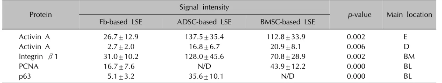
Table 1. Comparison of the level of protein expression by image analysis
Protein Signal intensity
p-value Main location Fb-based LSE ADSC-based LSE BMSC-based LSE
Activin A 26.7±12.9 137.5±35.4 112.8±33.9 0.002 E
Activin A 2.7±2.0 16.8±6.7 20.9±8.1 0.006 D
Integrin β1 31.0±10.2 128.0±45.6 70.8±28.9 0.002 BM
PCNA 16.7±7.6 N/D 43.9±12.2 0.000 BL
p63 5.1±3.2 35.6±10.1 N/D 0.000 BL
Values are presented as mean±standard deviation. The Kruskal-Wallis test was used to compare the signal intensity among the three LSE models. Fb: fibroblast, LSE: living skin equivalent, ADSC: adipose-derived stem cell, BMSC: bone marrow mesenchymal stem, PCNA: proliferating cell nuclear antigen, E: epidermis, D: dermis, BM: basement membrane, BL: basal layer, N/D: not detected.
National University Bundang Hospital Institutional Review Board (IRB no. 1305/202-004 and 1207/164-003).
Three different LSE models (fibroblast-based LSE, BMSC- based LSE, and ADSC-based LSE) were reconstructed ac- cording to the methods described previously6. For re- construction of the LSE models; ADSC, BMSC, and fibro- blast were added as dermal cells to prepare the dermal substitutes. After gelling of the dermal substitutes, the ker- atinocytes were seeded. Human keratinocytes and dermal fibroblasts then were isolated from foreskins obtained dur- ing child circumcision, and human BMSCs (PT-2501) and ADSCs (PT-5006) were purchased from Lonza, Inc., Walkersville, MD, USA.
To analyze the LSE, sections were stained with hematox- ylin and eosin. The level of activin A (sc-166503; Santa Cruz Biotechnology, Santa Cruz, CA, USA) expression was evaluated by immunofluorescence and integrin β1 (sc-6622; Santa Cruz Biotechnology) was stained to identi- fy the basement membrane. Proliferating cell nuclear anti- gen (PCNA, sc-7907; Santa Cruz Biotechnology) and p63 (sc-8431; Santa Cruz Biotechnology) were stained to eval- uate epidermal proliferation status. For the analysis, three non-overlapping areas were digitally captured using the same magnification, illumination and exposure time. To quantify the level of protein expression, we analyzed the signal intensity using Image Pro-Plus 6.0 (MediaCybernetics Inc., Rockville, MD, USA). Finally, mRNA sequencing was used to elucidate gene expression. A more than fourfold ratio was considered a significant difference in gene expression.
Histologically, epidermal stratification was present in all of the LSE models. Interestingly, we observed that MSC-based LSEs showed more cuboidal shaped keratino- cytes along the basal layer of the LSE (Fig. 1A∼C). When comparing the level of expression by image analysis, the difference of the means of signal intensity among the LSE models were statistically significant for all proteins (Table 1). Activin A was strongly expressed in the epidermis and
dermis of the MSC-based LSEs (Fig. 1E, F) compared to fi- broblast-based LSEs (Fig. 1D). In addition, MSC-based LSEs showed stronger integrin β1 expressions with a thick basement membrane compared to the fibroblast-based LSEs (Fig. 1G∼I). These features were especially prom- inent in the ADSC-based LSE. The pattern of PCNA and p63 expression were very interesting in that both were low in the fibroblast-based LSE. However, the opposite pattern of expression was observed in the remaining two types of LSEs (Fig. 1J∼O). In the ADSC-based LSE, most of basal cells were positive for p63 but not PCNA. Most of basal cells, however, were negative for p63 but were pos- itive for PCNA in the BMSC-based LSEs. Previously we re- ported similar patterns of PCNA and p63 expression in the ADSC-based LSE and hypothesized that these cells may not be in the state of proliferation although the potential of proliferation was maintained7. Our new results replicated the results from previous independent experiments sug- gesting that the ADSC has regulatory effects on stem cell characteristics of basal keratinocytes. Furthermore, by pro- viding an optimal niche intra-cellular environment, strong expression of integrin β1 was highly correlated with number of p63 cells. On the other hand, the BMSC-based LSE showed a completely different pattern that suggests al- most all basal cells were in the state of proliferation.
Among the three models, the BMSC-based model showed the thickest epidermis and highest number of PCNA pos- itive cells that was consistent with the histological features. Different functional roles suggest these cells could be used in clinical application.
To analyze the expression of these cells, mRNA sequenc- ing revealed that the inhibin βA gene was up-regulated (10.7-fold increase for BMSC and 8.5-fold increase for ADSC) but other genes in the activin subunits were not differentially expressed compared to fibroblasts. Kyoto Encyclopedia of Genes and Genomes pathway analysis al- so revealed that the gene activin A was the only up-regu- lated gene among the other activin subunits.
Brief Report
Vol. 30, No. 6, 2018 757 In the study, we used LSE models to investigate the effects
of MSCs during skin regeneration. The results showed that epidermal proliferation and basement membrane for- mation were improved by the presence of these cells. This improvement can be explained by the paracrine effects of stem cells and their derivatives to secrete various factors including antioxidant mediators and cytokines8. Among them, we postulated that activin A could be an important factor in the wound healing process. In a mouse model, activin A promoted healthy granular tissue formation, re- generated epidermal keratinocytes, and influenced kerati- nocyte migration4. In hair follicles, this pathway is also re- lated to the hair regeneration cycle9. Previous reports and our findings suggest that activin A serves an important function for maintaining homeostasis of skin tissue.
Activin A is mainly produced in keratinocytes and the MSC10, and we also verified that activin A was largely ex- pressed in both epidermal and dermal layers of the LSE. It seems that activin A produced from the MSCs in the der- mis affects the basal keratinocytes through paracrine ef- fects, and contributes to epidermal proliferation and kera- tinocyte migration.
In summary, it was concluded that activin A may have an important role in wound healing as a mediator in der- mal-epidermal interaction.
ACKNOWLEDGMENT
This study was supported by a grant of the Korean Health Technology R&D Project, Ministry of Health & Welfare, Republic of Korea (grant number. HI14C2040). This study was supported by the Technology Innovation Program, No. 10053020. Development of in-situ 3D bio-printing systems for wound-tailored skin regeneration funded By the Ministry of Trade, Industry & Energy (MI, Korea).
CONFLICTS OF INTEREST
The authors have nothing to disclose.
REFERENCES
1. Kørbling M, Estrov Z. Adult stem cells for tissue repair - a new therapeutic concept? N Engl J Med 2003;349:570-582.
2. de Kretser DM, O’Hehir RE, Hardy CL, Hedger MP. The roles of activin A and its binding protein, follistatin, in inflammation and tissue repair. Mol Cell Endocrinol 2012;
359:101-106.
3. Djouad F, Jackson WM, Bobick BE, Janjanin S, Song Y, Huang GT, et al. Activin A expression regulates multi- potency of mesenchymal progenitor cells. Stem Cell Res Ther 2010;1:11.
4. Bamberger C, Schärer A, Antsiferova M, Tychsen B, Pankow S, Müller M, et al. Activin controls skin morphogenesis and wound repair predominantly via stromal cells and in a concentration-dependent manner via keratinocytes. Am J Pathol 2005;167:733-747.
5. El Ghalbzouri A, Hensbergen P, Gibbs S, Kempenaar J, van der Schors R, Ponec M. Fibroblasts facilitate re-epithelialization in wounded human skin equivalents. Lab Invest 2004;84:
102-112.
6. Kim J, Jeong HS, Li H, Baek KJ, Kwon NS, Yun HY, et al.
Effects of Cervi cornus Colla (deer antler glue) in the reconstruction of a skin equivalent model. Arch Dermatol Res 2013;305:85-89.
7. Huh CH, Kim SY, Cho HJ, Kim DS, Lee WH, Kwon SB, et al.
Effects of mesenchymal stem cells in the reconstruction of skin equivalents. J Dermatol Sci 2007;46:217-220.
8. Wha Kim S, Lee IW, Cho HJ, Cho KH, Han Kim K, Chung JH, et al. Fibroblasts and ascorbate regulate epidermalization in reconstructed human epidermis. J Dermatol Sci 2002;
30:215-223.
9. Nakamura M, Matzuk MM, Gerstmayer B, Bosio A, Lauster R, Miyachi Y, et al. Control of pelage hair follicle develop- ment and cycling by complex interactions between follistatin and activin. FASEB J 2003;17:497-499.
10. Shao L, Frigon NL Jr, Sehy DW, Yu AL, Lofgren J, Schwall R, et al. Regulation of production of activin A in human marrow stromal cells and monocytes. Exp Hematol 1992;
20:1235-1242.