저작자표시-비영리-동일조건변경허락 2.0 대한민국 이용자는 아래의 조건을 따르는 경우에 한하여 자유롭게 l 이 저작물을 복제, 배포, 전송, 전시, 공연 및 방송할 수 있습니다. l 이차적 저작물을 작성할 수 있습니다. 다음과 같은 조건을 따라야 합니다: l 귀하는, 이 저작물의 재이용이나 배포의 경우, 이 저작물에 적용된 이용허락조건 을 명확하게 나타내어야 합니다. l 저작권자로부터 별도의 허가를 받으면 이러한 조건들은 적용되지 않습니다. 저작권법에 따른 이용자의 권리는 위의 내용에 의하여 영향을 받지 않습니다. 이것은 이용허락규약(Legal Code)을 이해하기 쉽게 요약한 것입니다. Disclaimer 저작자표시. 귀하는 원저작자를 표시하여야 합니다. 비영리. 귀하는 이 저작물을 영리 목적으로 이용할 수 없습니다. 동일조건변경허락. 귀하가 이 저작물을 개작, 변형 또는 가공했을 경우 에는, 이 저작물과 동일한 이용허락조건하에서만 배포할 수 있습니다.
Correlation of CD10 and EGFR Expression
in Phyllodes Tumors of The Breast
by
Ra Mi Kim
Major in Medicine
Department of General Surgery
The Graduate School, Ajou University
Correlation of CD10 and EGFR Expression
in Phyllodes Tumors of The Breast
by
Ra Mi Kim
A Dissertation Submitted to The Graduate School of
Ajou University in Partial Fulfillment of the Requirements for
The Degree of Master of Medicine
Supervised by
Young Sik Jung, M.D., Ph.D.
Major in Medicine
Department of General Surgery
The Graduate School, Ajou University
This Certifies that the dissertation
of Ra Mi Kim is approved
SUPERVISORY COMMITTEE
Young Sik Jung
Euy-Young Soh
Mison Chun
Ku Sang Kim
The Graduate School, Ajou University
i
- ABSTRACT -
Correlation of CD10 and EGFR Expression
in Phyllodes Tumors of The Breast
Background and objectives Phyllodes tumor of breast is an uncommon disease, with the ability to recur and metastasis. The specific parameters that define the degree of malignancy
and predict prognosis still not universally established. The aim of this study is to evaluate the
expression of CD10 and epidermal growth factor receptor (EGFR) of phyllodes tumors and to determine whether the degree of their expression is related to the clinical outcome and classification of phyllodes tumors.
Materials and Methods A total of 82 phyllodes tumors of the female breast were retrieved from our institution between December 1995 and July 2010. This study included 57 benign, 11 borderline and 14 malignant phyllodes tumors for CD10 and EGFR expression using immunohistochemistry (IHC). We investigated the correlation between expression, amplification of CD10 and EGFR, and the degree of malignancy and recurrence. We also we evaluated the relationship between the degree of malignancy and histological features including tumor margin, nuclear pleomorphism, stromal cellularity, stromal overgrowth and other categorical measurements.
Results All the 82 patients were from women, with the overall age range from 11 to 60 years (mean 36.59 ± 10.81 years). The tumor size ranged from 2.42 to 260mm (mean 46.93 ± 36.49 mm). Of these, seven patients were recurred. The age of patients was closely related with the degree of malignancy (p = 0.015). The correlations of the degree of malignancy with recurrence (p = 0.022) and histological parameters such as tumor margin status, stromal cellularity, mitotic activity, nuclear pleomorphism, stromal overgrowth was significant statically (p < 0.001). In the expression of CD10, there was a significant difference between benign, borderline and malignant phyllodes tumor (p = 0.041 between benign and malignant,
p = 0.017 between borderline and malignant respectively) except between benign and
borderline. While the difference in the IHC expression of EGFR between benign and malignant were significant (p = 0.027) and also between borderline and malignant were
ii
significant (p = 0.014), there was no different relation between benign and borderline cases. Conclusions The results of this study provide evidence that immunohistochemical CD10 and EGFR over-expression is significantly related with the degree of malignancy and pathogenesis of phyllodes tumors.
__________________________________________________________________________ Key words: Phyllodes Tumor, EGFR, CD10
iii
TABLE OF CONTENTS
ABSTRACT---i TABLE OF CONTENTS---iii LIST OF FIGURES---iv LIST OF TABLES---v I. INTRODUCTION---1II. MATERIALS AND METHODS---3
A. Case collection---3
B. Histology and grading---3
C. Tissue Microarray Construction---3
D. Immunohistochemistry---4
E. Statistical analyses---4
III. RESULTS---5
A. Clinical Manifestation---5
B. IHC expression for CD 10 and EGFR---6
IV. DISCUSSION---11
V. CONCLUSIONS---14
VI. REFERENCES---15
iv
LIST OF FIGURES
Fig. 1. IHC expression of CD10 and EFGR according to the degree of malignancy---8 Fig. 2. Relations between the percentage of EGFR stromal staining and the degree of
v
LIST OF TABLES
Table 1. Clinical features in phyllodes tumor according to degrees of malignancy---5 Table 2. Correlation with the histologic parameters and degree of tumor grade---6 Table 3. IHC expression of CD10 and EGFR in stromal cells in phyllodes tumors of differing
degrees of malignancy---7 Table 4. Characteristics of primary phyllodes tumors in recurrent cases---10
1
I. INTRODUCTION
Phyllodes tumors have been considered rare fibroepithelial neoplasms that comprise 0.3 to 1.5% of all breast lesions (Karim et al, 2009; Jara-Lazaro and Tan, 2009; Tse et al, 2005; Tse et al, 2009; Tse et al, 2010). The tumor was first fully characterized by Johannes Mu¨ ller in 1838. It is a biphasic breast tumor composed of epithelium and a spindle cell stroma (Won et al, 2007; Esposito et al, 2006).It usually occurs in middle-aged females, but can rarely occur in a younger age group and in men. The median and mean age of patients is 45 year, and the average size is 4–5 cm (Tse et al, 2005; Tse et al, 2009; Chen et al, 2000; Yonemori et al, 2006).
Clinically, phyllodes tumors resemble fibroadenomas however, in general the patient’s age for phyllodes tumor is approximately 15–20 years older than for fibroadenoma and present with a history of rapidly enlarging breast lump (Tse et al, 2010; Tsai et al, 2006). Macroscopically, they appear as lobulated round or oval masses with well-circumscribed borders and rarely contain calcifications. Slit-like spaces may be visible on the cut surface, which may appear slightly granular. Microscopically, the tumors show prominent stromal proliferation, so that the stroma abuts into the epithelial lined spaces, forming the slit-like spaces or leaf like pattern, hence the name phyllodes (Tse et al, 2010; Esposito et al, 2006). Currently, phyllodes tumors are classified into benign, borderline malignant and frankly malignant subtypes based on assessment of a combination of histological features including stromal cellular atypia, mitotic activity, stromal overgrowth, and tumor margins. However, various grading systems have been proposed, but none is universally accepted. The vast of majority of phyllodes tumors are benign, while the incidence of the malignant subtypes has been reported to range from 26% to 35% (Esposito et al, 2006; Chen et al, 2000; Yonemori et al, 2006; Kernisting et al, 2006). Local recurrence can occur in all phyllodes tumors and systemic recurrence may also develop in cases of borderline or frankly malignant phyllodes tumor (Tse et al, 2009; Tse et al, 2002). The clinical behavior of phyllodes tumors of the breast is difficult to predict. Although histological features of phyllodes tumor have been traditionally used as predictors of clinical outcome, specific parameters that define recurrence and prognosis are still not universally established (Jara-Lazaro and Tan, 2009; Tse et al, 2009). Therefore, any other factors have been required for proper treatment
2
strategies.
Previous several studies have been conducted to investigate the usefulness of biologic tumor markers to predict the clinical outcome and classification of phyllodes tumors (Yonemori et al, 2006; Tse et al, 2002 Tan et al, 2005). The markers evaluated include p53 expression, Ki67 index, microvessel density, vascular endothelial growth factor (VEGF) expression, HER2/neu, stromal CD10, CD34 expression and c-kit expression. However, these studies did not add substantially to the information already provided by standard histopathological analysis. Of these, CD10 was found to be consistently positive in normal breast myoepithelial cells and may be a useful adjuvant in assessing the malignancy of the fibroepithelial lesions of the breast (Yeon et al, 2008; Moritani et al, 2002). In a large study, the stroma of fibroadenomas was almost always CD10-negative, whereas phyllodes tumors, especially in malignant, were often CD10-positive (Tse et al, 2005). However, CD10 expression is not well documented in the breast and studies of CD10 expression in breast stromal cells are even rarer. Only few studies have assessed the use of CD10 expression in breast as a possible predictor of clinical outcome (Iwaya et al, 2002; Mechtersheimer et al, 1990). The EGFR (ErbB-1; HER1 in humans) is a cell-surface receptor for members of the epidermal growth factor family (EGF-family). EGFR is a member of the ErbB family of receptors and closely related with a subfamily of four receptors of tyrosine kinases: EGFR (ErbB-1), HER2/c-neu (ErbB-2), HER 3 (ErbB-3) and HER 4 (ErbB-4) (Tse et al, 2010). EGFR is now also established as a significant prognostic marker in many human cancers. For example, in non-small cell lung cancer and some head and neck carcinomas, EGFR has been used as specific marker. Targeted therapy against this marker has been established in these cancers (Dacic et al, 2002; Kleer et al, 2001; Niezabitowski et al, 2001; Tse et al, 2004). However, the EGFR repression has not been established in mammary phyllodes tumor.
The aim of the present study was to evaluate histopathologic characteristics of phyllodes tumor and access the IHC expression of CD10 and EGFR in tumors. And also it was to determine whether the degree of their expression in the stromal cells is related to the grade of the tumor in order to ascertain that malignant progression is associated with CD10 and EGFR expression and such differences may be diagnostically useful.
3
II. MATERIALS AND METHODS
A. Case collectionThe study population is consisted of all patients with phyllodes tumor of the breast diagnosed at our institution between December 1995 and July 2010. Clinicopathologic data of patients, slides and tumor blocks were obtained from a review of the medical records and histopathology archives. The histological sections were re-reviewed by a single pathologist for diagnosis. A total of 82 phyllodes tumors of the female breast were retrieved from the files.
B. Histology and grading
The diagnosis for benign and malignant phyllodes tumor was based on the criteria of stromal cellularity, cellular atypia and mitotic activity by a hematoxylineosin (H&E)-stained slide by one pathologist. All the slides were reviewed for the following histological parameters:(1) stromal cellularity; (2) nuclear pleomorphism (3) stromal overgrowth; (4) mitotic rate; and (5) margin of the tumor (whether infiltrative or rounded). Parameters (1) and (2) were graded as low/mild, moderate or severe; stromal overgrowth was graded as present or absent; and the mitotic count was expressed as the number of mitotic figures per 10 high-power fields (400, Nikon Labophot, field area 0.19mm2). The diagnosis of benign phyllodes tumor was made when there was low cellularity, no stromal overgrowth, mild pleomorphism, a rounded margin and a mitotic count of 2 or less per 10 high power fields. Malignant phyllodes tumor was diagnosed when the mitotic count was 5 or more per 10 high-power fields together with stromal overgrowth and an infiltrative margin. Phyllodes tumor of borderline malignancy was diagnosed when the criteria for malignant phyllodes tumor were not totally fulfilled.
C. Tissue Microarray Construction
A tissue microarray of 82 phyllodes tumors of the breast was constructed according to standard protocols using a dedicated tissue microarray instrument (Beecher Instruments, Silver Spring, MD, USA) (Packeisen et al, 2003). Six cores of 0.6mm in diameter were punched out of the donor block and placed at a distance of 0.2mm in the recipient block. For
4
the localization of representative tumor areas including epithelial and stromal component, H&E sections were prepared from each original tumor block.
D. Immunohistochemistry
For IHC detection of CD10, staining procedure was performed using a labeled streptavidin-biotin-peroxidase method. The staining intensity was graded as 0 (no staining), or low, moderate, or strong. The percentage of stromal cells stained was also assessed. A specimen was considered positive if more than 10% of stromal cells exhibited moderate to strong CD10 expression. For IHC detection of EGFR, endogenous peroxidase activity was blocked for 30 min in a methanol solution containing 0.3% hydrogen peroxide after deparaffination and rehydration. After antigen retrieval, specimens were allowed to cool for 30min, then incubated overnight at 4°C with primary antibody. EGFR expression was detected using biotinylated rabbit anti-mouse antibody. The signal was amplified by avidin-biotin complex formation and developed with diaminobenzidine followed by haematoxylin counter staining. Before the slides were mounted, all sections were dehydrated in alcohol and xylene. For evaluation of EGFR expression, all slides were examined by light microscopy. Membranous staining of tumor cells was scored from 0 to 3(1 weak, 2 moderate, 3 strong). Regional stromal cells showing a moderate membranous staining or above were counted as positive for EGFR over-expression. The sections were also scored for percentage of stromal cells showing staining. The percentage could range from 0% to 100%. Staining with 50% area or more, moderate to strong intensity are counted as strong positive.
E. Statistical analyses
Statistical analysis and tests were performed with SPSS Version 12.0. The t-test and one– way ANOVA were used to determine the correlation between expression, amplification of CD10, EGFR and the degree of malignancy, recurrence. And also we evaluated the relationship between the degree of malignancy and histological features including tumor margin, nuclear pleomorphism, stromal cellularity, stromal overgrowth and other categorical measurements. In all cases only p values less than 0.05 were considered as significant.
5
III. RESULTS
A. Clinical manifestation82 patients with breast phyllodes tumors, including 57 benign, 11 borderline and 14 malignant, were evaluated. All the cases were from women, with the mean age 36.59 ± 10.81 years (range 11 – 60 years). The mean tumor size was 46.93 ± 36.49 mm (range 2.42 – 260mm). Of these, seven tumors were recurred later. Two tumors of the seven tumors were initially benign, three cases were borderline and the other two tumors were malignant. One of the malignant phyllodes tumor developed distant metastases. In the benign group, the mean age was 34.33 ± 9.8 years (range 11 – 49 years) and the mean of tumor size was 40.59 ± 25.78mm (range 2.42 – 130mm). In the borderline group, the mean age was 41.36 ± 11.33 years (range 12 – 50years) and the mean of tumor size was 44.45 ± 29.69mm (range 9 – 120mm). In the malignant group the mean age was 42 ± 12years (range 19 – 60years) and the mean of tumor size was 74.71 ± 61.17mm (range 20 – 260mm) (Table1).
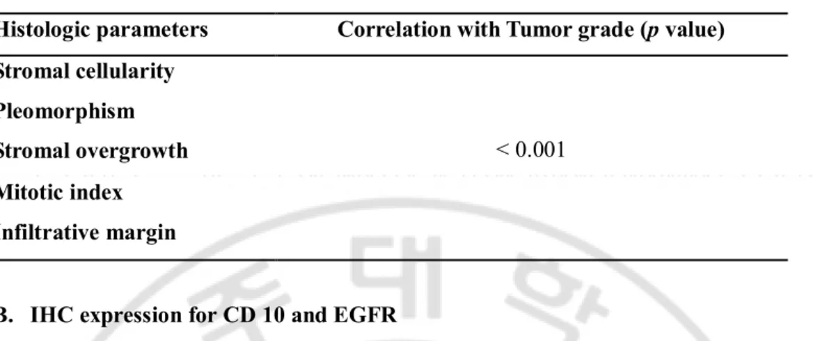
Significant difference could be seen in the age and the degree of malignancy (p = 0.015). The recurrence showed significant correlation with the degree of malignancy (p = 0.022). The relation between the degree of malignancy and the five histological parameters were also significant (All p < 0.001) (Table 2).
Table 1. Clinical features in phyllodes tumor according to degrees of malignancy.
Total Benign Borderline Malignancy p – value Age(year) 36.59±10.81 (11 – 60) 34.33±9.8 (11 – 49) 41.36±11.33 (12 – 50) 42 ±12 (19 – 60) 0.015 Size(mm) 46.93±36.49 (2.42 – 260) 40.59± 25.78 (2.42 – 130) 44.45±29.69 (9 – 120) 74.71±61.17 (20 – 260) 0.160 Recurrence n (%) 7 (8.5) 2 (3.5) 3 (27.3) 2 (14.3) 0.022
6
Table 2. Correlation with the histologic parameters and degree of tumor grade. Histologic parameters Correlation with Tumor grade (p value) Stromal cellularity < 0.001 Pleomorphism Stromal overgrowth Mitotic index Infiltrative margin
B. IHC expression for CD 10 and EGFR
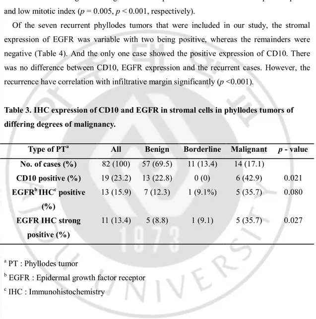
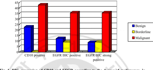
In the entire series, CD10 expression was noted in the stromal cells in 19 of total cases (23.2%), including 13 of 57 cases of benign (22.8%), none of borderline and six of 14 cases of malignant (42.9%). There was a significant difference between benign, borderline and malignant phyllodes tumors (p = 0.041 between benign and malignant, p = 0.017 between borderline and malignant). However, no correlation was showed between benign and borderline cases (Table 3) (Fig 1). IHC expression of EGFR was noted in 13 of total cases (15.9%) ; Seven cases of 57 benign (12.3%) in which five cases were strong positive (8.8%), one case of 11 borderline (9.1%) which was strong positive, five cases of 14 malignant (35.7%) which were all strong positive. There was a significant difference between benign and malignant phyllodes tumors (p = 0.027) and also significant difference was noted in strong positivity between benign and malignant phyllodes tumors (p = 0.021). And there was difference between borderline and malignant cases (p = 0.014). While the difference in the IHC expression of EGFR between benign, borderline and malignant were significant, no different relation between benign and borderline cases.
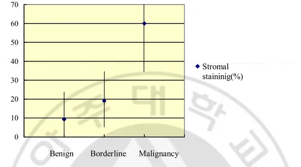
There was an increasing trend of EGFR stromal staining with increasing degree of malignancy. The differences in the percentage of stromal cells with EGFR expression between benign, borderline and malignant tumors were significant (p = 0.001 between benign and borderline tumors, p = 0.021 between borderline and malignant tumors, p < 0.001 between benign and malignant tumors) (Fig 2). The relation between percentage of stromal cells with EGFR expression and the five histological parameters were also significant. For infiltrative margins, EGFR expressions were higher than cases with round margin (p = 0.011). For cellularity, cases with low cellularity showed significantly lower EGFR expression than
7
cases with intermediate or severe cellularity (p < 0.001). For the stromal overgrowth status, cases of overgrowth presence significantly showed higher EGFR expression than cases of overgrowth absence (p <0.001). Similarly, cases with severe pleomorphism and high mitotic index also showed significant higher EGFR expression than cases with low pleomorphism and low mitotic index (p = 0.005, p < 0.001, respectively).
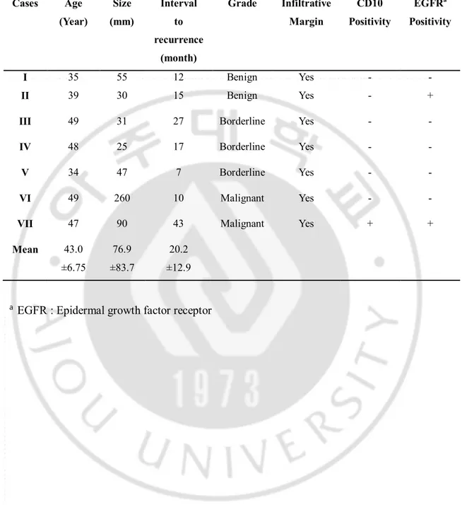
Of the seven recurrent phyllodes tumors that were included in our study, the stromal expression of EGFR was variable with two being positive, whereas the remainders were negative (Table 4). And the only one case showed the positive expression of CD10. There was no difference between CD10, EGFR expression and the recurrent cases. However, the recurrence have correlation with infiltrative margin significantly (p <0.001).
Table 3. IHC expression of CD10 and EGFR in stromal cells in phyllodes tumors of differing degrees of malignancy.
a PT : Phyllodes tumor
b EGFR : Epidermal growth factor receptor c IHC : Immunohistochemistry
Type of PTa All Benign Borderline Malignant p - value
No. of cases (%) 82 (100) 57 (69.5) 11 (13.4) 14 (17.1)
CD10 positive (%) 19 (23.2) 13 (22.8) 0 (0) 6 (42.9) 0.021 EGFRb IHCc positive
(%)
13 (15.9) 7 (12.3) 1 (9.1%) 5 (35.7) 0.080 EGFR IHC strong
positive (%)
8 0 5 10 15 20 25 30 35 40 45
CD10 positive EGFR IHC positive EGFR IHC strong positive
Benign Borderline Malignant
Fig. 1. IHC expression of CD10 and EFGR according to the degree of malignancy. In CD 10 expression, a significant difference in between benign and malignant p = 0.041 and borderline and malignant p = 0.017. However, no correlation was showed between benign and borderline. In EGFR expression, a significant difference between benign and malignant (p = 0.027) and also significant difference was noted in strong positivity between benign and malignant (p = 0.021). And also, the relation between borderline and malignant was significantly different including strong positive cases (all p = 0.014).
9 0 10 20 30 40 50 60 70 0 1 2 3 4 Stromal staininig(%)
Fig. 2. Relations between the percentage of EGFR stromal staining and the degree of malignancy. The percentage of stromal cells with EGFR expression between benign, borderline and malignant tumors were significant (p = 0.001 between benign and borderline tumors, p < 0.021 between borderline and malignant tumors, p < 0.001 between benign and malignant tumors).
10
Table 4. Characteristics of primary phyllodes tumors in recurrent cases.
Cases Age (Year) Size (mm) Interval to recurrence (month) Grade Infiltrative Margin CD10 Positivity EGFRa Positivity I 35 55 12 Benign Yes - - II 39 30 15 Benign Yes - +
III 49 31 27 Borderline Yes - -
IV 48 25 17 Borderline Yes - -
V 34 47 7 Borderline Yes - -
VI 49 260 10 Malignant Yes - -
VII 47 90 43 Malignant Yes + +
Mean 43.0 ±6.75 76.9 ±83.7 20.2 ±12.9
11
IV. DISCUSSION
Although phyllodes tumors typically behave in benign feature, up to 35% of patients die from histological malignancy. In malignant phyllodes tumors, standard treatment includes mastectomy or wide local excision. Unlike invasive ductal carcinoma, axillary lymph node dissection is usually not performed, because the rate of lymph node metastases is less than 1% (Kapiris et al, 2001).However, phyllodes tumors have high recurrence rate, overall 21%, and the percentage breakdown of recurrence by grade of primary tumor are 17%, 25% and 27% for benign, borderline and malignant phyllodes tumor respectively, reported by the World Health Organization (WHO) (Tavassoli et al, 2003). In addition, distant metastasis may also be preceded by local recurrence. And the role of postoperative radiotherapy and chemotherapy has not been fully established in the treatment of phyllodes tumors. Therefore, it is important to classify the tumors according to grades for predicting the recurrence and prognosis. However, there was no definite relationship between the histologic grades of the primary and the recurrence, although most of the recurrent cases were of similar to higher histologic grade. Some studies have shown that stromal overgrowth, infiltrating margins (Feakins et al, 1999), high mitotic rate (Tse et al, 2002) and degree of stromal atypia (Pietruszka et al, 1978) are important predictors of recurrence and prognosis. Of these histological parameters, margin status is the most consistent indicator of local recurrence (Moffat et al, 1995) and our study also shows the same results; the recurrence have close correlation with infiltrative margin significantly (p <0.001).
However, many other authors have found no correlation between histologic characteristics and tumor recurrence (Karim et al, 2009; Tse et al, 2002; Barth et al, 1999; Mokbel et al, 1999). Such results highlight the need for markers that can more reliably predict patient outcome. Up to now, correlations between expressions of p53, Ki67, c-kit, EGFR and stromal CD10, CD34 with tumor grade have been described. Of these, we investigated CD10 and EGFR. The CD10 or the common acute lymphoblastic leukemia antigen (CALLA) is a cell surface neutral endopeptidase, and is expressed by lymphoid precursor cells and normal myoepithelial cells of the mammary glands (Greaves et al, 1975; Greaves et al, 1983). Some studies suggested that increased CD10 expression may reflect the metastatic potential of high-grade lesions of breast phyllodes tumor (Tse et al, 2005; Tsai et al, 2006). One study
12
assessed prognostic significance of stromal CD10 expression in ductal carcinoma in situ (DCIS) cases. The study suggested that CD10 expression correlated with a higher tumor grade and estrogen receptor negative status in DCIS. Therefore, there was a trend toward higher nuclear grade with CD10 expression and further the trial defined CD10 as a marker of DCIS recurrence (Witkiewicz et al, 2010). One recent study showed that the expression of CD10 can be used to predict the occurrence of distant metastasis in phyllodes tumors of the breast (Al-Masri et al, 2011). Otherwise, other authors suggested the effectiveness and utility of stromal CD10 IHC staining for tumor grading was questionable because of the low sensitivity. They said that the combination of CD10 and smooth muscle actin or other factors will provide more information about malignant potential of phyllodes tumor (Yeon et al, 2008; Moritani et al, 2002). One large study investigated for CD10 expression in the stromal cells, a progressive increase in CD10 expression was seen from fibroadenoma to frankly malignant phyllodes tumors (Tse et al, 2005). Similarly, in current study, we found that CD10 expression was low in benign and was higher in malignant tumors. The trend showed significant difference except in the correlation between benign and borderline. Because the borderline cases were much smaller than benign cases and both was similar in histological findings. The relation with metastasis, recurrence between the expressions of CD10 was not defined in our study. The detailed mechanism of CD10 expression and tumorigenesis has been still unknown. However, our results may imply the aggressive potential of tumor behavior with CD10 expression. Therefore, CD10 may be useful in assisting in the diagnosis of benign and frankly malignant phyllodes tumors.
The EGFR is a cell-surface tyrosine kinase receptor for members of the epidermal growth factor family of extracellular protein ligands. Specific mutations causing over-expression of EGFR have been correlated with activation of the receptor that could lead to uncontrolled cell division and subsequent cancer (Lynch et al, 2004). In breast cancer, EGFR appears to be involved in the pathogenesis and progression. About twenty years ago, one study investigated immunohistochemically for expression of epidermal growth factor receptor (EGFR) in 213 breast tumors. They concluded that in contrast to the normal state, EGFR-expression is a rather rare phenomenon in breast carcinoma cells, positively correlated with a declining grade of differentiation (p = 0.025) (Moller et al, 1989). However, still, the evaluation of EGFR as a potential biomarker in phyllodes tumors has not been well
13
established. In a large recent study analyzing 453 phyllodes tumors, the expression of EGFR in phyllodes tumors indicated by immunostaining, progressively increased from benign to malignant tumors (Tse et al, 2009). The results were that the overall positive rate for EGFR was 16.2%, 30.6% and 56% for benign, borderline, malignant and frankly malignant phyllodes tumors, respectively. And FISH demonstrated EGFR gene amplification in 8% of immunohistochemically positive cases. Other author EGFR over-expression was detected in 19%: in 12.5% of benign, in 10% of borderline and 63% of all malignant phyllodes tumors. There are significant correlations between tumor grade on the one hand and EGFR over-expression (Kernisting et al, 2006). Other study investigated the relation of over-expressions of Ki67, BM28, p53 and EGFR family members in neoplastic cells with malignancy and unfavorable clinical course. The study suggested that the expression of EGFR increased with increasing malignancy (Suo et al, 2000). In current study, there was a significant difference in the IHC expression of EGFR between benign, borderline and malignant except in the correlation between benign and borderline. And also the differences in the percentage of stromal cells with EGFR expression between benign, borderline and malignant tumors were all significant. These findings are similar to the result seen in recent above studies. However, we did not investigate the amplification of EGFR in FISH. If the results of FISH would be added, our study could clarify usefulness of EGFR in phyllodes tumors. Therefore further studies are needed. In addition, all five histological parameters showed significant correlation with EGFR expression and the degree of tumor malignancy in our study and so, it could be supporting further evidence about the role of EGFR in phyllodes tumor formation.
From this study, CD10 and EGFR have significant efficacy for discriminating between benign and malignant breast phyllodes tumors although they were not related with recurrence. And also combination with the historical features of the tumors aids in establishment of the correct diagnosis and elevate diagnostic sensitivity in questionable cases.
14
V. CONCLUSIONS
In our study, there was an increasing trend for CD10 and EGFR IHC expression in relation to the degree of malignancy. And the overall percentage of EGFR staining phyllodes tumors is related with degree of malignancy and histological parameters. Additionally tumor margin of five histological parameters had closely relation with tumor recurrence. However, this study did not show the correlation with recurrence and the expression of CD10 and EGFR. Despite the small number of cases included in this study, we believe our findings show that these biologic marker and histological parameters can be used for evaluation of breast phyllodes tumors.
15
VI. REFERENCES
1. Al-Masri, M, Darwazeh G, Sawalhi S, Mughrabi A, Sughayer M, Al-Shatti M: Phyllodes Tumor of the Breast: Role of CD10 in Predicting Metastasis. Ann Surg
Oncol 2076-2086, 2011
2. Barth RJ, Jr: Histologic features predict local recurrence after breast conserving therapy of phyllodes tumors. Breast Cancer Res Treat 57(3): 291-295, 1999
3. Chen CM, Chen CJ, Chang CL, Shyu JS, Hsieh HF, Harn HJ: CD34, CD117, and actin expression in phyllodes tumor of the breast. J Surg Res 94(2): 84-91, 2000 4. Dacic S, Kounelis S, Kouri E, Jones MW: Immunohistochemical profile of
cystosarcoma phyllodes of the breast: a study of 23 cases. Breast J 8(6): 376-381, 2002
5. Esposito NN, Mohan D, Brufsky A, Lin Y, Kapali M, Dabbs DJ: Phyllodes tumor: a clinicopathologic and immunohistochemical study of 30 cases. Arch Pathol Lab
Med 130(10): 1516-1521, 2006
6. Feakins RM, Mulcahy HE, Nickols CD, Wells CA: p53 expression in phyllodes tumours is associated with histological features of malignancy but does not predict outcome. Histopathology 35(2): 162-169, 1999
7. Greaves M, Brown G, Rapson N: Antisera to acute lymphoblastic leukaemia cells.
Clin Immunol Immunopathol 4: 67-84, 1975
8. Greaves M, Hariri G, RA N: Selective expression of the common acute lymphoblastic leukaemia (gp 100) antigen on immature lymphoid cells and their malignant counterparts. Blood 61: 628-639, 1983
9. Iwaya K, Ogawa H, Izumi M, Kuroda M, Mukai K: Stromal expression of CD10 in invasive breast carcinoma: a new predictor of clinical outcome. Virchows Arch 440(6): 589-593, 2002
10. Jara-Lazaro AR, Tan PH: Molecular pathogenesis of progression and recurrence in breast phyllodes tumors. Am J Transl Res 1(1): 23-34, 2009
11. Kapiris I, Nasiri N, A'Hern R, Healy V, Gui GP: Outcome and predictive factors of local recurrence and distant metastases following primary surgical treatment of high-grade malignant phyllodes tumours of the breast. Eur J Surg Oncol 27(8):
723-16
730, 2001
12. Karim RZ, Gerega SK, Yang YH, Soillae A,Carmalt H, Scolyer RA, Lee CS: Phyllodes tumours of the breast: a clinicopathological analysis of 65 cases from a single institution. Breast 18(3): 165-170, 2009
13. Kersting C, Kuijper A, Schmidt H, Packeisen J, Liedtke C, Tidow N, Gustmann C, Hinrichs B, Wülfing P, Tio J, Boecker W, van Diest P, Brandt B, Buerger H: Amplifications of the epidermal growth factor receptor gene (egfr) are common in phyllodes tumors of the breast and are associated with tumor progression. Lab Invest 86(1): 54-61, 2006
14. Kleer CG, Giordano TJ, Braun T, Oberman HA: Pathologic, immunohistochemical, and molecular features of benign and malignant phyllodes tumors of the breast. Mod
Pathol 14(3): 185-190, 2001
15. Lynch TJ, Bell DW, Sordella R, Gurubhagavatula S, Okimoto RA, Brannigan BW, Harris PL, Haserlat SM, Supko JG, Haluska FG, Louis DN, Christiani DC, Settleman J, Haber DA: Activating mutations in the epidermal growth factor receptor underlying responsiveness of non-small-cell lung cancer to gefitinib. N
Engl J Med 350(21): 2129-2139, 2004
16. Mechtersheimer G KK, Born IA, Möller P: Antigenic profile of mammary fibroadenoma and cystosarcoma phyllodes. A study using antibodies to estrogen and progesterone receptors and to a panel of cell surface molecules. Pathol Res Pract 186: 427-438, 1990
17. Moffat CJ, Pinder SE, Dixon AR, Elston CW, Blamey RW, Ellis IO: Phyllodes tumours of the breast: a clinicopathological review of thirty-two cases.
Histopathology 27(3): 205-218, 1995
18. Mokbel K, Price RK, Mostafa A, Wells CA, Carpenter R: Phyllodes tumour of the breast: a retrospective analysis of 30 cases. Breast 8(5): 278-281, 1999
19. Möller P, Mechtersheimer G, Kaufmann M, Moldenhauer G, Momburg F, Mattfeldt T, Otto HF: Expression of epidermal growth factor receptor in benign and malignant primary tumours of the breast. Virchows Arch A Pathol Anat Histopathol 414(2): 157-164, 1989
17
Availability of CD10 immunohistochemistry as a marker of breast myoepithelial cells on paraffin sections. Mod Pathol 15(4): 397-405, 2002
21. Niezabitowski A, Lackowska B, Rys J, Kruczak A, Kowalska T, Mitus J, Reinfuss M, Markiewicz D: Prognostic evaluation of proliferative activity and DNA content in the phyllodes tumor of the breast: immunohistochemical and flow cytometric study of 118 cases. Breast Cancer Res Treat 65(1): 77-85, 2001
22. Packeisen J, Korsching E, Herbst H, Boecker W, Buerger H: Demystified tissue microarray technology. Mol Pathol 56(4): 198-204, 2003
23. Pietruszka M, Barnes L: Cystosarcoma phyllodes: a clinicopathologic analysis of 42 cases. Cancer 41(5): 1974-1983, 1978
24. Suo Z, Nesland JM: Phyllodes tumor of the breast: EGFR family expression and relation to clinicopathological features. Ultrastruct Pathol 24(6): 371-381, 2000 25. Tan PH: 2005 Galloway Memorial Lecture: Breast phyllodes tumours--morphology
and beyond. Ann Acad Med Singapore 34(11): 671-677, 2005
26. Tavassoli F, Devilee P: Pathology and genetics: tumours of the breast and female genital tract. IARC 4, 2003
27. Tsai WC, Jin JS, Yu JC, Sheu LF: CD10, actin, and vimentin expression in breast phyllodes tumors correlates with tumor grades of the WHO grading system. Int J
Surg Pathol 14(2): 127-131, 2006
28. Tse GM, Lui PC, Lee CS, Kung FY, Scolyer RA, Law BK, Lau Ts, Karim R, Putti TC: Stromal expression of vascular endothelial growth factor correlates with tumor grade and microvessel density in mammary phyllodes tumors: a multicenter study of 185 cases. Hum Pathol 35(9): 1053-1057, 2004
29. Tse GM, Lui PC, Vong JS, Lau KM, Putti TC, Karim R, Scolyer RA, Lee CS, Yu AM, Ng DC, Tse AK, Tan PH: Increased epidermal growth factor receptor (EGFR) expression in malignant mammary phyllodes tumors. Breast Cancer Res Treat 114(3): 441-448, 2009
30. Tse GM, Niu Y, Shi HJ: Phyllodes tumor of the breast: an update. Breast Cancer 17(1): 29-34, 2010
31. Tse GM, Putti TC, Kung FY, Scolyer RA, Law BK, Lau TS, Lee CS: Increased p53 protein expression in malignant mammary phyllodes tumors. Mod Pathol 15(7):
18
734-740, 2002
32. Tse GM, Tsang AK, Putti TC, Scolyer RA, Lui PC, Law BK, Karim RZ, Lee CS: Stromal CD10 expression in mammary fibroadenomas and phyllodes tumours. J
Clin Pathol 58(2): 185-189, 2005
33. Witkiewicz, AK, Freydin B, Chervoneva I, Potoczek M, Rizzo W, Rui H, Schwartz GF, Lisanti MP: Stromal CD10 and SPARC expression in ductal carcinoma in situ (DCIS) patients predicts disease recurrence. Cancer Biol Ther 10(4): 391-396, 2010 34. Won RD, Wan JC, Han L: The Clinical and, Pathologic Risk Factors for Local
Recurrence of Phyllodes Tumor. J Breast Cancer 10(1): 85-89, 2007
35. Yeon S, Hye-Kyoung Y: Immunohistochemical Phenotypes of Phyllodes Tumor of the Breast. The Korean Journal of Pathology 42:151-6, 2008
36. Yonemori K, Hasegawa T, Shimizu C, Shibata T, Matsumoto K, Kouno T, Ando M, Katsumata N, Fujiwara Y: Correlation of p53 and MIB-1 expression with both the systemic recurrence and survival in cases of phyllodes tumors of the breast. Pathol
19 - 국문 요약 –
유방의 엽상종양에서 CD10과 EGFR의 발현 분석
아주대학교 대학원 의학과 김 라 미 (지도 교수: 정 용 식) 연구 배경 및 목적: 유방 엽상 종양은 유방에서 발생되는 매우 드문 질환으로서 재발 및 전이를 잘 하는 특성을 가지고 있다. 그러나 유방 엽상종양의 악성 정도와 임상학적인 예후를 예상할 수 있는 특정한 인자는 아직 정확히 밝혀진 바는 없다. 따라서 본 연구의 목적은 유방 엽상종양에서의 CD10과 EGFR의 발현도를 분석하여 이들의 발현과 종양의 악성 정도와의 관계를 평가하고 또한 엽상종양의 분류 및 임상적인 경과와의 관계를 입증하기 위한 것이었다. 재료 및 방법: 1995년 12월부터 2010년 6월까지 본 병원에서 유방 엽상종양으로 수술을 받은 82명의 환자를 대상으로 연구를 진행하였다. 이중 57명은 양성, 11명은 경계성, 14명은 악성 엽상종양으로 분류되었으며 CD10과 EGFR의 발현도를 평가하여 악성도 및 재발과의 관계를 분석하였다. 또한 엽상종양의 조직학적 특성과 악성도의 관계도 함께 평가하였다 결과: 환자들의 평균 나이는 36.59 ± 10.81살 이었으며 범위는 11살에서 60살이었다. 종양의 크기는 평균 46.93 ± 36.49mm였으며 범위는 2.42mm에서 260mm였다 82명의 환자 중 7명은 재발하였다. 나이가 많을수록 엽상종양은 악성일 가능성이 높아진다는 것이 통계적으로 유의한 차이를 보였다. (p = 0.022) 또한 엽상 종양의 조직학적인 특징과 악성도 및 재발과의 관계에서도 유의한 차이를 보였다. (p < 0.001). CD10과 EGFR의 발현도가 양성과 악성 그리고 경계성과 악성 사이에서 유의한 차이를 보였다 (p = 0.041 양성과 악성, p = 0.017 경계성과 악성) 그러나 양성과 경계성 사이에서는 유의한 차이를 보이지 않았다.20
결론: 유방의 엽상종양에서 CD10과 EGFR은 악성으로 갈수록 발현도가 높아지는
통계학적으로 유의한 차이를 보인다.
__________________________________________________________________________ 핵심어: 유방의 엽상 종양, EGFR, CD10