Introduction
Sea cucumbers are echinoderms belonging to the class Holothuroidea. Halothurian are found on the sea floor of deep seas[1], and are characterized by a cylindrical body and rough skin. Sea cucumbers are utilized as a traditional health food and for medicinal purposes in Korea, China, Japan, and other Southeast Asian countri es. Previous studies suggest that sea cucumbers are an effective anticancer [2-5], antiangiogenic, antihypertens ion, anti-inflammatory, and anticoagulant [6-9] agent. Therefore, the sea cucumbers are believed to be a sourc e of high value elements in the functional foods and nutraceutical industry [6]. Among the species of sea cucumbers, Stichopus japonica is classified based on
it color into three groups, namely red, green, and black. These color types exhibit differences in morphology, physiology, and ecology [10]. Moreover, the color affec t the taste and the market price of this species [11]. Red sea cucumber is quantitatively and qualitatively hi gher, and is the most favored among the other color types in Korea [12, 13]. Red sea cucumbers can be found in abundance offshore of the Jeju Island due to its suitable habitat with average water temperature of 15.7°C-18.7°C and gravel bed offshore topography. Th erefore, for this study, we collected a red sea cucumber (S. japonicus) from the Jeju Island and investigated its anticancer activity.
Colorectal cancer (CRC) is the most commonly diagnos ed cancer [4] and the fourth most common cancer causi
* Corresponding author Phone: +82 64 798 6101
E-mail: [email protected]
This is an open-access journal distributed under the terms of the Creative Commons Attribution Non-Commercial License
(http://creativecommons.org/licenses/by-nc/4.0/)
https://doi.org/10.15433/ksmb.2020.12.2.091 ISSN 2383-5400 (Online)
Red Sea Cucumber (Stichopus japonicus) Suppresses Cancer Progression by Promoting the ROS-Me
diated Inhibition of the MAPK Pathway
Jusnseong Kim1, Eun-A Kim1, Nalae Kang1, Youn Kyung Choi2, Soo-Jin Heo1,3* 1
Jeju Marine Research Center, Korea Institute of Ocean Science and Technology (KIOST), Jeju 63349, Korea
2
Department of Medicine, School of Medicine, Jeju National University, 102 Jejudaehakno, Jeju 63243, Korea
3
Department of Biology, University of Science and Technology (UST), Daejeon, South Chungcheong 34113, Korea (Received 3 July 2020, Revised 11 December 2020, Accepted 24 December 2020)
Abstract Stichopus japonicas (red sea cucumbers) inhabit the coastal sea surrounding Jeju
Island, South Korea, and are thought to have various medicinal properties. In this study, we investigated the anticancer activity of a red sea cucumber (S. japonicus) collected from Jeju Island. We obtained the red sea cucumber extract (RSCE), and observed that it inhibited the tumor cell growth and increased reactive oxygen species (ROS) production associated with the induction of apoptosis through the mitogen-activated protein kinase (MAPK) pathway in murine colon carcinoma cells (CT-26). Treatment with RSCE and N-acetylcysteine, which is a ROS scavenger, increased ROS production and apoptosis via the regulation by the MAPK pathway on the ERK and JNK compared with the nontreated group. Therefore, RSCE pro-motes ROS-mediated suppression of the ERK and JNK activation, and subsequently inhibits cancer progression, suggesting that RSCE may be beneficial in treating colon carcinoma.
ng death worldwide [14]. The incidence of obesity and overweight, smoking, stress, lack of exercise, and exces sive alcohol consumption increase the risk of CRC [15]. Although CRC is a treatable cancer, its mortality rate continues to increase. Current treatment options are surgery, chemotherapy, radiotherapy, and targeted thera py. However, these therapies cause secondary complica tions, including metastasis and reduced immune compet ence, and may not prevent the recurrence of tumors [16, 17]. Developing anticancer drugs from natural pro duct materials may improve the effectiveness of these therapies and may eliminate the need for harsh treatmen t measures with concomitant negative effects. In the present study, we confirmed that the red sea cucumber extracts [RSCE] is an effective treatment for CRC [18].
Materials and Methods
1.1 Materials
Red sea cucumber was collected from the sea surroun ding the Jeju Island, South Korea August of 2017. The sample was washed three times with tap water to remov e salt. This was followed by rinsing with fresh water and vacuum drying at 25°C-30°C for 72 h. The vacuum -dried sample was homogenized with a grinder prior to extraction.
1.2 Extraction of red sea cucumber
Dried red sea cucumber powder was extracted with 70% methanol at room temperature until its color was lost. A liquid layer was obtained via filtration, and the filtrate was concentrated using an evaporator under red uced pressure. The concentrated filtrate was dissolved in dimethylsulfoxide (DMSO) and used in experiments.
1.3 Cell cultures
The murine colon cancer cells CT26 and normal cells RIE-1 were purchased from the Korean Cell Line Bank (Seoul, South Korea). The cells were cultured in RPMI medium (Welgene) with 10% fetal bovine serum and 1% antibiotics. The CT26 and RIE-1 cells were culture d every 2 days, and incubated in an atmosphere of 5% CO2 at 37°C.
1.4 Cell viability
Cell viability was measured using the MTT assay. The CT26 cells and RIE-1 cells were seeded on 24-well plates at a concentration of 1.5 × 10⁵ cells/ml. After 16 h, the cells were treated with different concentrations of RSCE (50, 100, and 200 μg/ml), and then incubated for 48 h at 37°C in an incubator in a humidified atmosp here of 5% CO2. MTT solution (5 mg/ml) was added at 50 μl per well for 3 h at 37 °C. Formazan crystals in each well were dissolved in DMSO. The intensity of purple formazan was determined by measuring the absorbance at 540 nm using a microplate reader.
1.5 Measurement of ROS production
The CT26 cells were seeded in 60-mm dishes and co-treated with RSCE (50, 100, and 200 μg/ml) and H2DCFDA dye in the absence at 37°C. After 1 h, the cells were harvested and ROS production was detected using a flow cytometer (BD Accuri C6; BD Bioscience s, San Diego, CA, USA). The data were analyzed using a flow cytometer with measurement in the FL1 channel. For the inhibition of ROS production, the cells were pretreated with N-acetyl-L-cysteine (NAC, 2.5 mM, Sig ma-Aldrich, St. Louis, MO, USA) for 1 h before RSCE and H2DCFDA co-treatment.
1.6 Measurement of apoptosis
The CT26 cells were seeded in 60-mm dishes and trea ted with RSCE (50, 100, and 200 μg/ml) for 48 h, in the absence at 37°C. Thereafter, the cells were stained with annexin V-FITC (BD Bioscience, San Jose, CA, USA), and placed in the dark at room temperature for 15 min, followed by incubation with 7AAD in the dark at room temperature for 15 min. Annexin V- and 7AAD -positive cells were detected using a flow cytometer (BD Accuri C6; BD Biosciences, San Diego, CA, US A). The data were analyzed using a flow cytometer wit h measurements in the FL1 and FL3 channels.
1.7 Western blotting
The CT26 cells were seeded in 6-well plates at a conc entration of 1.5 × 10⁵ cells/ml. After 16 h, the cells were treated with various concentrations of RSCE (50, 100,
and 200 μg/ml), and then incubated for 24 h or 15 min at 37 °C in an incubator in a humidified atmosphere of 5% CO2. After incubation, the cells were harvested and washed twice with cold PBS. The cells were lyzed with RIPA buffer, and equal amounts of protein in total cell lysate were run on 8%-12% sodium dodecyl sulfate polyacrylamide gel electrophoresis and transferred to a nitrocellulose membrane. The membrane was blocked and blotted with the relevant primary antibodies. Anti-a ctin, -Bcl2, -p-ERK, -ERK and -p-JNK antibodies were purchased from Santa Cruz Biotechnology (Santa, CA, USA). Anti-AKT, -JNK1, -p-AKT, -PARP, p-STAT3, and -STAT3 antibodies were obtained from Cell Signal ing (Danvers, MA, USA). The protein bands were visua lized using the ECL Western blotting detection kit (BIO -RAD, USA) and Olympus FV10i Self-Contained conf ocal laser system.
1.8 Statistics
All data are expressed as means ± S.D. Significant differences among the groups were determined using the unpaired Student’s t-test. The P-values of <0.5, <0.
1, < 0.001 were considered statistically significant.
Results
2.1 Effects of RSCE induced apoptosis in
can-cer cells
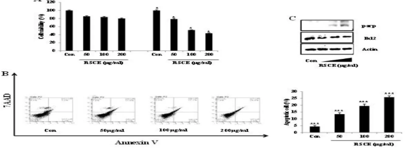
The CT26 and RIE-1 cells were treated with different concentrations (0, 50, 100, and 200 µg/ml) of RSCE for 48 h. RSCE significantly suppressed growth in the CT26 cells, but did not affect RIE-1 cells’ viabilities (Figure 1A). To examine if RSCE inhibited cell growth by inducing apoptotic cell death, we investigated the expression of the extent of annexin V staining and apop tosis-related proteins using flow cytometry and Western blot, respectively. RSCE dose-dependently increased th e presence of apoptotic cells as shown by annexin V and 7AAD double-positive cell staining (Figure 1B). Moreover, RSCE increased the expression of PARP an d decreased the expression of Bcl2 (Figure 1C). Theref ore, RSCE decreased the growth of cancer cells by incr easing apoptosis.
Figure 1. RSCE induced apoptotic cell death: (A) Cells were seeded in 24-well plates and then treated with different concentration
s of RSCE (0, 50, 100, and 200 μg/ml) and DMSO for 48 h. Significant differences among the groups were identified using the unpaired Student’s t-test. * P <0.5, ** P <0.1, and *** P < 0.001 were considered statistically significant. (B) CT26 cells were treated with RSCE (0, 50, 100, and 200 μg/ml) for 24 h, and then harvested. Cells were stained with annexin V and 7AAD in a binding buffer at room temperature in the dark. The stained cells were detected using FACSCalibur. The graph shows the examples of annexin V only-positive cells (early apoptotic cells) and annexin V and 7AAD double-positive cells (late apoptosis cells) from the total stained cells. * P <0.5, ** P <0.1, and *** P < 0.001 were considered statistically significant. (C) The CT26 cells were treated with RSCE (0, 50, 100, and 200 μg/ml) for 24 h, and whole lysates were analyzed using Western blot with PARP and Bcl2 antibodies. Actin was used as the internal control.
2.2 RSCE induces apoptosis via MAPK pathway
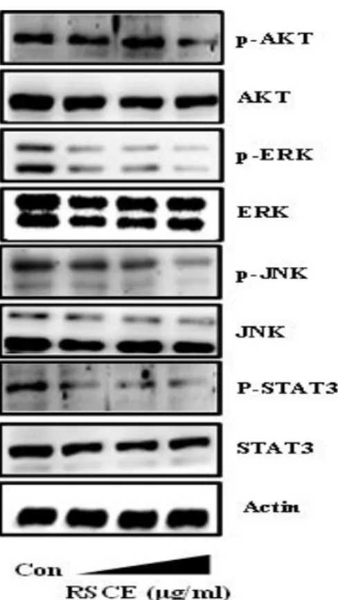
To investigate the anticancer effects of RSCE, we identi fied which intracellular signaling pathways were involve d. The CT26 cells were treated with different concentrati ons (0, 50, 100, and 200 µg/ml) of RSCE for 15 min and then subjected to Western blotting. RSCE considerab ly decreased the phosphorylation of the AKT, ERK, JNK and STAT3 in a dose-dependent manner (Figure 2).
Figure 2. RSCE inhibits the cell signaling pathway: (A) The
CT26 cells were treated with RSCE (0, 50, 100, and 200 μg/ml) for 15 min, and the whole lysates were analyzed using Western blot with anti-p-AKT, -AKT, -p-ERK, -ERK, - p-JN K, -JNK, -p-STAT3 and -STAT3 antibodies. Actin was used as the internal control.
2.3 RSCE increases ROS production in cancer
cells
The regulation of ROS production plays an important role in cell proliferation and apoptosis. The CT26 cells were treated with different concentrations (0, 50, 100,
and 200 µg/ml) of RSCE to confirm ROS production. RSCE increased ROS production by 4.5%, 8.2%, and 18.0% in 50, 100, and 200 µg/ml respectively, compare d with the control (Figure 3A). Further, we used co-trea tment with RSCE and NAC to confirm ROS productio n. NAC, which is well-known ROS scavenger suppress ed RSCE-induced ROS production as shown in Figure 3B. Thus, RSCE regulates ROS production in cancer cells.
Figure 3. RSCE increases ROS production in cancer cells:
(A) The CT26 cells were co-treated with (0, 50, 100, and 200 μg/ml) of RSCE and H2DCFDA dye for 1 h at 37°C. ROS production was detected using FACSCalibur. The graph shows H2DCFDA-positive cells from the total cells. (B) The cells were pretreated for 1 h with or without NAC, followed by exposure to 200 μg/ml of RSCE and H2DCFDA dye for 1 h at 37°C. * P <0.5, ** P <0.1, *** P < 0.001 were consider ed statistically significant.
2.4 RSCE induces ROS generation causes
MAPK-kinase pathway and apoptosis
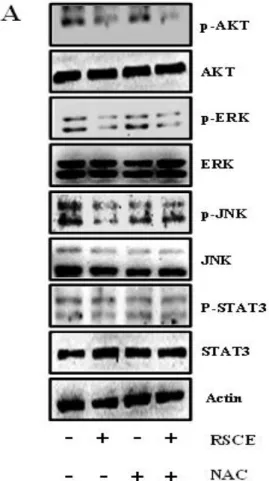
ROS production inhibits the MAPK pathway. Therefo re, we investigated the MAPK expression in cells after co-treatment with RSCE and NAC. We observed that RSCE-mediated reduction in the phosphorylated MAP K pathway kinases (ERK and JNK) was restored after treatment with NAC (Figure 4A). Therefore, RSCE ind uces apoptosis through ROS production by the MAPK pathway.
Figure 4. RSCE-induced ROS production causes the MAP
kinases’ inhibition and apoptosis: (A) The CT26 cells were pretreated with NAC (2.5 mM) for 1 h and then exposed to RSCE (200 μg/ml) for 15 min. p-AKT, AKT, p-ERK, ER K, p-JNK, JNK, p-STAT3 and STAT3 protein expression lev els were analyzed using Western blot. Actin was used as the loading control. * P <0.5, ** P <0.1, and *** P < 0.001 were considered statistically significant.
Discussion
In this study we confirmed the potential anticancer properties of red sea cucumber from the Jeju Island, and showed that the components of red sea cucumber produce cell death in the CT26 colorectal cancer cells. Advancements in the treatment of colorectal cancer usi ng various methods are ongoing, and currently the focu s is on isolates from natural products because of their anticancer properties (2, 19). Further, it is necessary to develop new anticancer drugs with high stability and excellent anticancer effects (20). Many natural compou nds appear to inhibit the development of cancer cells via ROS production (21). ROS damages DNA and lead s to gene mutations, and once damaged, the cells that are not removed become stem cells and finally cancer cells (22, 23). Therefore, inhibiting the production of ROS can prevent cancer cell proliferation. Our results showed that RSCE can regulate ROS levels and enhanc e apoptosis in cancer cells. ROS production, which caus es mitochondrial oxidative stress, involves and regulate s intracellular signaling pathways. Higher levels of RO S contribute to apoptosis through impaired receptors and mitochondrial oxidative stress pathways, mitochon dria dysfunction, and impaired cerebral energy metabol ism (24-26). The phosphorylation of specific proteins (MAPK) in the cell affects cancer cell death (27, 28). RSCE led to ROS-mediated apoptosis by down-regulati on of the ERK and JNK phosphorylation. The MAPKs are serine/threonine protein kinases that play a major role in signal transduction in the cell nucleus (29). Studi es have shown that ROS is involved in the regulation of various signaling pathways, including the MAPK pat hway and transcription factors. The ERK signaling path way modulates various processes, such as survival, diff erentiation, proliferation, and migration, depending on the particular cell type (30, 31). The JNK inhibitors have been considered for cancer treatment because they can interfere with DNA repair in response to genotoxic drugs (32). Both ERK and JNK can affect cell survival in response to oxidant damage (33, 34). RSCE inhibits the activation of the ERK and JNK proteins associated
with apoptosis, suggesting its involvement in the signali ng initiation of mitochondrial-mediated cell death in the CT26 cells (Figure 2). Further, NAC inhibited RSCE induced apoptosis through the MAPK pathway.
Conclusion
Thus, RSCE induces apoptosis via the ROS-mediated MAPK pathway in CRC. Therefore, RSCE, a vacuum- dried extract of a red sea cucumber from the Jeju Islan d, may provide an important option for future treatment of CRC.
Acknowledgements
This research was supported by a research grants from the Korea Institute of Ocean Science and Technology (PE99822) and was partially supported by the National Research Foundation of Korea(NRF) grant funded by the Korea government(MSIT)( NO. NRF_2017R1C1B 2011398).
References
1. Ahmedin Jemal, Freddile Bray, Melissa M Center, Jacques Ferlay, Elizabeth Ward and David Forman. 2011. Global cancer statistics. CA: a cancer journal for clinicians 61(2): 69-90.
2. Allal Ouhtit, Rajiv Lochan Gaur, Mohamed Abdrabo h, Shubha K. Ireland, Prakash N Rao, Shailaja G Raj, Hamad Al-Riyami, Somya Shanmuganathan, Ish ita Gupta, Subramanyam N Murthy, Andrew Hollenb ach and Madhwa HG Raj. 2013. Simultaneous inhibit ion of cell-cycle, proliferation, survival, metastatic pathways and induction of apoptosis in breast cancer cells by a phytochemical super-cocktail: Genes that underpin its mode of action. Journal of Cancer 4(9): 703.
3. Atsushi Matsuzawa and Hidenori Ichijo. 2005. Stress -responsive protein kinases in redox-regulated apopt
osis signaling. Antioxidants & redox signaling 7(3-4): 472-481.
4. Bobbili V.V. Pardhasaradhi, A. Mubarak Ali, A. Leel a Kumari, Pallu Reddanna and Ashok Khar. 2003. Phycocyanin-mediated apoptosis in ak-5 tumor cells involves down-regulation of bcl-2 and generation of ros. Molecular cancer therapeutics 2(11): 1165-1170. 5. Chong Sun and Rene Bernards. 2014. Feedback and
redundancy in receptor tyrosine kinase signaling: Rel evance to cancer therapies. Trends in biochemical sci ences 39(10): 465-474.
6. Coutney L. Robertson, Susanna Scafidi, Mary C. Mc Kenna and Gary Fiskum. 2009. Mitochondrial mecha nisms of cell death and neuroprotection in pediatric ischemic and traumatic brain injury. Experimental ne urology 218(2): 371-380.
7. Dennis J. Goussetis and Leonidas C. Platanias. 2010. Arsenic trioxide and the pi3k/akt pathway in chronic lymphocytic leukemia. Clinical Cancer Research: cli ncanres. 1496. subyr.
8. Emile Tan, Henry Tilney, Mike Thompson, Jason Smith, Paris P. Tekkis and Britain AoCoG. 2007. The united kingdom national bowel cancer project–e pidemiology and surgical risk in the elderly. Europea n journal of cancer 43(15): 2285-2294.
9. Fang Tian, Xiongwen Zhang, Yungyuang Tong, Yan ghua Yi, Shilong Zhang, Ling Li, Peng Sun, Liping Lin and Jian Ding. 2005. Pe, a new sulfated saponin from sea cucumber, exhibits anti-angiogenic and anti -tumor activities in vitro and in vivo. Cancer biology & therapy 4(8): 874-882.
10. Haider Raza, Annie John and Sheela Benedict. 201 1. Acetylsalicylic acid-induced oxidative stress, cell cycle arrest, apoptosis and mitochondrial dysfunctio n in human hepatoma hepg2 cells. European journal of pharmacology 668(1-2): 15-24.
11. Hua Li Xu, Xiao Feng Yu, Shao Chun Qu, Xiang Ru Qu and Yan Fang Jiang. 2012. Juglone, from juglans mandshruica maxim, inhibits growth and in duces apoptosis in human leukemia cell hl-60 throu gh a reactive oxygen species-dependent mechanis m. Food and chemical toxicology 50(3-4): 590-596.
12. Irina Vasilevskaya and Peter J. O’Dwyer. 2003. Rol e of jun and jun kinase in resistance of cancer cells to therapy. Drug resistance updates 6(3): 147-156. 13. Jin Hyang Yang. 2005. The effect of foot reflexolog y on nausea, vomiting and fatigue of breast cancer patients undergoing chemotherapy. Journal of Kore an Academy of Nursing 35(1): 177-185.
14. Jung-Ha Kang, Ki-Hwan Yu, Jung-Youn Park, Chu l-Min An, Je-Cheon Jun and Sang-Jun Lee. 2011. Allele-specific pcr genotyping of the hsp70 gene po lymorphism discriminating the green and red color variants sea cucumber (apostichopus japonicus). Jou rnal of genetics and genomics 38(8): 351-355. 15. Katherine L Dunn, Paula S Espino, Bojan Drobic,
Shihua He and James R Davie. 2005. The ras-mapk signal transduction pathway, cancer and chromatin remodeling. Biochemistry and Cell Biology 83(1): 1-14.
16. Kyung-Uk Park, Jae-Yong Kim and Kwon-Il Seo. 2009. Antioxidative and cytotoxicity activities again st human colon cancer cells exhibited by edible crud e saponins from soybean cake. THE KOREAN SO CIETY OF FOOD PRESERVATION 16(5): 754-75 8.
17. Laura Poillet-Perez, Gilles Despouy, Regis Delage-Mourroux and Michael Boyer-Guittaut M. 2015. Int erplay between ros and autophagy in cancer cells, from tumor initiation to cancer therapy. Redox biol ogy 4 184-192.
18. Li L and Li Q. 2010. Effects of stocking density, temperature, and salinity on larval survival and gro wth of the red race of the sea cucumber apostichopu s japonicus (selenka). Aquaculture international 18 (3): 447-460.
19. MAnami Kan-No and Akihiro Kijima. 2003. Geneti c differentiation among three color variants of japan ese sea cucumber stichopus japonicus. Fisheries sci ence 69(4): 806-812.
20. Nahed El-Najjar, Manal Chatila, Hiba Moukadem, Heikki Vuorela, Matthias Ocker, Muktheshwar Gan desiri, Regine Schneider-Stock and Hala Gali-Muht asib.2010. Reactive oxygen species mediate thymoq
uinone-induced apoptosis and activate erk and jnk signaling. Apoptosis 15(2): 183-195.
21. Naveena B. Janakiram, Altaf Mohammed and Chint halapally V. Rao. 2015. Sea cucumbers metabolites as potent anti-cancer agents. Marine drugs 13(5): 2909-2923.
22. Paul D. Ray, Bo-Wen Huang and Yoshiaki Tsuji. 2012. Reactive oxygen species (ros) homeostasis an d redox regulation in cellular signaling. Cellular sig nalling 24(5): 981-990.
23. Pawson DL. 1980. Holothuroidea. Studies in Geolo gy, Notes for a Short Course 3(175-189.
24. Ryan B. Corcoran, Hiromichi Ebi, Alexa B. Turke, Erin M. Coffee, Michya Nishino, Alexandria P. Co gdill, Ronald D. Brown, Patricia Della Pelle, Dora Dias-Santagata and Kenneth E. Hung, Keith T. Flah erty, Adriano piris, Jennifer A wargo, Jeffrey Settle man, Mari Mino-Kenudson and Jeffrey A. Engelma n. 2012. Egfr-mediated re-activation of mapk signal ing contributes to insensitivity of braf mutant colore ctal cancers to raf inhibition with vemurafenib. Can cer discovery: CD-11-0341.
25. Saito M, Kunisaki N, Urano N and Kimura. 2002. Collagen as the major edible component of sea cucu mber (stichopus japonicus). Journal of food science 67(4): 1319-1322.
26. Samir Attoub, Kholoud Arafat, Tamam Khalaf, Sha hrazad Sulaiman and Rabah Iratni. 2018. Frondosid e A Enhances the anti-cancer effects of oxaliplatin and 5-Fluorouracil on colon cancer cells. Nutrients 10(5). 560.
27. Sara Bordbar, Farooq Anwar and Nazamid Saari. 2011. High-value components and bioactives from sea cucumbers for functional foods—a review. Mari ne drugs 9(10): 1761-1805.
28. Shan Deng, Bing Hu, Hong-Mei An, Qin Du, Ling Xu, Ke-Ping Shen, Xiu-Feng Shi, Meng-Meng Wei and Yang Wu. 2013. Teng-long-bu-zhong-tang, a chinese herbal formula, enhances anticancer effects of 5-fluorouracil in ct26 colon carcinoma. BMC co mplementary and alternative medicine 13(1): 128. 29. Shiguo Chen, Changhu Xue, Liang Yin, Qingjuan
Tang, Guangli Yu and Wengang Chai. 2011. Comp arison of structures and anticoagulant activities of fucosylated chondroitin sulfates from different sea cucumbers. Carbohydrate Polymers 83(2): 688-696. 30. Shin-Hwar Wu, Liang-Wen Hang, Jai-Sing Yang,
Hung-Yi Chen, Hui-Yi Lin, Jo-Hua Chiang, Chi-Ch eng Lu, Jiun-Long Yang, Tung-Yuan Lai, Yang-Chi ng Ko and Jing-Gung Chung. 2010. Curcumin indu ces apoptosis in human non-small cell lung cancer nci-h460 cells through er stress and caspase cascade -and mitochondria-dependent pathways. Anticancer research 30(6): 2125-2133.
31. Siran Yu, Xuewei Ye, Lu Chen, Xin Xie, Qian Zho u, Xiao-Yuan Lian and Zhizhen Zhang. 2015. Cyto toxic and anti-colorectal tumor effects of sulfated saponins from sea cucumber holothuria moebii. Phy tomedicine 22(12): 1112-1119.
32. U von Freeden-Jeffry, P Vieira, L A Lucian, T McN eil, S E Burdach and R Murray. 1995. Lymphopenia in interleukin (IL)-7 gene-deleted mice identifies iL -7 as a nonredundant cytokine. Journal of Experime ntal Medicine 181(4): 1519-1526.
33. Xiao-Fang Che, Shota Moriya, Chun-Lei Zheng, Akihisa Abe, Akio Tomoda and Keisuke Miyazaw a. 2013. 2-aminophenoxazine-3-one-induced apopt osis via generation of reactive oxygen species follo wed by c-jun n-terminal kinase activation in the human glioblastoma cell line ln229. International journal of oncology 43(5): 1456-1466.