Effects of MMP-2 activation and FSH or LH Hormone
Supplementation on Embryo Development in In Vitro
Fertilization of Porcine
Sang Hwan Kim1 and Jong Taek Yoon1,2, *
1Institute of Genetic Engineering, Hankyong National University, Anseoung, Gyeonggi-do 456-749, Korea
2Department of Animal Life Science, Hankyong National University, Anseoung, Gyeonggi-do 456-749, Korea
Abstract
The purpose of this study was to analyze whether FSH and LH hormone treatment directly or indirectly affect embryo development in embryonic development. To determine this, we compared the development of embryonic cells through the expression pattern of MMPs. As a result, 33.8% of blastocysts were formed in FSH added group, 20.8% in LH added group and 10% in FSH + LH added group. In addition, the activity of MMP-9 was highly detected in the FSH-added group, and the expression of Casp-3 was much lower than that of the other groups. These results suggest that the addition of FSH seems to increase the activity of MMP-9 in embryonic cells, and that LH, on the contrary, may activate MMP-2 activity. In addition, the expression level of MMP-2 in the FSH-added group was high in the Trophoblast cell group and in the LH-added group, the hormone ideal secretion might affect the development of the embryonic cell.
Received 18 December 2018
Revised 19 December 2018
Accepted 20 December 2018 Key Words : MMPs, TIMPs, FSH and LH, Embryo, Porcine
* The authors contributed equally to this work
INTRODUCTION
In vitro fertilization of pigs and development of embryos is a very important study in reproduction and production of transgenic animals (Abeydeera 2001). The study development of in vitro fertilization system of pigs has resulted in the production of many embryos (Yoshioka et al., 2001), but the production of in vitro fertilized eggs is still very lowly compared with the production of in vitro fertilized eggs (Kikuchi et al., 2008). Especially in the production of successful in vitro fertilized eggs, the action of FSH and LH hormone expressed in early oocyte maturation is very important. The role of FSH hormone is to regulate the activity of cumulus cells in the maturation of the oocyte, to affect the cytoplasm and nuclear maturation of the oocyte and to make a functional change in the hormone to form estrogen biosynthesis, it is important to play a role in the final differentiation of the oocyte (Hsueh et al., 2010). In particular, several studies have demonstrated that FSH plays a role in the maturation of early oocytes (development of cumulus cell and nuclear maturation) and in the development of cumulus cells (Roy and Greenwald 1989; Cain et al., 1995), and is known to act on the formation of primary follicles and preantral follicles, other studies did not improve the growth of preantral follicles (Li et al., 1995; Boland et al., 1993). The action of LH hormones is accommodated in granulosa cells and cavernosal cells from large antral follicles (Bao et al., 1997) and can play many roles in the development of follicles, but most studies focus on the physiological function of follicles. Regarding the maturation of oocytes, the important function of FSH and LH is that FSH affects the development of oocytes, but does not directly participate in nuclear maturation (Suss et al., 1988). In the end, hormones involved in the nuclear maturity of the oocytes can be seen to be formed by the action of the LH (Dominko et al., 1992). Studies by Keefer et al., 1993 have shown that the action of LH in the maturation of porcine oocytes will play an important role in the final blastocyst production. Previous studies have focused on the effects of FSH and LH on the maturation of oocytes, but there is little information on the effects of FSH and LH on development of embryos. Therefore, this study was conducted to investigate the effects of apoptosis and MMPs (Matrix metalloproteinases) and TIMPs (Tissue inhibitor of metalloproteinase) on embryo development in zygotes cultured on in vitro culture media supplemented with FSH or LH hormone.
MATERIALS AND METHODS
1. Oocytes maturation
Pig ovaries were collected from gilts at a local slaughterhouse and transported within 2 h to the laboratory in preincubated saline solution (with 100 IU/mL penicillin G and 100 μg/mL streptomycin) at 30 35 °C. Follicular fluid from follicles 3 to 6 mm in diameter was aspirated using an 18-gauge needle attached to a 10 ml disposable syringe. With the aid of a dissecting microscope, all naked oocytes, cumulus-oocyte complexes (COCs), and debris were removed from the aspirate and washed twice in phosphate buffered saline (PBS). Grade 1(oocytes with cumulus cells and good cytoplasm) oocytes in the cumulus oocyte complexes were collected, and placed under a microscope after they were washed and prepared in Hepes-buffered tissue culture medium-199 (TCM-199; Gibco, MD, USA : 50 μg/mL genatamycin (SK chemical, Geyonggi, Korea), 0.3% (w/v) fatty acid free bovine serum albumin (BSA; Sigma, MO, USA)). After preparation of the oocytes in TCM-199, they were washed once with preincubated IVM medium (2.5 μg/mL FSH (Sigma, MO, USA), 1 μg/mL estradiol-17β (Sigma, MO, USA), 20ng/mL epidermal growth factor (Sigma, MO, USA) and 50 μg/mL gentamycin (SK chemical, Geyonggi, Korea) with 10%fetal bovine serum (FBS, Gibco, MD, USA)) and checked for the affinity of final oocytes and binding ability of cumulus cells. They were transferred into about 500 uL of IVM medium in a 4-well dish (20 to 25 oocytes in each well) and matured in vitro at 39°C in a 5% carbon dioxide incubator at 40 h.
2. In-vitro fertilization
After maturation for 24 h in vitro, small COCs were removed from mature medium (IVM-M199) and washed three times in TL-Hepes (Bio-Whittaker, Walkersville, MD, USA) and divided into 20-25 groups. Then wash three times in the in-vitro fertilization medium (BO : Brackett and Zuelke 1993) and transfer the mineral oil (Becton Dickinson, Franklin Lake, New Jersey, USA) to 50 μL of fertilized medium in a Petri dish. The dishes were kept at 5% CO2 in air at 39 ° C. Sperm used for fertilization were washed twice by centrifugation at 453 x g for 8 minutes. Subsequently, sperm pellets were re-suspended in the appropriate sperm wash media to a volume of 250 μL. After final washing, sperm motility, and concentration was determined. Thirty microliters (30 μL) of the re-suspended sperm was added to each fertilization drop, giving a total concentration of 1 × 10⁷ sperm/mL in each of the
IVF media. Oocytes were then incubated with the washed sperm for 6 h in 5% CO2 in air at 39 °C.
3. In-vitro culture
At the completion of IVF, presumptive zygotes were denuded of remaining cumulus cells and loosely bound sperm, washed in NCSU-32, placed in 50 µl droplets of NCSU-32 (Four hormone treatment groups : 1) Hormones non-treatment group, 2) FSH (2.5IU/ml), 3) LH (2.5IU/ml), 4) FSH and LH (each : 2.5IU/ml)), and incubated in 6% CO2, 5% O2 and 89% N2 at 38.5ºC. On Day 4 of IVC, cleavage was assessed and the NCSU-32 was supplemented with 10% fetal bovine serum (FBS; heat inactivated, Australian origin; Gibco). Blastocyst formation was assessed on Day 7 of IVC.
4. Gelatin Zymography
Gelatinase activity was localized by in situ zymography following a previously described method (Khandoker et al., 2001; Nemori and Tachikawa 1999). Used GN film (Fuji Photo Film Co., Ltd, Tokyo, Japan) coated with a gelatin base emulsion was employed to detect and localize the gelatinase activity of the underlying tissue. Seven-micron-thick, unfixed ovarian cryosections were mounted onto the coated film, followed by incubation for 24 h at 37°C and staining with bearish scarlet (BS; Chroma-Gesellschaft, mbH & Co., Munster, Germany) and hematoxylin. Gelatinase activity was detected and localized when a specific pattern of gelatin digestion was indicated by a white color caused by the weaker staining of BS.
5. ELISA
For western blots and ELISA, total protein was extracted from ovarian tissues using Pro-Prep solution (Intron) according to the manufacturer’s instructions. Total protein was quantified using a Bradford protein assay kit (Bio-Rad). For quantification of specific protein from the culture medium and cell proteins, samples diluted in assay buffer were used to coat a 96-well ELISA plate overnight at 4°C. The plate was then washed twice using washing buffer (1× PBS with 2.5% Triton X-100), and blocked using 1% BSA blocking solution at RT for 3 h. Primary antibodies (MMP-2 (ab78796, Abcam, Cambridge, UK), MMP-9(Santa Cruz Biotechnology Inc., Texas USA), TIMP-2 (sc-9905, Santa Cruz Biotechnology Inc., Texas USA), TIMP-3 (sc-6836, Santa Cruz Biotechnology Inc., Texas USA), Casp-3, PCNA, FSH-r and LH-r) were detected overnight at 4°C. After
washing, immune reactions were detected using secondary antibodies (anti-rabbit, (sc-2054, Santa Cruz Biotechnology Inc., Texas USA) and anti-mouse (sc-2054 and sc-2031, Santa Cruz Biotechnology Inc., Texas USA)) for 2 h, and substrate solution (BD) was added to initiate the reaction. The reaction was stopped with 1 M NH2SO4, and absorbency was measured at 450 nm. 6. Western Blot
Samples containing 30 μg of protein were separated by SDS-PAGE (in duplicate) on a 12% SDS-polyacrylamide gel and transferred to an Immuno-Blot PVDF membrane (Bio-Rad, CA, USA). The membrane was blocked using blocking buffer consisting of 3% BSA in TBST (0.1% Tween 20, 50 mM Tris-HCl (pH 7.6), 200 mM NaCl) for 3 h at RT. Following this, the membrane was washed 3 times for 15 min with washing buffer (TBST), followed by overnight incubation at 4°C with primary antibodies (TIMP-2, TIMP-3 and β-actin(Santa Cruz Biotechnology Inc., Texas, USA)) (diluted 1:1000 in blocking buffer). The membrane was then washed thrice with TBST buffer for 15 min each, and incubated for 2 h with HRP-conjugated secondary antibodies (diluted 1:5000 in blocking buffer). Detection was carried out using an ECL detection kit with 5 min incubation in a dark room. The detection reagent was drained, and the membrane was exposed to a sheet of diagnostic film in a film cassette for 1 to 30 min.
7. Immunofluorescence
We cultured samples (Blastocyst) on sterilized glass coverslips and fixed them with 4 % paraformaldehyde, followed by blocking with 0.1 % BSA in PBS. Dehydration and permeabilization were performed by freezing the slides at 20 °C in 5 mM 0.1 % Triton X-100 in PBS. After blocking with 3 % BSA in PBS, slides were incubated with an antibody against the active form of MMP-2, MMP-9, TIMP-2, and TIMP-3 at 1:150 dilutions. The slides were washed and incubated with anti-rabbit and anti-mouse IgG conjugated to Alexa Fluor 488 or Alexa Fluor 594 (Molecular Probes). Nuclei were counterstained with 1 ug/mL Hoechst 33258, and slides were mounted using fluorescent mounting medium (Dako, Carpinteria, CA). Images were acquired using Olympus AX70 fluorescence microscope fitted with a CCD color camera.
8. Statistical analysis
using the Statistical Analysis System (SAS Institute, version 9.4, Cary, NC, USA). Differences among treatments were determined by using Duncan’s multiple range tests. The statistical significance was established at p < 0.05.
RESULTS
1. Development of blastocysts and expression of MMPs by hormone addition during in vitro culture
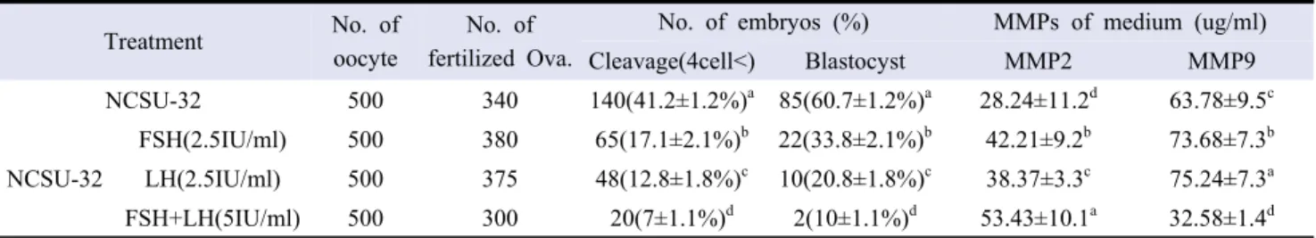
The results of analysis of embryonic development and MMPs expression in FSH, LH and FSH + LH supplemented groups after in vitro fertilization are the same as those of Table 1. In our results, the development of more than 4 cells of total embryos was 17.1 ± 2.1% in FSH treated group, 12.8 ± 1.8% in LH and 7 ± 1.1% in FSH + LH. The development of blastocysts according to the above results was 33.8 ± 2.1% in FSH group, which was the highest (p <0.05). The concentration of MMPs was significantly
higher in FSH + LH (53.43 ± 10.1 μl/ml) in MMP-2 and 38.37 ± 3.3 μl/ml in LH, the lowest in hormone group. MMP-9 was higher in the LH group (75.24 ± 7.3ul/ml) than the other groups, but not significantly different from the FSH group.
2. Expression of MMPs and TIMPs in embryos
The results of analysis of MMPs and TIMPs in mature embryos in each hormone treated group are shown in Figure 1. Expression of MMPs was similar to that of Table 1. However, the activity of MMPs was slightly different, and the activity of MMP-9 was confirmed in all treatment groups except FSH + LH group. However, the activity of MMP-2 was the lowest in the control group and very high in the LH-treated group. In the case of TIMPs which are inhibitors of MMPs, TIMP-3, which is an inhibitor of MMP-9, was significantly higher in FSH + LH group and TIMP-2 was relatively higher in control group and very low in LH group.
Treatment No. of
oocyte
No. of fertilized Ova.
No. of embryos (%) MMPs of medium (ug/ml)
Cleavage(4cell<) Blastocyst MMP2 MMP9 NCSU-32 500 340 140(41.2±1.2%)a 85(60.7±1.2%)a 28.24±11.2d 63.78±9.5c NCSU-32 FSH(2.5IU/ml) 500 380 65(17.1±2.1%)b 22(33.8±2.1%)b 42.21±9.2b 73.68±7.3b LH(2.5IU/ml) 500 375 48(12.8±1.8%)c 10(20.8±1.8%)c 38.37±3.3c 75.24±7.3a FSH+LH(5IU/ml) 500 300 20(7±1.1%)d 2(10±1.1%)d 53.43±10.1a 32.58±1.4d
*FSH : Follicle stimulation hormone, LH : Luteinizing Hormone, MMPs : Matrix metalloproteinases.
Table 1. Mean (%) fertilization rates and embryo development on the IVC media
Figure 1. Expression of activation MMPs and TIMPs. A : Zymorgraphy, B : Western blot, C : ELISA analysis, 1) Hormone non-treat group, 2) FSH group, 3) LH group, 4) FSH+LH group. a,b,c,dDifferent letters within the same column represent a significant difference (p<0.05).
3. Expression of Casp-3, PCNA and hormone-receptor in embryos
In the present study, expression of Casp-3 was significantly higher in the LH group than in the other treatment groups, and PCNA was highly expressed in the FSH group. FSH-receptor was significantly higher in FSH group, but it gradually decreased to LH group and FSH + LH group, but it was found that FSH + LH group was gradually expressed in LH-receptor group (Figure 2).
4. Expression of MMP-2 and Casp-3 in blastocysts developed in each hormone-treated group
The expression of MMP-2 and CAsp-3 in blastocysts developed in each hormone-treated group is shown in Figure 3. As a result, the expression of MMP-2 was found to be expressed around the trophoblast cell of the blastocyst. In the control group not added to the hormone, the expression of MMP-2 was very low, but the expression of MMP-2 was high in the hormone-added control group. In the FSH group, expression was higher in the periphery
of the blastocyst but in the LH group and FSH + LH group, the expression was higher in the inner cell mess section. Expression patterns of Casp-3 were also similar to those of MMP-2 expression. In the inner cell mess of LH and FSH + LH, expression was higher than that of the other groups.
DISCUSSION
The purpose of this study was to analyze the effects of hormone addition on the blastocyst development during in vitro maturation of pig embryos. In the development of porcine oocytes, according to Anderiesz et al., 2000, the combination of LH and FSH suggests that LH and FSH-induced effects on the oocyte nutrition and appropriate embryonic developmental environment control the intercellular signaling pathways. It can improve embryo development. Cortvrindt et al., 1998 also reported that FSH and LH hormones affect the ability of the oocyte to synthesize proteins and affect cell differentiation (Moor et al., 1985). However, the mechanism by which the addition of FSH
Figure 2. ELISA assay for Casp-3, PCNA, FSH-r and LH-r protein expression in porcine embryo. a,b,c,dDifferent letters within
and LH plays a direct role in embryonic development is unknown. In our study, we observed that the incidence of blastocysts was significantly different in the in vitro culture of FSH, LH, FSH and LH in NCSU-32 culture medium. This suggests that the addition of hormones does not play a positive role in embryo development and is different from the results presented by Anderiesz et al., 2000. In particular, the effects of MMPs on the development of embryonic cells were analyzed, suggesting that LH stimulation expresses both increased MMPs and increased Casp-3 in embryonic cells. However, the results of FSH were different. The expression of MMP-2 was found in the trophoblast cell and the expression of Casp-3 was significantly lower than that of the other treatment groups. These results are similar to the results of Wen-Jui et al., 2015, which shows that embryonic development affects embryonic development depending on the presence or absence of MMPs. In addition, as in the study of Imai et al., 2002, as the expression of MMPs in the cytoplasm has a positive effect on cell division, the differences in the expression and activity of MMPs in this study may clearly affect embryo development. However, it is not known exactly how FSH and LH hormone affect the expression mechanism of MMP-2 and MMP-9, and it cannot be concluded that it directly affects embryo development. However, this study suggests that the
treatment of FSH and LH may have some effect on embryonic development. Therefore, this study will be an important basic data on the effect of Inappropriate secretion of hormone on embryo development.
ACKNOWLEDGMENTS
This work was supported by a research grant from Hankyong National University for a academic exchange program in 2016.
REFERENCES
Abeydeera LR. In vitro fertilization and embryo development in pigs. Reprod Suppl 2001; 58: 159 173.
Anderiesz A, Ferraretti AP, Magli C, Fiorentino A, Fortini D, Gianaroli L. 2000. Effect of recombinant human gonadotrophins on human, bovine and murine oocyte meiosis, fertilization and embryonic development in vitro. Hum Reprod. 15:1140
1148.
Bao B, Garverick HA, Smith GW, Smith MF, Salfen BE, Youngquist RS. 1997. Changes in messenger ribonucleic acid encoding
Figure 3. Immunofluorescence analysis of MMPs and TIMPs proteins in porcine embryo. A-1) Hormone non-treat group, A-2) FSH group, A-3) LH group, A-4) FSH+LH group.
luteinizing hormone receptor, cytochrome p450-side chain cleavage, and aromatase are associated with recruitment and selection of bovine ovarian follicles. Biol Reprod. 56:1158 1168.
Boland NI, Humpherson PG, Leese H, Gosden RG. 1993. Pattern of lactate production and steroidogenesis during growth and maturation of mouse ovarian follicles in vitro. Biol Reprod. 48:798 806.
Brackett BG, Zuelke KA. 1993. Analysis of factors involved in the in vitro production of bovine embryos. Theriogenology. 39:43-64.
Cain LS, Chatterjee A, Collins TJ. 1995. In vitro folliculogenesis of rat preantral follicles. Endocrinology. 136:3369 3377. Cortvrindt R, Hu Y, Smitz J. 1998. Recombinant luteinizing
hormone as a survival and differentiation factor increases oocyte maturation in recombinant follicle stimulating hormone- supplemented mouse preantral follicle culture. Hum Reprod. 13:1292 1302.
Dominko T and First NL. 1992. Kinetics of bovine oocyte maturation allows selection for developmental competence and is affected by gonadotropins. Theriogenology. 37:203.
Hsueh AJ, Adashi EY, Jones PB, Welsh TH. 1984. Hormonal regulation of the differentiation of cultured ovarian granulose cells. Endocr Rev. 5:76 127.
Imai K, Khandoker MA, Yonai M, Takahashi T, Sato T, Ito A, 2003. Matrix metalloproteinases-2 and -9 activities in bovine follicular fluid of different-sized follicles: relationship to intra-follocular inhibin and steroid concentrations. Domestic Animal Endocrinology. 24: 171-183.
Keefer CL, Stice SL, and Dobrinsky J. 1993. Effect of Follicle- Stimulating Hormone and Luteinizing Hormone During Bovine In Vitro Maturation on Development Following In Vitro Fertilization and Nuclear Transfer. MOLECULAR REPRODUCTION AND DEVELOPMENT 36:469-474.
Khandoker MA, Imai K, Takahashi T and Hashizume K. 2001. Role of gelatinase on follicular atresia in the bovine ovary. Biol Reprod 65: 726-732.
Kikuchi K, Kashiwazaki N, Nagai T, Nakai M, Somfai T, Noguchi J, Kaneko H. 2008. Selected aspects of advanced porcine reproductive technology. Reprod Domest 43(2): 401 406. Li R, Phillips DM, Mather JP. 1995. Activin promotes ovarian
follicle development in vitro. Endocrinology. 136:849 856. McGee EA, Spears N, Minami S, Hsu S, Chun SY, Billig H.
1997. Preantral ovarian follicles in serum-free culture: suppression of apoptosis following activation of the cGMP pathway and stimulation of growth and differentiation by FSH. Endocrinology. 138:2417 2424.
Moor RM, Osborn JC, Crosby IM. 1985. Gonadotrophin-induced abnormalities in sheep oocytes after superovulation. J Reprod Fertil. 74:167 172.
Nemori R and Tachikawa T. 1999. A review for in situ zymographymethod for localization of protease activities in a tissue. Tissue Cult Eng 25: 361-364.
Roy SK, Greenwald GS. 1989. Hormonal requirements for the growth and differentiation of hamster preantral follicles in long-term culture. J Reprod Fertil. 87:103 114.
Suss U, Wuthrich K, and Stranzinger G. 1988. Chromosome configurations and time sequence of the first meiotic division in bovine oocytes matured in vitro. Biol Reprod 38:871-880. Wen-Jui Yang, Fon-Chang Liu, Jih-Sheng Hsieh, Ching-Hung
Chen, Shun-Yu Hsiao and Chih Sheng Lin. 2015. Matrix metalloproteinase 2 level in human follicular fluid is a reliable marker of human oocyte maturation in in vitro fertilization and intracytoplasmic sperm injection cycles. Reprod Biol Endocrinol. 13(102) doi: [10.1186/s12958-015-0099-8]. Yoshioka K, Noguchi M, Suzuki C. 2012. Production of piglets
from in vitro-produced embryos following non-surgical transfer. Anim. Reprod. Sci. 131: 23 29.