저작자표시 2.0 대한민국 이용자는 아래의 조건을 따르는 경우에 한하여 자유롭게 l 이 저작물을 복제, 배포, 전송, 전시, 공연 및 방송할 수 있습니다. l 이차적 저작물을 작성할 수 있습니다. l 이 저작물을 영리 목적으로 이용할 수 있습니다. 다음과 같은 조건을 따라야 합니다: l 귀하는, 이 저작물의 재이용이나 배포의 경우, 이 저작물에 적용된 이용허락조건 을 명확하게 나타내어야 합니다. l 저작권자로부터 별도의 허가를 받으면 이러한 조건들은 적용되지 않습니다. 저작권법에 따른 이용자의 권리는 위의 내용에 의하여 영향을 받지 않습니다. 이것은 이용허락규약(Legal Code)을 이해하기 쉽게 요약한 것입니다. Disclaimer 저작자표시. 귀하는 원저작자를 표시하여야 합니다.
Doctoral Thesis in Medicine
Surgical Strategy of Hepatocellular
Carcinoma with Bile Duct Tumor
Thrombus
Ajou University Graduate School
Major in Medicine
Surgical Strategy of Hepatocellular
Carcinoma with Bile Duct Tumor
Thrombus
Hee-Jung Wang, M.D., Ph.D., Advisor
I submit this thesis as doctoral thesis in medicine.
August, 2016
Ajou University Graduate School
Major in Medicine
i
- ABSTRACT -
Surgical Strategy of Hepatocellular Carcinoma with Bile Duct
Tumor Thrombus
Objective: The long-term outcomes of resection for hepatocellular carcinoma (HCC)
with bile duct tumor thrombus (BDTT) have not been well assessed. This study intended to research the surgical strategy of HCC patients with BDTT.
Methods: From February 1994 to December 2012, 877 HCC patients underwent
hepatic resection in Ajou University Hospital. Thirty (3.5%) HCC patients with BDTT (Ueda type 3 or 4) were included and retrospective reviewed in this study.
Results: Totally, 20 patients underwent ipsilateral hemihepatectomy. They were
divided into two groups: cases underwent hemihepatectomy with extrahepatic bile duct resection (Group 1: n=10) and with only removal of BDTT (Group 2: n=10). Their 1, 3, 5-year overall survival rates were 75.0%, 50.0% and 27.8%, respectively. The 1, 3, and 5-year survival rates of Group 1 were 90.0%, 80.0% and 45.7%, and those of Group 2 were 50.0%, 20.0%, and 10.0%, respectively. (p=0.014) The 1, 3, and 5-year recurrence free survival rates of Group 1 were 90.0%, 70.0% and 42.0%, and those of Group 2 were 36.0%, 36.0% and 0%, respectively. Ipsilateral hemihepatectomy with
ii
thrombectomy, infiltrative growth pattern were found as independent prognostic factors for recurrence free survival by multivariate analysis. Ipsilateral hemihepatectomy with thrombectomy, infiltrative growth pattern and high ICG R15 were found as independent prognostic factors for overall survival by multivariate analysis.
Conclusion: We suggested that the adequate surgical procedure for HCC patients with
bile duct tumor thrombus should comprise of ipsilateral hemihepatectomy with caudate lobectomy and extrahepatic bile duct resection.
Key words: Hepatocellular carcinoma, Bile duct tumor thrombus, Surgical treatment.
iii
TABLE OF CONTENTS
ABSTRACT ··· ⅰ TABLE OF CONTENTS ··· ⅲ LIST OF FIGURES ··· ⅳ LIST OF TABLES ··· ⅴ ABBREVIATION ··· ⅵ . Ⅰ INTRODUCTION ··· 1 . Ⅱ METHODS ··· 3 . Ⅲ RESULTS ··· 5 . Ⅳ DISCUSSION ··· 11 . Ⅴ CONCLUSION ··· 15 REFERENCES ··· 16iv
LIST OF FIGURES
Fig. 1. Ueda classification of hepatocellular carcinoma with bile duct tumor thrombus classified according to thrombus location
Fig. 2. Actuarial survival curve after hemihepatectomy for hepatocellular carcinoma with grossly bile duct invasion
Fig.3 (A) Actuarial survival curves of hemihepatectomy with thrombectomy group and hemihepatectomy with extrahepatic bile duct resection
Fig.3 (B) Actuarial recurrence free survival curves of hemihepatectomy with thrombectomy group and hemihepatectomy with extrahepatic bile duct resection group
Fig 4 The description of recurrence sites for 15 recurrence cases
Fig 5 (A) One bile duct tumor thrombi case with a skipped bile duct invasion. Some fine fibrous tissues with minute oozing without any residual tumor thrombi.
Fig 5 (B) One BDTT case with a skipped BD invasion. Some fine fibrous tissues with minute oozing without any residual tumor thrombi.
Fig 5 (C) Histologic examination of the fibrous bridge structure. A focus of skipped tumor invasion (BDE, bile duct epithelium)
v
LIST OF TABLES
Table 1. Clinical and pathology profiles of Extrahepatic BDR group and Thrombectomy group Clinicopathological characteristics of HCC tissues
Table 2. Univariate and Multivariate analyses in relation to RFS and OS for HCC patients with B3/B4, using cox proportional hazards model.
vi
ABBREVIATION
α-FP: alpha-fetoprotein BDR: bile duct resection
BDTT: bile duct tumor thrombus CBD: common bile duct
CI: confidence intervals CL: caudate lobectomy Eg: expanding growth type
HBsAg: hepatitis B surface antigen HCC: hepatocellular carcinoma HR: hazard ratio
ICG R15: Indocyanine Green Retention Rate at 15min Ig: infiltrative growth type
LC: liver cirrhosis
McVI: microvascular invasion OS: overall survival
RFS: recurrence free survival RL: right lobectomy
1
-I. INTRODUCTION
Jaundice is present in 19 to 40 % of patients with hepatocellular carcinoma (HCC). Among the common causes is decompensation of underlying liver cirrhosis or extensive destruction of liver parenchyma by tumor. However, bile duct tumor invasion or bile duct tumor thrombi (BDTT), hemobilia and compression of bile duct by tumor may, also, cause jaundice. Lin [1] classified such cases of HCC as “icteric hepatocellular carcinoma”. The icteric HCC has been rarely reported in the past. In 1947, Mallory and colleagues [2], first, reported twelve cases of icteric HCC, caused by tumor invasion to extrahepatic bile duct. Edmondson [3] encountered common bile duct tumor thrombus causing icteric HCC in 1950. In 1956, a case reported by Frocher and Creed [4] described a HCC patient presenting jaundice and right upper quadrant abdominal discomfort.
In 1975, Lin [1] reported eight cases of icteric HCC among 408 HCC patients, and in 1979, Tsuzuki [5] reported successful resection of 20 icteric HCCs. According to Lau [6] in 1990, the icteric type HCC manifests in 3%, and Ueda [7] reported only 1.66% of HCC patients had jaundice. The mean survival after diagnosis of icteric HCC is shorter compared with conventional HCC, as reports by Kojiro [8] and Lau [6]; 16 and 35 days. This is owing to the lower diagnostic rate. The patients present to the hospital with jaundice, the differential
2
-diagnosis from bile duct cancer, bile duct stone, and hepatic hilar cancer is not easy. Recently, even the magnetic resonance imaging (MRI) in diagnosing of HCC with bile duct tumor thrombi has improved preoperative accuracy on HCC diagnosis [9]. However, the resection rate is markedly low, because, in many cases, the tumor presents near liver hilum, and especially at caudate lobe [5,10]. The consensus is not unanimity as to the treatment of the icteric HCC.
In 1999, we reported that the most appropriate curative treatment for icteric HCC is the hemihepatectomy with caudate lobectomy and extrahepatic bile duct resection. However, If the HCC extends to the vessels and to the contralateral lobe, or if the liver function is very poor, external drainage of bile duct or biliary stent insertion followed by hepatic artery embolization is preferred to limited hepatectomy or removal of BDTT through
choledochotomy [11]. In this article, however, we would like to discuss the adequate extent of liver resection in curative resection of icteric HCC. Three questions are still under debate: Is hemihepatectomy mandatory? Is extrahepatic bile duct resection mandatory? Should liver transplantation be considered if primary tumor meets Milan criteria? Through our experience and review of the literature, we would like to answer to these questions.
3
-II. Methods
Between February 1994 and December 2012, 877 HCC patients underwent hepatic resection in our hospital; their clinical data were prospectively collected. In this study, we focus on the icteric HCC (B3 or B4). According to Ueda and colleagues [7], the most common cause of icteric HCC is BDTT. They classified icteric HCC into 4 types; Type 1: BDTT locates in the secondary branch of the bile duct tree. Type 2: BDTT extends to the first branch of the bile duct tree. Type3a: The BDTT extends to the common hepatic duct. 3b: an implanted tumor growing in the common bile duct (CBD), type IV: floating tumor debris from the ruptured tumor in CBD (Fig. 1).
Fig. 1. Ueda classification of hepatocellular carcinoma with bile duct tumor thrombus classified according to thrombus location
4
Totally, thirty cases who with gross BDTT (B3 or B4) were retrospective reviewed. Ten of thirty patients were excluded in this study by following reasons: HCC
invasion in major portal vein and/or hepatic vein (n = 8), combined HCC and CCC (n = 2). Finally, 20 patients who received ipsilateral hemihepatectomy with radical treatment intention were enrolled in this study. They were divided into two groups: patients who underwent ipsilateral hemihepatectomy with extrahepatic bile duct resection (Group 1: n=10) and ipsilateral hemihepatectomy with removal of BDTT by throbectomy (Group 2: n=10). These patients were followed up until September 2015 or patient death.
Any statistical difference among the groups was analyzed with the unpaired t-test or Chi-square test. Overall survivals were calculated using the Kaplan-Meier method. Univariate and multivariate analysis for the risk factor of recurrence free survival and overall survival were performed with Cox regression. Statistical significance was defined as p<0.05. SPSS version 12.0 (SPSS Inc., Chicago, IL, USA) was used for all statistical analyses.
5
-III. RESULTS
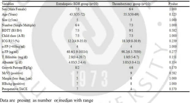
The clinical and pathology data of the 20 patients underwent ipsilateral
hemihepatectomy which were divided into extrahepatic bile duct resection group (group 1) and thrombectomy (group 2) are showed in Table1. Even though the
infiltrative growth type were found more frequently in thrombectomy group (6 in 10), the result did not reach statistical significance (p=0.170). And there were no
differences between the two groups for all the other variables in Table1.
Table 1. Clinical and pathology profiles of Extrahepatic BDR group and Thrombectomy group
Data are present as number or median with range
Abbreviation: α-FP: alpha fetoprotein, BDR: bile duct resection, BDTT: bile duct tumor thrombi, HBsAg: hepatitis b surface antigen, ICG R15: indocyanine green retention rate at 15min, McVI: micro vascular invasion, TACE: transarterial chemoembolization, Eg/Ig:expanding growth type/infiltrative growth type.
In this study, the median survival time was 39 months (range 2-233). The 1, 3 and 5-year overall survival rates were 75.0%, 50.0%, and 27.8%, respectively.(Fig. 2)
6
-Fig.2 Actuarial survival curve after hemihepatectomy for hepatocellular carcinoma with grossly bile duct invasion
The 1, 3, and 5-year survival rates of Group 1 were 90%, 80%, and 45.7%, and those of Group 2 were 50%, 20%, and 10%, respectively. (p=0.014) (Fig. 3A) The 1, 3, and 5-year recurrence free survival rates of Group 1 were 90.0%, 70.0% and 42.0%, and those of Group 2 were 36.0%, 36.0% and 0%, respectively. (p=0.023) (Fig. 3B)
A B
Fig.3 (A) Actuarial survival curves of hemihepatectomy with thrombectomy group and hemihepatectomy with extrahepatic bile duct resection
7
-(B) Actuarial recurrence free survival curves of hemihepatectomy with thrombectomy group and hemihepatectomy with extrahepatic bile duct resection
There were 15 patients who experienced recurrence during the follow up period, 7 cases in extrahepatic BDR group and 8 cases in thrombectomy group (Fig. 4).
Fig 4 The description of recurrence sites for 15 recurrence cases
In thrombectomy group, 6 of 8 cases were recurrence within 12 months. However, 6 in 7 cases were recurrence beyond 12 months in extrahepatic BDR group. The frequency of postoperative recurrence in the remnant bile duct was: 30% (3/10) after hemihepatectomy with thrombectomy, and 0% (0/10) after hemihepatectomy with extrahepatic bile duct resection.The most important cause of recurrence is thought to be the microscopic skipped invasion of BDTT into the bile duct wall. The key factor of a skipped invasion seems to be tightness of contact between bile duct wall and bile duct tumor thrombi.Shiomi et al.[12] reported that the bile duct tumor thrombi invaded microscopically the bile duct wall in 1 of the 5 patients (20%) who underwent bile duct resection and reconstruction.
8
-Fig 5 (A) One bile duct tumor thrombi case with a skipped bile duct invasion. Some fine fibrous tissues with minute oozing without any residual tumor thrombi.
(B) One BDTT case with a skipped BD invasion. Some fine fibrous tissues with minute oozing without any residual tumor thrombi.
(C) Histologic examination of the fibrous bridge structure. A focus of skipped tumor invasion (BDE, bile duct epithelium)
One case of skipped metastasis was shown in Fig. 5A/5B. After removal of BDTT, there were some fibrous tissues with minute oozing without any residual tumor thrombi in the bile duct. Series of wedge biopsies of bile duct wall was done. Fig. 5C shows the results of histological examination of bile duct wall including fibrous
9
-tissues. There were the microscopic residual tumor thrombi (left), and we can see clear cell nest in the HCC foci (right).
As shown in Fig 4, the recurrence pattern was significant difference between two groups. And the median recurrence time for thrombectomy group was significant short than extrahepatic BDR group (4.5months VS 47months, P=0.042).
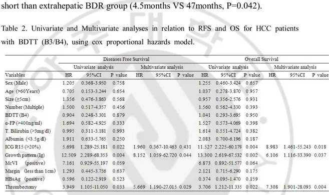
Table 2. Univariate and Multivariate analyses in relation to RFS and OS for HCC patients with BDTT (B3/B4), using cox proportional hazards model.
Abbreviation: α-FP: alpha fetoprotein, BDR: bile duct resection, BDTT: bile duct tumor thrombi, CI: confidence intervals, HR: hazard ratio, HBsAg: hepatitis b surface antigen, Ig: infiltrative growth type, McVI: micro vascular invasion, RFS: recurrences free survival, OS: overall survival.
Table 2 shown that the univariate and multivariate analysis for recurrence free survival and overall survival by cox regression. Infiltrative growth pattern (hazard ratio: HR, 8.512 [range, 1.509-62.720], P value: 0.044), ipsilateral hemihepatectomy with thrombectomy (HR, 5.669 [range, 1.190-27.015], P value: 0.029) were found as independent prognostic factors for recurrence free survival by multivariate analysis. Infiltrative growth pattern (HR, 6.106 [range, 1.116-33.390], P value: 0.037),
- 10 -
ipsilateral hemihepatectomy with thrombectomy (HR, 7.308 [range, 1.901-28.093], P value: 0.004) and high ICG R15 (HR, 8.983 [range, 1.461-55.243], P value: 0.018) were found as independent prognostic factors for overall survival by multivariate analysis.
In this study, we have four long-term survivors after hemihepatectomy with
extrahepatic bile duct resection for icteric HCC. (Table 3) The survival period ranged from 108 to 233 months after surgery and the recurrence free survival time ranged from 47 to 233. They had 4 common characteristics; first, Young patients with relatively early stage of primary tumor and preserve good liver function. Secondly, their preoperative AFP values were, somewhat, lower. Thirdly, all of the four patients were performed with hemihepatectomy with caudate lobectomy and
extrahepatic BD resection, and lastly, the tumor growth pattern of the four cases was expanding growth type.
Table 3. Clinical profiles and common characteristics of four long-term survivors
Abbreviation: α-FP: alpha fetoprotein, BDR: bile duct resection, CL: caudate lobectomy, ICG R15: indocyanine green retention rate at 15min, LC: liver cirrhosis, OS: overall survival, RL: right lobectomy, RFS: recurrences free survival.
- 11 -
Ⅳ
. DISCUSSION
The first technical problem as to whether the minor hepatectomy is feasible in the treatment of icteric HCCs is still under debate. There may be two different options; "to perform major or minor hepatectomy according to liver function" versus "to perform hemihepatectomy as a minimum-required surgery according to liver function". We believe we should perform hemihepatectomy as a minimum-required surgery according to liver function because of our early painful experience (3 patients who underwent minor hepatic resection with only removal of BDTT were suffer from recurrence in the intrahepatic bile duct). The pathologic base maybe skipped metastasis. That means the infiltrative growth type tumor thrombi was infiltrative invasion the bile duct. If the liver function is very limited, other non-surgical modalities must be considered, instead. Huang et al.[13] reported that palliative treatment strategies for patients with poor liver function, including TACE and/or RCT showed a beneficial effect in improving the survival time, the median survival time was 13.4 months.
The second concern is whether to remove the BDTT through a choledochotomy during surgical resection or to perform hemihepatectomy with caudate lobectomy and extrahepatic bile duct resection. The authors [12,14,15] who favor of the previous viewpoint reported that there were no significant differences between two groups in 5-year survival rate. Therefore, they concluded that it is not necessary to perform extrahepatic bile duct resection. On the other hands, some authors [11, 16] reported contrary results that perform hemihepatectomy with caudate lobectomy and extrahepatic bile duct resection could significantly increase the 5-year survival rate.
- 12 -
Of 4,308 HCC patients surgically treated from four Korean institutions, this single-arm retrospective study included 73 patients (1.7%) who underwent resection for HCC with BDTT. According to Ueda classification, BDTT was type 2 in 34 cases (46.6%) and type 3 in 39 cases (53.4%). Systematic hepatectomy was
performed in 69 patients (94.5%), and concurrent bile duct resection was performed in 31 patients (42.5%). Patient survival rates were 76.5% at 1 year, 41.4% at 3 years, 32.0% at 5 years, and 17.0% at 10 years. Recurrence rates were 42.9% at 1 year, 70.6% at 3 years, 77.3% at 5 years, and 81.1% at 10 years. Results of univariate survival analysis showed that maximal tumor size, bile duct resection, and surgical curability were significant risk factors for survival, and surgical curability was a significant risk factor for recurrence. Multivariate analysis did not reveal any independent risk factors. HCC patients with BDTT achieved relatively favorable long-term results after resection; therefore this study proposed that extensive surgery should be recommended when complete resection is anticipated. In this study, there is a problem; gross portal vein invasion in 24 (32.9%). We think it needs further study for icteric HCC patients without gross portal invasion because gross portal vein invasion is so strong prognostic factor in HCC [17,20].
In the present study, we exclude 6 cases of gross portal vein invasion and 2 cases of gross hepatic vein invasion. The patients in group 1 which performed with extensive surgery by ipsilateral hemihepatectomy with caudate lobectomy and extrahepatic bile duct resection have a significantly longer recurrence free survival time. This extensive surgery procedure can achieve a R0 resection that the recurrence free survival rate was higher, its 1, 3, and 5-year recurrence free survival rate was
- 13 -
90.0%, 70.0% and 42.0%, respectively. Additionally, the recurrent sites were not located at the bile duct in all the 7 recurrent cases, 3 at liver, 2 at lung, one at bone and one at abdominal lymph node. The patients with the tumor recurrence on the liver were accessibility received subsequent treatment, such as TACE, reresection and/or transplantation. Finally, their 1, 3, and 5-year survival rate was 90%, 80%, and 45.7%, respectively. On the contrary, there were 8 cases were experienced recurrence in the group 2, 3 cases recurrence at bile duct and 5 at the liver.
Unfortunately, the 3 cases recurrence at bile duct was died within 6 months due to the liver failure caused by obstruction on bile duct and invasive tumor growth after operation. And the 1, 3, and 5-year survival rate of group 2 was 36.0%, 36.0% and 0%, respectively. Our multivariate analysis demonstrated that remove of BDTT by thrombectomy and infiltrative growth type were independent risk factors for tumor recurrence. And they combined high ICG R15 (more than 20%) were independent risk factors for overall survival.
The last technical debate is "Should icteric HCC be an indication for liver transplantation?". It is still under debate, because, so far, there were only three reports with total 19 cases.[15, 17, 18] One reports that 4 patients (80%) died of HCC recurrence within 3 years[17]. The other reports have the same conclusion on high risk of HCC recurrence post-transplantation.[18] Therefore, liver transplantation for HCC patients with BDTT still carries a high risk of HCC recurrence, thus
requiring further study. How do we perform bile duct reconstruction after liver transplantation for icteric HCC? Duct-to-duct anastomosis versus
- 14 -
extrahepatic BDR or not. Peng et al. [15] asserted extrahepatic bile duct resection followed by hepaticojejunostomy. On the contrary, Lee et al. [19] reported the followings; after the extent of BDTT was manually evaluated, they performed a bile duct dissection, stapling the distal portion of BDTT with TA stapler. They examined the resection margin of the bile duct for malignant cells and reconstructed the bile duct with hepaticojejunostomy in case of insufficient length for duct-to-duct anastomosis. It is also debatable, however, due to the theory of tumor skipped metastasis the hepaticojejunostomy after extrahepatic BDR may be a more rational approach.
Therefore, whatever liver transplantation or ipsilateral hemihepatectomy with caudate lobectomy, the extrahepatic BDR should be performed for icteric HCC patients.
- 15 -
.
Ⅴ
CONCLUSION
The incidence of icteric HCC is very rare, and its prognosis is poor. Although surgical resection is the only option for curative treatment, there are still many debates in technical aspects of surgical treatment of icteric HCC.
Ipsilateral hemihepatectomy with caudate lobectomy and extrahepatic bile duct resection should be performed for the patients who preserve good liver function and with expanding growth type.
Three variables were found as independent risk factors for overall survival by multivariable analysis in our study. They were infiltrative growth type, high ICG R15 (more than 20%) and remove the BDTT by thrombectomy.
The limitation of the present study is small cases number, and no liver transplantation cases. In the future, the international multicenter collaboration research in this subject may play an important role in forming global consensus.
- 16 -
REFERENCES
1. Lin TY. Tumor of the liver. In: Bockus HL, editor. Gastroenterology. Philadelphia: WB Saunders; 1976. p.522-33.
2. Clark W, Schulz MD, et al. [Hepatoma, with invasion of cystic duct and metastasis to third lumbar vertebra]. N Engl J Med 1947;237:673-6.
3. Edmondson HA. Tumors of the liver and intrahepatic bile ducts. Washington: Armed Forces Institute of Pathology; 1958.
4. Creed DL, Fisher ER. Clot formation in the common duct; an unusual manifestation of primary hepatic carcinoma. AMA Arch Surg 1956;73:261-5. 5. Tsuzuki T, Ogata Y, Iida S, Kasajima M, Takahashi S. Hepatoma with obstructive
jaundice due to the migration of a tumor mass in the biliary tract: report of a successful resection. Surgery 1979;85:593-8.
6. Lau WY, Leung JW, Li AK. Management of hepatocellular carcinoma presenting as obstructive jaundice. Am J Surg 1990;160:280-2.
7. Ueda M, Takeuchi T, Takayasu T, Takahashi K, Okamoto S, Tanaka A, et al. Classification and surgical treatment of hepatocellular carcinoma (HCC) with bile duct thrombi. Hepatogastroenterology 1994;41:349-54.
8. Kojiro M, Kawabata K, Kawano Y, Shirai F, Takemoto N, Nakashima T. Hepatocellular carcinoma presenting as intrabile duct tumor growth: a clinicopathologic study of 24 cases. Cancer 1982;49:2144-7.
9. Liu Q, Chen J, Li H, Liang B, Zhang L, Hu T. Hepatocellular carcinoma with bile duct tumor thrombi: correlation of magnetic resonance imaging features to histopathologic manifestations. Eur J Radiol 2010;76:103-9.
- 17 -
10. Taguchi H, Ogino T, Miyata A, Munehisa T. [Bile duct invasion of hepatocellular carcinoma]. Nihon Shokakibyo Gakkai Zasshi 1983;80:2259-68.
11. Wang HJ, Kim JH, Kim JH, Kim WH, Kim MW. Hepatocellular carcinoma with tumor thrombi in the bile duct. Hepatogastroenterology 1999;46:2495-9.
12. Shiomi M, Kamiya J, Nagino M, Uesaka K, Sano T, Hayakawa N, et al. Hepatocellular carcinoma with biliary tumor thrombi: aggressive operative approach after appropriate preoperative management. Surgery 2001;129:692-8. 13. Huang JF,Wang LY, Lin ZYet al. Incidence and clinical outcome of icteric type
hepatocellular carcinoma. J. Gastroenterol. Hepatol 2002; 17: 190–95.
14. Satoh S, Ikai I, Honda G, Okabe H, Takeyama O, Yamamoto Y, et al. Clinicopathologic evaluation of hepatocellular carcinoma with bile duct thrombi. Surgery 2000;128:779-83.
15. Peng SY, Wang JW, Liu YB, Cai XJ, Deng GL, Xu B, et al. Surgical intervention for obstructive jaundice due to biliary tumor thrombus in hepatocellular carcinoma. World J Surg 2004;28:43-6.
16. Mok KT, Chang HT, Liu SI, Jou NW, Tsai CC, Wang BW. Surgical treatment of hepatocellular carcinoma with biliary tumor thrombi. Int Surg 1996;81:284-8. 17. Moon DB, Hwang S, Wang HJ, Yun SS, Kim KS, Lee YJ, et al. Surgical
outcomes of hepatocellular carcinoma with bile duct tumor thrombus: a Korean multicenter study. World J Surg 2013;37:443-51.
18. TY. Ha, S. Hwang, DD. Moon, CS. Ahn, KH. Kim, GW. Song, et al. Long-term Survival Analysis of Liver Transplantation for Hepatocellular Carcinoma With Bile Duct Tumor Thrombus. Transplant Proc 2014;46:774-7.
- 18 -
19. Lee KW, Park JW, Park JB, Kim SJ, Choi SH, Heo JS, et al. Liver transplantation for hepatocellular carcinoma with bile duct thrombi. Transplant Proc 2006;38:2093-4.
20. Xuguang Hu, Wei Mao, Yongkeun Park, Weiguang Xu, Bongwan Kim, Heejung Wang, et al. Blood Neutrophil to Lymphocyte ratio predicts tumor recurrence in patients with hepatocellular carcinoma within Milan criteria after hepatectomy. Yonsei Medical Journal 2016;90:3.


