Analysis of mitochondrial toxicity
induced by antiviral treatment
for chronic hepatitis
Yoon Ok Jang
The Graduate School
Yonsei University
Analysis of mitochondrial toxicity
induced by antiviral treatment
for chronic hepatitis
Directed by Professor Soon Koo Baik
A Dissertation
Submitted to the Department of Medicine
and the Graduate School of Yonsei University
in partial fulfillment of the
requirements for the degree of
Master of Medical Science
Yoon Ok Jang
This certifies that the dissertation of
Yoon Ok Jang is approved.
Thesis Supervisor : Prof. Soon Koo Baik
Thesis Committee Member : Kyu-Sang Park
Thesis Committee Member : Moon Young Kim
The Graduate School
Yonsei University
ACKNOWLEDGEMENTS
First of all, I would like to express my sincere gratitude to my supervisor, professor Soon Koo Baik, who gave me a great opportunity to challenge the degree of Medical Science during the past several years. He has advised me with his heartful encouragement and generous support. Without his patient instruction, insightful criticism and excellent guidance, the completion of this thesis would not be possible.
I would also like to express my heartfelt gratitude to the members of my dissertation committee, professor Kyu-Sang Park, Moon Young Kim, for their generous personality, invaluable advice and thoughtful suggestions.
Especially, I gratefully thank professor Sang Ok Kwon, Jae Woo Kim, for their generous support and encouragement.
I gratefully thank professor Chan Mug Ahn, Dr. Xianglan Quan, Ranjan Das, Shanhua Xu, for great contributions to my research successfully. I would like to thank the members of department of internal medicine and laboratory, friends, for their helps, concerns and supports.
Last but not least, I feel a deep sense of gratitude to my family, especially my mother, for their love, support and encouragement, which gave me the strength and courage to mount greater heights and pursue my dreams.
Yoon Ok Jang
July 2011
i
TABLE OF CONTENTS
LIST OF FIGURES
······························································································iii
LIST OF TABLE
···································································································iv
ABSTRACT
·············································································································v
I
. INTRODUCTION
······························································································ 1II
. MATERIALS AND METHODS
·································································· 10 1. Cell culture and drugs ························································································ 10 2. Quantitative PCR ······························································································· 11 3. MTT assay ········································································································· 13 4. ATP and lactate measurements ········································································· 13 5. Insulin measurement ·························································································· 13 6. Statistical analysis ······························································································ 14III.
RESULTS
········································································································· 15 1. Effects of clevudine on mtDNA copy number and mRNA levels of mtDNAencoded genes ····································································································· 15 2. Mitochondrial dysfunction induced by clevudine ·············································· 18
3. Inhibition of glucose-stimulated insulin secretion by clevudine ························ 22
ii
V
. CONCLUSION
································································································ 28REFERENCE
········································································································ 29iii
LIST OF FIGURES
Figure 1. Hepatitis B virus replication cycle ························································· 3 Figure 2. Mitochondrial biogenesis ······································································ 6 Figure 3. The chemical structure of Clevudine ······················································ 9 Figure 4. Effects of clevudine on mitochondrial DNA copy number and mRNA levels of mitochondria-related genes in INS-1E cells ···················································· 16 Figure 5. Effects of clevudine on mitochondrial DNA copy number and mRNA levels of mitochondria-related genes in HepG2 cells ····················································17 Figure 6. High-dose clevudine decreased MTT intensity in INS-1E cells ··············· 19 Figure 7. Effects of clevudine on cytosolic ATP content in INS-1E cells. ·············· 20 Figure 8. Effects of clevudine on lactate production from INS-1E cells ················· 21 Figure 9. Clevudine inhibited glucose-stimulated insulin secretion ······················· 23
iv
LIST OF TABLE
v
ABSTRACT
Analysis of mitochondrial toxicity induced
by antiviral treatment for chronic hepatitis
Yoon Ok Jang
Dept. of Medicine
The graduate School
Yonsei University
Clevudine is a nucleoside analog reverse transcriptase inhibitor that exhibits potent antiviral activity against hepatitis B virus (HBV) without serious side effects. However, mitochondrial myopathy has been observed in patients with chronic HBV infection taking clevudine. Moreover, the development of diabetes was recently reported in patients receiving long-term treatment with clevudine.
In this study, we investigated the effects of clevudine on mitochondrial function and insulin release in a rat clonal β-cell line, INS-1E. We measured the levels of mitochondrial DNA (mtDNA) and cytochrome c oxidase-1 (Cox1) mRNA transcribed from mtDNA using quantitative PCR.
vi
Both INS-1Ecells and HepG2cells, which originated from human hepatoma, showed dose-dependent decreases in mtDNA copy number and Cox1 mRNA level following culture with clevudine (10mM-1mM) for 4wks. INS-1E cells treated with clevudine had reduced total mitochondrial activities, lower cytosolic ATP contents, and enhanced lactate production. Insulin release in response to glucose application was markedly decreased in clevudine-treated INS-1E cells, which might be a consequence of mitochondrial dysfunction.
Our data suggest that high-dose treatment with clevudine induces mitochondrial dysfunction caused by the depletion of mtDNA and impairs glucose-stimulated insulin secretion in insulin-releasing cells. These findings partly explain the development of diabetes in patients receiving clevudine who might have a high susceptibility to mitochondrial toxicity.
Key words : clevudine, mitochondrial DNA, mitochondrial dysfunction, glucose-stimulated insulin secretion
1
Analysis of mitochondrial toxicity induced
by antiviral treatment for chronic hepatitis
Yoon Ok Jang
Department of Medicine
The graduate School, Yonsei university
(Directed by Professor Soon Koo Baik)
I. INTRODUCTION
Chronic hepatitis B virus (HBV) infection, affecting 350 million people worldwide, is associated with significant morbidity and mortality. Approximately 15 – 40% of individuals with chronic HBV infection will develop chronic hepatitis B that has potential of evolving into cirrhosis, decompensated liver disease or hepatocellular carcinoma1.
2
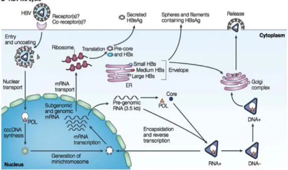
As carriers of HBV serve as reservoirs for vertical and horizontal transmissions, the prevalence of carriers and the number of acute and chronic liver diseases place HBV infection among the most frequent and important transmissible diseases of the hepatobiliary system2,3,38 (Fig 1).
The selection of an anti-HBV therapy is critical in reducing HBV DNA replication. Implementation of effective treatment in the early stages of disease may reduce the burden of chronic HBV infection by reducing progression to more advanced stages of liver disease2,4.
3
Figure 1. Life cycle of hepatitis B virus (Debika and
Chloe, 2010).
cccDNA, covalently closed circular DNA; ER, endoplasmic reticulum; HBeAg, hepatitis B e antigen; HBsAg, hepatitis B surface antigen; HBx, HBV X protein; mRNA, messenger RNA; POL, polymerase.
4
Several antiviral drugs including immunomodulators, interferon-α (IFN-α) and nucleoside analog reverse transcriptase inhibitors (NRTIs) including entecavir, lamivudine, adefovir and telbivudine have been developed and prescribed for HBV infection.
NRTIs undergo intracellular and intramitochondrial phosphorylation into active triphosphates that are capable of inhibiting HIV reverse transcriptase (RT)5. However,
these drugs have side effects such as lipodystrophy, neuropathy, myopathy, and liver steatosis, all of which are related to mitochondrial toxicity.
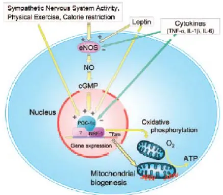
Mitochondrial biogenesis is a complex process requiring the coordinated expression and assembly of 1500 proteins encoded by both the nuclear and mitochondrial genomes6,7. Mitochondrial genes involved in mitochondrial biogenesis, including
peroxisome proliferator-activated receptor co-activator 1 (PGC-1), nuclear respiratory factor-1 (NRF-1), and mitochondrial transcription factor A (Tfam) are upregulated as a consequence, leading to increased mtochondrial biogenesis8 (Fig 2).
Cytochrome c oxidase (COX1) is an enzyme complex of the mitochondrial respiratory chain that is a reliable indicator of mitochondrial activity9. PGC-1 is
regarded as a crucial regulator of mitochondrial biogenesis by virtue of its ability to co-activate and augment the expression and activity of several transcription factors that in turn bind to the promoters of distinct sets of nuclear-encoded mitochondrial genes6,10. PGC-1 is also indirectly involved in regulating the expression of
mitochondrial DNA (mtDNA) transcription via increased expression of Tfam as executed by its co-activation of NRF-111-13.
5
In this manner, PGC-1 may act as a central regulator of the adaptive response to exercise by coordinating metabolic gene expression in both the nuclear and mitochondrial genomes.
6
7
In vitro and in vivo studies have shown that some NRTIs inhibit DNA polymerase-r,
a nuclear-encoded polymerase important for mitochondrial DNA (mtDNA)
replication4,14. Depletion of mtDNA induced by NRTIs may attenuate mitochondrial oxidative phosphorylation, which could limit their clinical use.
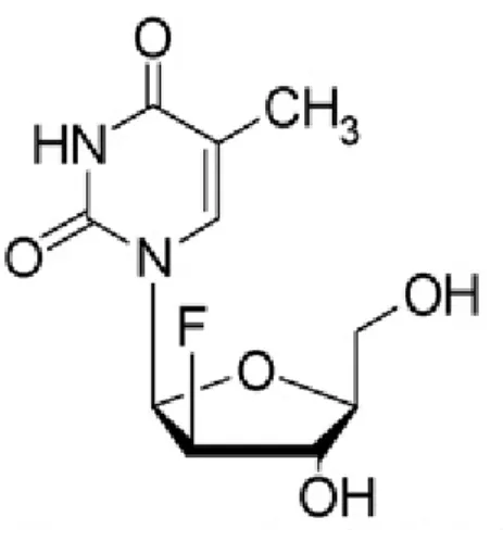
Clevudine (1-(2-deoxy-2-fluoro-β-L-arabinofuranosyl) thymine) is a L-thymidine analogue with potent activity against HBV as well as Epstein–Barr virus in vitro39
(Fig. 3). Clevudine has a long half-life, low toxicity and no effect on mitochondrial structure or DNA function and inhibits the DNA-dependent DNA activity of HBV polymerase, as well as reverse transcription and priming2,15-17. However, long-term therapy for more than one year results in the development of considerable drug resistance and skeletal myopathy2,17-19.
Muscle biopsies from patients with myopathy as a complication of clevudine treatment revealed severe necrosis with cytochrome c oxidase (COX)-negative ragged red fibers, the typical phenotype of mitochondrial myopathy2,20. Clevudine-induced
myopathy developed in approximately 4-5% of patients and was usually reversible after discontinuation of clevudine19.It is well known that mitochondria play a critical
role in nutrient-stimulated insulin secretion, as well as in insulin actions at target cells
21. Recently, a patient who developed diabetes mellitus after clevudine treatment was
reported22.
We hypothesized that the mitochondrial dysfunction invoked by clevudine treatment could be a precipitating factor in diabetogenesis. Until now, the majority of in vitro studies for antiviral agent toxicities have been performed in different cell types, yielding conflicting results23-25.
8
Insulin-secreting cells are highly specialized fuel sensors that maintain blood glucose level in the body by monitoring the ATP/ADP ratio, which is strictly regulated by mitochondrial oxidative phosphorylation. Thus, insulin-secreting cells are an appropriate model system for identification of mitochondrial toxicity and its functional consequences following antiviral therapy.
In this study, we investigated the effects of clevudine exposure on mtDNA content, mitochondrial function, and metabolism-secretion coupling in insulin-releasing cells to elucidate the mechanism underlying the reversible diabetes observed in clevudine-treated patients.
9
Figure 3. The chemical structure of Clevudine (Ashoke S et al., 2008).
10
II. MATERIALS AND METHODS
1. Cell culture and drugs
INS-1E cells received from Prof. Claes B. Wollheim (University of Geneva, Switzerland) were cultured in complete medium composed of RPMI 1640 (Invitrogen, Carlsbad, CA, USA) supplemented with 10% fetal calf serum, 1mM sodium pyruvate, 50 mM 2-mercaptoethanol, 2 mM glutamine, 10 mM HEPES, 100 units/ml penicillin, and 100 mg/ml streptomycin. HepG2 cells were grown in DMEM medium (Invitrogen) containing 5.6 mM glucose, 4 mM L-glutamine and 1 mM sodium pyruvate. Cells maintained free-floating at 37℃ in an atmosphere of 5% CO2 and
95% air in a humidified incubator.
Clevudine was purified from Revovir® tablets (Bukwang Pharm. Co., Seoul, Korea). The amount of harvested clevudine was analyzed using HPLC (Agilent G1315B UV Diode array detector, AD, Santa Clara, CA, USA). A single peak with the expected amount of clevudine was measured based on the known weight of one tablet.
11
2. Quantitative PCR
Total DNA or RNA was isolated and purified from INS-1E and HepG2 cells using DNeasy or RNeasy kits (Qiagen, Valencia, CA, USA), respectively.
DNA and RNA concentration were determined using Ultrospec 2100 pro UV/Visible Spectrophotometer (Amersham Bioscience, Freiburg, Germany).
For cDNA synthesis, reverse transcription (RT) was performed with 1ug RNA, random hexamers (Takara, Kyoto, Japan) using reverse transcriptase (Promega, Madison, WI, USA). Samples were incubated at 42℃ for 60min, 99℃ for 5min, 4℃ for 6min.
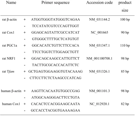
For PCR amplification, sequence-specific oligonucleotide primers for the genes of interest were designed (Bioneer, Daejeon, Korea) based on rat and human sequences in the GenBank database (Table 1).
Quantitative real-time PCR using SYBR Green PCR Master Mix (Applied Biosystems, Foster City, CA, USA) was performed in an ABI PRISM 7900HT Sequence Detection System (Applied Biosystems) according to the manufacturer’s protocol. All amplifications were followed by melting curve analysis.
Data were obtained as Ct values (the cycle number at which logarithmic PCR plots cross a calculated threshold line) according to the manufacturer`s guidelines and used to determine △Ct values (Ct of target gene - Ct of housekeeping gene) as raw data for gene expression. Fold change in gene expression was determined by subtracting △Ct values for clevudine treated samples from their control. The resulting △△Ct
12
Table 1. Primers for quantitative PCR
Name
Primer sequence
Accession code product
size
rat β-actin + ATGGTGGGTATGGGTCAGAA NM_031144.2 100 bp
- TCCATATCGTCCCAGTTGGT
rat Cox1 + GGAGCAGTATTCGCCATCAT NC_001665 90 bp
- GTGGGCTTTTGCTCATGTGT
rat PGC1a + GGCACATCTGTTCTTCCACA NM_031347.1 110 bp
- TTCCTGGTCTTGGAGCTGTT
rat NRF1 + GGACAGCAAGCCATTGTTCT NM_001100708.1 98 bp
- TACTTGCGCACCACATTCTC
rat Tfam + GCTGAGTGGAAGGTGTACAAAG NM_031326.1 85 bp
- CTTCCTTCTCTAAGCCCATCAG
human β-actin + AAGTTCACAATGTGGCCGAG NM_001101.3 98 bp
- ATGGCAAGGGACTTCCTGTA
human Cox1 + CACACTCCACGGAAGCAATA NC_012920.1 82 bp
13
3. MTT assay
3-(4,5-dimethylhioazol-2-yl)-2,5-diphenyltetrazolium bromide (MTT) was purchased from Sigma (St. Louis, MO, USA). INS-1E cells seeded onto a 96-well plate were incubated with MTT (50 mg/well) for 2 hrs, and then the medium was discarded and replaced with dimethylsulphoxide (100 ml/well)26. The absorbance of
each well was measured at 570 nm using an enzyme-linked immunosorbent assay (ELISA) reader, after background subtraction at 650 nm.
4. ATP and lactate measurements
INS-1E cells seeded onto 24-well plates (3 x 10⁵cells/well) were preincubated with
glucose-free medium for 2hrs prior to incubation with KRBH solution (135mMNaCl, 3.6mMKCl, 2mMNaHCO3, 0.5mMNaH2PO4, 0.5mMMgSO4, 1.5mMCaCl2,
10mMHEPES, pH7.4) containing 2.8mM glucose for 30min. The cells were then stimulated for 15min with KRBH buffer at a low (2.8mM) or high (16.7mM) glucose concentration. The ATP content in the cell lysate and the lactate level in the cell supernatant were measured as described previously26. Measurement of the protein
concentration in cell lysates was performed using the Bradford assay.
5. Insulin measurement
INS-1E cells were seeded and cultured as for ATP and lactate measurement. For insulin measurement, 0.1% bovine serum albumin was included in the KRBH solution, and the cells were stimulated with low or high concentrations of glucose for
14
30 min, as described previously26. Insulin levels in supernatant and cell extracts were
measured using a rat insulin enzyme immunoassay kit (Shibayagi Co., Gunma, Japan).
6. Statistical analysis
Data are presented as mean ± SEM (standard error of the mean), and the statistical significance was determined using Student’s t test. A P values of less than 0.05 was considered to be statistically significant.
15
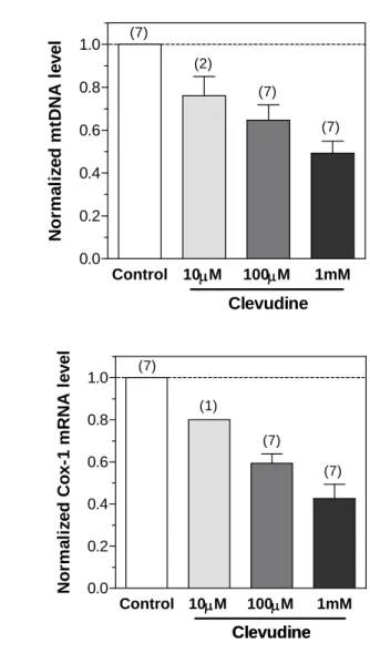
IV. RESULTS
1. Effects of clevudine on mtDNA copy number and mRNA level of
mtDNA encoded genes
INS-1E cells were cultured with different concentrations of clevudine for 4wks and the in vitro effects on mtDNA replication and translation were measured. Treatment with clevudine (10mM to 1mM) reduced the mtDNA copy number in a dose-dependent manner (Figure 4A). The mRNA levels of mtDNA-encoded Cox-1 were also dose-dependently attenuated by clevudine (Figure 4B). Treatment with 1mM clevudine reduced the mtDNA copy number and mRNA levels to less than half of those in the controls.
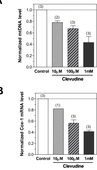
We next examined the effects of clevudine on the levels of mtDNA and mtRNA in the human hepatoma cell line HepG2, major target cells of insulin action. Clevudine showed suppressive effects on mtDNA replication and transcriptionin HepG2 cells, similar to the effect in INS-1Ecells (Figures 5A and 5B).
We also observed upregulation of PPAR- coactivator 1 (PGC-1, 4.12-fold, n=3), mitochondrial transcription factor A (Tfam, 1.33-fold, n=3), and nuclear respiratory factor 1 (NRF1, 1.25-fold, n=3) in clevudine (100uM)-treated INS-1E cells. Upregulation of these transcription factors could be a nuclear response to mitochondrial dysfunction4.
16
A
B
Figure 4. Effects of clevudine on mitochondrial DNA copy number and
mRNA levels of mitochondria-related genes in INS-1E cells.
Mitochondrial DNA copy number and mRNA level of the mtDNA-encoded Cox-1 gene were measured using quantitative PCR. Clevudine reduced the expressions of mtDNA (A) and Cox-1 mRNA (B) in the rat clonal β–cell line INS-1E cell in a dose-dependent manner. Data presented are mean ± SEM. The number of experiments is shown in parenthesis. Control 10M 100M 1mM 0.0 0.2 0.4 0.6 0.8 1.0 Clevudine (7) (7) (2) (7) Nor m a li z e d m tDNA l e v e l Control 10M 100M 1mM 0.0 0.2 0.4 0.6 0.8 1.0 Clevudine Clevudine (7) (7) (1) (7) N o rm a li z e d C o x -1 m R N A l evel17
A
B
Figure 5. Effects of clevudine on mitochondrial DNA copy number and
mRNA levels of mitochondria-related genes in HepG2 cells.
Mitochondrial DNA copy number and mRNA level of the mtDNA-encoded Cox-1 gene were measured using quantitative PCR. Clevudine reduced the expressions of mtDNA (A) and Cox-1 mRNA (B) in the human hepatoma cell line HepG2 cells in a dose-dependent manner. Data presented are mean ± SEM. The number of experiments is shown in parenthesis. Control 10M 100M 1mM 0.0 0.2 0.4 0.6 0.8 1.0 Clevudine Clevudine (3) (3) (2) (3) No rm a li z e d m tDNA level Control 10M 100M 1mM 0.0 0.2 0.4 0.6 0.8 1.0 Clevudine Clevudine (3) (1) (3) (3) No rm a li z e d Co x -1 m R NA l e v e l18
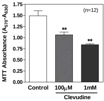
2. Mitochondrial dysfunction induced by clevudine
The amount of formazan reaction product formed in the MTT assay reflects the total mitochondrial enzymatic activity in each well. INS-1E cells treated with or without clevudine for 4 wks were seeded 48 hrs before the MTT assay. Exposure to clevudine decreased the MTT absorbance (71% by 100 mM and 56% by 1 mM), implying a reduction in cell number, as well as mitochondrial activity (Figure 6).
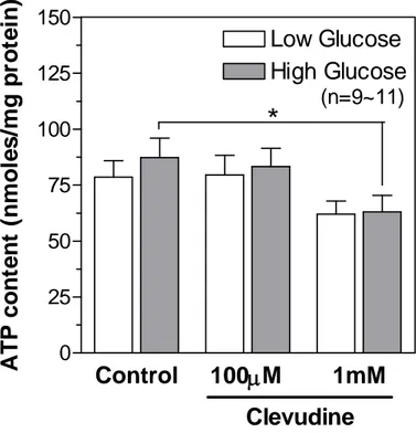
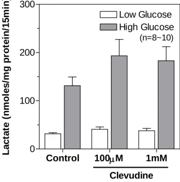
We measured the cellular contents of ATP in control and clevudine-treated INS-1E cells using a bioluminescence method after incubation with low (2.8 mM) or high (16.7 mM) concentrations of glucose for 15 min. As shown in Figure 7, cells cultured with 1 mM clevudine had a low cytosolic ATP level in both low and high glucose conditions. However, treatment with 100 mM clevudine did not affect the ATP content (Figure 7), even though it significantly decreased the MTT signal (Figure 6). Lactate production from INS-1E cells was markedly elevated by incubation with glucose for 15 min (Figure 8). The glucose-induced lactate production was modestly increased in cells treated with clevudine (100 mM and 1 mM) compared with that in the control cells (Figure 8).
19
Figure 6. High-dose clevudine decreased MTT intensity in INS-1E cells.
Clevudine reduced the MTT signal, which reflects total mitochondrial activity within each well. Data are presented as means ± SEM and ** denotes p<0.01.Control
100
M
1mM
0.00 0.25 0.50 0.75 1.00 1.25 1.50 1.75Clevudine
**
**
(n=12)M
T
T
A
b
sor
b
ance (
A
57 0-A
63 0)
20
Figure 7. Effects of clevudine on cytosolic ATP content in INS-1E cells.
C
ellular contents of ATP in control and clevudine-treated INS-1E cells were measured by using a bioluminescence method after incubation with low (2.8 mM) or high (16.7 mM) concentrations of glucose for 15 min.C
levudine decreased the cytosolic ATP level in both low and high glucose conditions. Data are presented as means ± SEM and * denotes p<0.05.Control
100
M
1mM
0
25
50
75
100
125
150
Clevudine
Low Glucose
High Glucose
*
(n=9~11)
A
T
P cont
ent
(
n
m
o
le
s/
m
g
pr
ot
ei
n)
21
Figure 8. Effects of clevudine on lactate production from INS-1E cells.
The glucose-induced lactate production was modestly increased in cells treated with clevudine (100 mM and 1 mM) compared with that in the control cells. Data are presented as means ± SEM.Control
100
M
1mM
0
100
200
300
Low Glucose
High Glucose
Clevudine
(n=8~10)L
a
ct
at
e (
n
m
o
les/
m
g
p
ro
tei
n/
15m
in
)
22
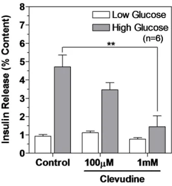
3. Inhibition of glucose-stimulated insulin secretion by clevudine
To identify whether clevudine-induced mitochondrial dysfunction affects insulin secretory activity, we measured the released and cellular contents of insulin via an enzyme immunoassay. The cellular insulin contents were not significantly different between control (816±167 ng/well) and clevudine (1 mM)-treated INS-1E cells (769±170 ng/well). After incubation for 30 min with low (2.8 mM) or high (16.7 mM) concentrations of glucose, the released insulin was normalized to the cellular content and expressed as a percentage of the content released.
We observed that high concentration glucose stimulated the release of insulin by 5.1-fold in control cells but by only 3.1-fold and 1.9-fold in cells treated with 100 mM and 1 mM clevudine, respectively (Figure 9). There was no difference in % insulin releases induced by low concentration glucose between the control and clevudine-treated groups (Figure 9).
23
Figure 9. Clevudine inhibited glucose-stimulated insulin secretion.
Released insulin and cellular insulin content were measured using an enzymatic immunoassay after incubation with Krebs buffer containing 2.8 mM or 16.7 mM glucose for 15 min. Insulin release was normalized to the insulin content. **denotes p<0.01.24
IV. Discussion
In pancreatic β-cells, mitochondria are of particular importance in the regulation of insulin secretion because they produce ATP as well as other coupling factors which link nutrient metabolism and insulin exocytosis21. mtDNA-depleted β-cell lines show
complete absence of nutrient-stimulated insulin secretion27. Patients with mtDNA
mutations develop diabetes, accounting for up to 1% of the total number of diabetic patients28. Moreover, postmortem islets from type 2 diabetes patients display
functional deterioration of mitochondria29. Therefore, factors that disturb the
mitochondrial function in pancreatic β-cells might affect metabolism-secretion coupling and diabetogenesis.
The present study showed that the effective anti-HBV agent clevudine has a negative effect on the copy number and transcription of mtDNA in insulin-releasing cells and hepatoma cells. The reduced expressions of mtDNA-encoded proteins lead to attenuation of mitochondrial function. In insulin-releasing cells, clevudine-induced mitochondrial dysfunction can elicit defective insulin secretion in response to substrates for mitochondrial metabolism. To our knowledge, this is the first demonstration that an antiviral agent can impair nutrient-stimulated insulin secretion as a result of mitochondrial dysfunction. Because of their high dependency on mitochondrial function in metabolism-secretion coupling, insulin-secreting cells provide a useful model to investigate the functional consequences of drug-induced mitochondrial toxicity.
25
NRTIs are widely used to treat various viral diseases such as acquired immunodeficiency syndrome (AIDS) and hepatitis B30. However, in vitro studies
showed that NRTIs can alter mtDNA content by inhibiting DNA polymerase-r31.
Moreover, myopathy accompanied by mtDNA depletion has been reported in NRTI-treated patients14. Clevudine treatment has also been associated with the development
of mitochondrial complications. In contrast to early studies5, depletion of mtDNA in
skeletal muscle has been observed in patients treated with clevudine2,32. Typical histological features of mitochondrial myopathy and abnormal mitochondria morphology were displayed in tissues from patients with increased lactate dehydrogenase and lactate levels18,20. Although the incidence of clevudine-induced
myopathy was reported to be low (~5%)19, a substantial proportion (~14.5%) of
clevudine-treated patients have been found to experience symptoms, signs, and laboratory abnormalities relevant to clevudine-induced myopathy33.
To directly confirm the effects of clevudine on mitochondrial function, we cultured cells with medium containing different concentrations of clevudine for 4 wks. Clevudine markedly decreased the MTT signal and cellular ATP content, consistent with depletion of mtDNA. We also observed some compensatory responses to reduced mtDNA copy number and its functional consequences. First, PGC-1a and its downstream transcriptional factors, NRF-1 and Tfam, were upregulated by clevudine. Second, lactate production was modestly increased in association with diminished ATP content. Pancreatic β-cells and clonal β-cell lines are known to have very low lactate dehydrogenase levels, which contribute to their dependency on mitochondrial function. The increase in lactate production observed in our study also demonstrates
26
that clevudine imposes selective defects on mitochondria rather than overall cytotoxicity.
Niu et al34. suggested that the intracellular level of the triphosphate form of
clevudine in cells exposed to 1 mM extracellular clevudine approximates the plasma level in patients receiving a 30 mg dose. Our results indicated that impairments in mitochondrial function and insulin secretion are elicited only by high concentrations of clevudine (>100mM). This means that clevudine would minimally affect mitochondrial function within the therapeutic concentration range. It is noteworthy, however, that mutations or polymorphisms of DNA polymerase-g were identified in NRTI-treated patients with mitochondrial complications35. This suggests that genetic
alterations in DNA polymerase-g are not normally deleterious, but that certain conditions such as NRTI treatment may push mitochondrial activity below the clinical threshold, causing pathogenic dysfunction36. Differences in genetic susceptibility to
mitochondrial toxicity could be one explanation for why a limited proportion of patients receiving clevudine have complications including myopathy. Clevudine-induced depletion of mtDNA is not restricted to insulin-secreting cells but is also observed in cultured hepatoma cells or muscle tissue from patients2,32. Mitochondrial
dysfunction in insulin target tissues such as liver and muscle could result in insulin resistance and diabetes37. In addition to defects in insulin secretion, decreased sensitivity in insulin target cells can also participate in diabetogenesis in patients receiving clevudine who might have a high susceptibility to mitochondrial toxicity. Further studies concerning the effects of NRTIs on mitochondrial function in different
27
cell types may help us understand these intractable complications and develop novel antiviral agents.
28
Ⅴ
. CONCLUSION
Both INS-1E cells and HepG2 cells, which originated from human hepatoma, showed dose-dependent decreases in mtDNA copy number and Cox1 mRNA level following culture with clevudine (10mM-1mM) for 4wks. INS-1E cells treated with clevudine had reduced total mitochondrial activities, lower cytosolic ATP contents, and enhanced lactate production. Insulin release in response to glucose application was markedly decreased in clevudine-treated INS-1E cells, which might be a consequence of mitochondrial dysfunction. These results indicate that high-dose treatment with clevudine induces mitochondrial dysfunction caused by the depletion of mtDNA and impairs glucose-stimulated insulin secretion in insulin-releasing cells.
29
REFERENCE
1. Tarik Asselah, Olivier Lada, Rami Moucari & Patrick Marcellin. Clevudine: a promising therapy for the treatment of chronic hepatitis B. Drugs. 2008, 17:1963-74.
2. Seok JI, Lee DK, Lee CH, Park MS, Kim SY, Kim HS, Jo HY, Kim DS. Long-term therapy with clevudine for chronic hepatitis B can be associated with myopathy characterized by depletion of mitochondrial DNA.
Hepatology. 2009, 49:2080-6.
3. Enzo Nisoli, Emilio Clementi, Michele O. Carruba, Salvador Moncada Defective. Mitochondrial Biogenesis A Hallmark of the High Cardiovascular Risk in the Metabolic Syndrome? Circ Res. 2007, 100:795-806.
4. Mallon PW, Unemori P, Sedwell R, Morey A, Rafferty M, Williams K, Chisholm D, Samaras K, Emery S, Kelleher A, Cooper DA, Carr A. In vivo, nucleoside reverse-transcriptase inhibitors alter expression of both mitochondrial and lipid metabolism genes in the absence of depletion of mitochondrial DNA. J Infect Dis. 2005, 191: 1686-96.
30
5. Balakrishna Pai S, Liu SH, Zhu YL, Chu CK, Cheng YC. Inhibition of hepatitis B virus by a novel L-nucleoside, 2'-fluoro-5-methyl-beta-L-arabinofuranosyl uracil. Antimicrob Agents Ch. 1996, 40:380-6.
6. Richard C. Scarpulla. Transcriptional Paradigms in Mammalian Mitochondrial Biogenesis and Function. Physiol Rev. 2008, 88:611–38.
7. Calvo S, Jain M, Xie X, Sheth SA, Chang B, Goldberger OA, Spinazzola A, Zeviani M, Carr SA, Mootha VK. Systematic identification of human mitochondrial disease genes through integrative genomics. Nat Genet. 2006, 38:576-82.
8. Enzo Nisoli, Emilio Clementi, Michele O. Carruba, Salvador Moncada. Defective Mitochondrial Biogenesis A Hallmark of the High Cardiovascular Risk in the Metabolic Syndrome? Circ Res. 2007, 100:795-806.
9. Mary Maj, Niroshan Sriskandarajah, Vinci Hung, Ikennah Browne, Bhavank Shah, Anita Weadge, Nicola L. Jamieson, Michael Tropak, Jessie M. Cameron, Jane B. Addis, Brian H. Robinson. Identification of drug candidates which increase cytochrome c oxidase activity in deficient patient fibroblasts. Mitochondrion. 2011, 11:264–72.
10. Christoph Handschin, Bruce M. Spiegelman. The role of exercise and PGC1α in inflammation and chronic disease. Nature. 2008, 454:463-69.
31
11. Yon Sik Choi,a Shukho Kim,b Hong Kyu Lee,c Ki-Up Lee,a and Youngmi Kim Pak. In vitro methylation of nuclear respiratory factor-1 binding site suppresses the promoter activity of mitochondrial transcription factor A.
Biochem Biophy Res Co. 2004, 314:118–22.
12. Joseph V, Virbasius, Richard C. Scarpulla. Activation of the human mitochondrial transcription factor A gene by nuclear respiratory factors: A potential regulatory link between nuclear and mitochondrial gene expression in organelle biogenesis. Proc Natl Acad Sci USA. 1994, 91:1309-13.
13. Isabella Irrcher, Peter J. Adhihetty, Treacey Sheehan, Anna-Maria Joseph, and David A. Hood. PPARr coactivator-1a expression during thyroid hormoneand contractile activity-induced mitochondrial adaptations. Am J
Physiol Cell Physiol. 2003, 284:C1669–C77.
14. Lewis W, Day BJ, Copeland WC. Mitochondrial toxicity of NRTI antiviral drugs: an integrated cellular perspective. Nat Rev Drug Discov. 2003, 2:812-22.
15. Jennifer wright witcher, F. Douglas bouditor, Betty H, Baldwin, Mary A, Ascenzi, Bud C, Tennant, Jin F, Du, Chung K. CHU. Pharmacokinetics of 1-(2-Fluoro-5-Methyl-b-L-Arabinofuranosyl) Uracil in Woodchucks.
32
16. Yao GQ, Liu SH, Chou E, Kukhanova M, Chu CK, Cheng YC. Inhibition of Epstein-Barr virus replication by a novel L-nucleoside, 2'-fluoro-5-methyl-beta-L-arabinofuranosyluracil. Biochem Pharmacol. 1996, 51: 941-7.
17. Kwon SY, Park YK, Ahn SH, Cho ES, Choe WH, Lee CH, Kim BK, Ko SY, Choi HS, Park ES, Shin GC, Kim KH. Identification and characterization of clevudine-resistant mutants of hepatitis B virus isolated from chronic hepatitis B patients. J Virol. 2010, 84:4494-503.
18. Tak WY, Park SY, Jung MK, Jeon SW, Cho CM, Kweon YO, Kim SK, Choi YH. Mitochondrial myopathy caused by clevudine therapy in chronic hepatitis B patients. Hepatol Res. 2009, 39:944-7.
19. Jang JH, Kim JW, Jeong SH, Myung HJ, Kim HS, Park YS, Lee SH, Hwang JH, Kim N, Lee DH. Clevudine for chronic hepatitis B: antiviral response, predictors of response, and development of myopathy. J Viral Hepat. 2011, 18: 84-90.
20. Tak WY, Park SY, Cho CM, Jung MK, Jeon SW, Kweon YO, Park JY, Sohn YK. Clinical, biochemical, and pathological characteristics of clevudine-associated myopathy. J Hepatol. 2010, 53:261-6.
21. Wiederkehr A, Wollheim CB. Minireview: implication of mitochondria in insulin secretion and action. Endocrinology. 2006, 147:2643-9.
33
22. Kim GW, Lee MY, Kim SY, Kim JH, Lee JH, Chung CH. Clevudine induced diabetes mellitus in a patient with chronic hepatitis B. Korean J Med, 2010, 79:569-72.
23. Nolan D, Hammond E, Martin A, Taylor L, Herrmann S, McKinnon E, Metcalf C, Latham B, Mallal S. Mitochondrial DNA depletion and morphologic changes in adipocytes associated with nucleoside reverse transcriptase inhibitor therapy. AIDS. 2003, 17:1329-38.
24. Miura T, Goto M, Hosoya N, Odawara T, Kitamura Y, Nakamura T, Iwamoto A. Depletion of mitochondrial DNA in HIV-1-infected patients and its amelioration by antiretroviral therapy. J Med Virol. 2003, 70:497-15.
25. Stankov MV, Lucke T, Das AM, Schmidt RE, Behrens GM. Relationship of mitochondrial DNA depletion and respiratory chain activity in preadipocytes treated with nucleoside reverse transcriptase inhibitors. Antivir Ther. 2007, 12:205-16.
26. Park KS, Wiederkehr A, Kirkpatrick C, Mattenberger Y, Martinou JC, Marchetti P, Demaurex N, Wollheim CB. Selective actions of mitochondrial fission/fusion genes on metabolism-secretion coupling in insulin-releasing cells. J Biol Chem. 2008, 283:33347-56.
34
27. Kennedy ED, Maechler P, Wollheim CB. Effects of depletion of mitochondrial DNA in metabolism secretion coupling in INS-1 cells.
Diabetes. 1998, 47:374-80.
28. Maassen JA, Janssen GM, t Hart LM. Molecular mechanisms of mitochondrial diabetes (MIDD). Ann Med. 2005, 37:213-21.
29. Deng S, Vatamaniuk M, Huang X, Doliba N, Lian MM, Frank A, Velidedeoglu E, Desai NM, Koeberlein B, Wolf B, Barker CF, Naji A, Matschinsky FM, Markmann JF. Structural and functional abnormalities in the islets isolated from type 2 diabetic subjects. Diabetes. 2004, 53:624-32.
30. Pinti M, Salomoni P, Cossarizza A. Anti-HIV drugs and the mitochondria.
Biochim Biophys Acta. 2006, 1757:700-7.
31. Lim SE, Copeland WC. Differential incorporation and removal of antiviral deoxynucleotides by human DNA polymerase gamma. J Biol Chem. 2001, 276:23616-23.
32. Fleischer RD, Lok AS. Myopathy and neuropathy associated with nucleos(t)ide analog therapy for hepatitis B. J Hepatol. 2009, 51:787-91.
33. Kim HJ, Park DI, Park JH, Cho YK, Sohn CI, Jeon WK, Kim BI. Comparison between clevudine and entecavir treatment for antiviral-naive patients with chronic hepatitis B. Liver Int. 2010, 30:834-40.
35
34. Niu C, Murakami E, Furman PA. Clevudine is efficiently phosphorylated to the active triphosphate form in primary human hepatocytes. Antivir Ther. 2008, 13:263-9.
35. Yamanaka H, Gatanaga H, Kosalaraksa P, Matsuoka-Aizawa S, Takahashi T, Kimura S, Oka S. Novel mutation of human DNA polymerase gamma associated with mitochondrial toxicity induced by anti-HIV treatment. J
Infect Dis. 2007, 195:1419-25.
36. Chan SS, Copeland WC. DNA polymerase gamma and mitochondrial disease: understanding the consequence of POLG mutations. Biochim
Biophys Acta. 2009, 1787:312-9.
37. Wang CH, Wang CC, Wei YH. Mitochondrial dysfunction in insulin insensitivity: implication of mitochondrial role in type 2 diabetes. Ann N Y
Acad Sci. 2010, 1201:157-65.
38. Debika Bhattacharya, Chloe L. Thio. Review of Hepatitis B Therapeutics.
Clinical Infectious Diseases. 2010, 51(10):1201–08.
39. Ashoke Sharon, Chung K. Chu. Understanding the molecular basis of HBV drug resistance by molecular modeling. Antiviral Research. 2008, 80:339– 353.
36
ABSTRACT IN KOREAN
만성간염치료를 위한 항바이러스 제제의
미토콘드리아 독성 분석
<지도교수 백 순 구>
연세대학교 대학원 의학과
장 윤 옥
만성 B 형 간염 바이러스는 간경화, 전격성 간부전, 간세포암과 같은 간질환의 주요 원인이다. B 형 간염 치료에는 대부분 바이러스 DNA 중합효소를 억제하는 nucleoside 유도체인 여러 항바이러스 제제가 사용되고 있으며, 클레부딘은 B 형 간염 바이러스에 대한 강력한 항바이러스 작용하는 nucleoside 유도체이다. 하지만 클레부딘을 장기간 복용한 만성 B 형 간염환자에서 근병증 및 당뇨병을 유발한다고 보고되었다.37
이에 본 연구에서는 클레부딘이 췌장 베타 세포주인 INS-1E 세포에서 미토콘드리아 기능과 인슐린 분비에 미치는 영향에 대해 연구하였다. Quantitative PCR 을 이용하여 미토콘드리아 DNA 로부터 미토콘드리아 복제수와 cytochrome c oxidase-1 (Cox1) mRNA 발현양을 분석하였다.
INS-1E 세포와 간암 세포주인 HepG2 세포에 클레부딘 (10um-1mM)을 적합한 DMEM 및 RPMI 배지에 4 주간 배양한 후 미토콘드리아 DNA 복제수와 Cox1 mRNA 발현양을 측정한 결과 용량이 증가할수록 이에 비례하여 유의하게 감소하였다. 클레부딘을 처리한 INS-1E 세포에서 ATP content 는 감소하고 (p<0.05), 젖산 생성양은 증가하며 (p<0.01), 인슐린 분비량은 감소됨을 확인하였다 (p<0.05). 이상의 실험결과들을 바탕으로 고농도의 클레부딘은 미토콘드리아 DNA 복제수 감소로 인해 미토콘드리아 기능부전을 일으키며, 결과적으로 인슐린 분비 세포에서 포도당 자극에 대한 인슐린 분비 능력의 손상을 초래함을 확인하였다. 이러한 결과는 미토콘드리아 독성에 취약한 사람에서 나타나는 클레부딘에 의한 당뇨발생의 기전을 부분적으로 설명하고 있다고 사료된다. 핵심단어 : 클레부딘, 미토콘드리아 DNA, 미토콘드리아 기능부전, 인슐린 분비