R E S E A R C H A R T I C L E
Open Access
Factors predicting the need for
hemorrhage control intervention in
patients with blunt pelvic trauma: a
retrospective study
Myoung Jun Kim, Jae Gil Lee and Seung Hwan Lee
*Abstract
Background: Blunt pelvic injuries are often associated with pelvic fractures and injuries to the rectum and genitourinary tract. Pelvic fractures can lead to life-threatening hemorrhage, which is a common cause of morbidity and mortality in trauma. Thus, early identification of patients with pelvic fractures at risk severe bleeding requiring urgent hemorrhage control is crucial. This study aimed to investigate early factors predicting the need for hemorrhage control in blunt pelvic trauma.
Methods: The medical records of 1760 trauma patients were reviewed retrospectively between January 2013 and June 2018. We enrolled 187 patients with pelvic fracture due to blunt trauma who were older than 15 years. The pelvic fracture pattern was classified according to the Orthopedic Trauma Association/Arbeitsgemeinschaft fur Osteosynthesefragen (OTA/AO) classification. A multivariate logistic regression model was used to determine independent predictors of the need for pelvic hemorrhage control intervention.
Results: The most common pelvic fracture pattern was type A (54.5%), followed by types B (36.9%) and C (8.6%). Of 187 patients, 48 (25.7%) required pelvic hemorrhage control intervention. Hemorrhage control interventions were most frequently performed in patients with type B fractures (54.2%). Multivariate logistic regression analysis revealed that type B (odds ratio [OR] = 4.024, 95% confidence interval [CI] = 1.666–9.720, p = 0.002) and C (OR = 7.077, 95% CI = 1.781–28.129, p = 0.005) fracture patterns, decreased body temperature (OR = 2.275, 95% CI = 0.134–0.567, p < 0.001), and elevated serum lactate level (OR = 1.234, 95% CI = 1.061–1.435, p = 0.006) were factors predicting the need for hemorrhage control intervention in patients with blunt pelvic trauma.
Conclusion: Patients with type B and C fracture patterns on the OTA/AO classification, hypothermia, or an elevated serum lactate level are at risk for bleeding and require pelvic hemorrhage control intervention.
Keywords: Pelvis, fracture, hemorrhage control Background
Pelvic injuries occur frequently, amounting to almost 9% of all blunt trauma patients [1]. Blunt pelvic injuries from high-energy mechanisms such as a fall from a height or road traffic collision are often associated with pelvic fractures and injuries to the rectum and genitouri-nary tract [1–4]. The seriousness of blunt pelvic frac-tures lies in the possible occurrence of retroperitoneal
hematomas and hemorrhagic shock [5, 6]. Most pelvic hemorrhage occurs from venous and fracture sites (85%) [7,8]. However, in the hemodynamically unstable patient with severe pelvic injury, arterial bleeding is frequent [4,
8]. The overall mortality rates of patients with pelvic ring fractures range from 8% to 13.5% [1, 9–11]. Pelvic bone fractures with hemodynamic instability are associ-ated with a higher incidence of pelvic vascular injury and hemorrhage, and the mortality rates are reported to be 30%-57% [10,12,13].
* Correspondence:seunghwan@yuhs.ac
Department of Surgery, Yonsei University College of Medicine, 50-1 Yonsei-ro, Seodaemun-gu, Seoul 03722, Republic of Korea
© The Author(s). 2018 Open Access This article is distributed under the terms of the Creative Commons Attribution 4.0 International License (http://creativecommons.org/licenses/by/4.0/), which permits unrestricted use, distribution, and reproduction in any medium, provided you give appropriate credit to the original author(s) and the source, provide a link to the Creative Commons license, and indicate if changes were made. The Creative Commons Public Domain Dedication waiver (http://creativecommons.org/publicdomain/zero/1.0/) applies to the data made available in this article, unless otherwise stated.
The recent evolution of rapid pelvic stabilization by external fixation or pelvic binding, and of hemostasis by angiographic embolization, resuscitative endovascular balloon occlusion, or preperitoneal pelvic packing has significantly decreased the mortality rates in devastating pelvic injuries [14–20]. However, early detection of bleeding is not easy in blunt pelvic fractures. Further-more, in blunt pelvic trauma with hemodynamic in-stability, it is difficult to achieve adequate hemostasis due to rapid exsanguination.
Therefore, early recognition of bleeding is important because it may increase the success rate of non-surgical treatment, such as angioembolization, and even if sur-gery is indicated, early sursur-gery has better prognosis. If hemorrhage and coagulopathy progress significantly, no treatment can be expected to produce good clinical out-comes [18, 19, 21]. Thus, the purpose of this study was to investigate early factors predicting the need for hemorrhage control intervention in patients with blunt pelvic trauma.
Methods
Patient enrollment and data collection
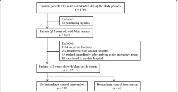
We conducted a retrospective observational study at a single center in an urban setting in Seoul, South Korea, from January 2013 to June 2018. Of 1760 trauma pa-tients ≥15 years, patients with penetrating injuries (n = 82) were excluded. In patients with blunt trauma (n = 1678), we also those with no pelvic fractures (n = 1184), those who had been managed or evaluated at other
hospitals (n = 252), those who died within minutes after arrival in the emergency room (n = 45), and/or those who were referred to other hospitals (n = 10). Therefore, the study was conducted with a total of 187 patients (Fig. 1). Patients were divided into those who did not undergo hemorrhage control intervention and those who did. Hemorrhage control intervention was defined as angioembolization, external fixation, or preperitoneal pelvic packing.
Clinical variables
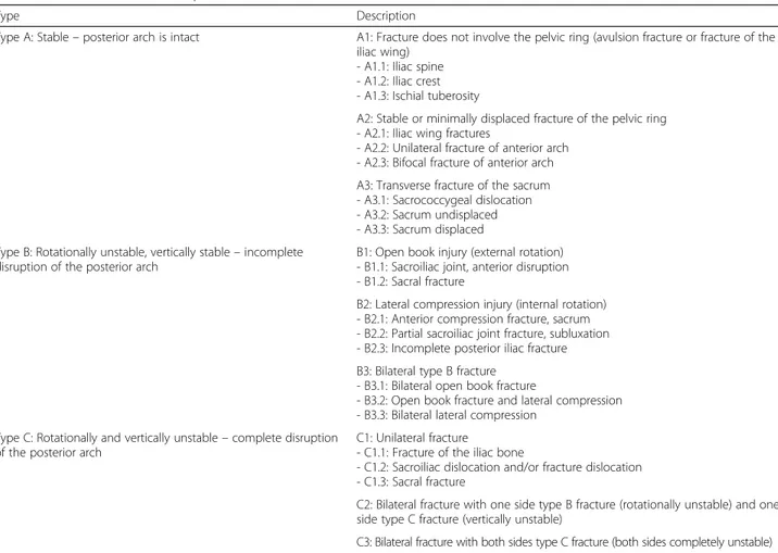
We analyzed the sex, age, injury mechanism, vital signs, Glasgow Coma Scale (GCS) score, current anticoagulant use, Abbreviated Injury Scale (AIS) score, Injury Severity Score (ISS), Revised Trauma Score (RTS), Trauma and Injury Severity Score (TRISS), Acute Physiology and Chronic Health Evaluation (APACHE) II score, and ar-terial blood values (pH, base excess, and lactate level). In addition, pelvic radiography of each patient was identi-fied, and patterns of pelvic fracture were classified as types A, B, and C according to the Orthopedic Trauma Association/Arbeitsgemeinschaft fur Osteosynthesefra-gen (OTA/AO) classification (Table1). The classification was based on the findings of trauma surgeons and orthopedic surgeons, which were additionally confirmed by surgical and radiological records.
Statistical analysis
Statistical analysis for investigated items was performed using SPSS® Statistics 23.0 (IBM, Armonk, NY, USA).
Categorical data are presented as numbers (%), and they were compared using the chi-square or Fisher’s exact tests. Continuous variables are expressed as mean ± stand-ard deviation or medians (the 25th and 75th quantiles), and the data were compared between groups using the Student t-test or Mann-WhitneyU test. Factors found to be significantly associated with the need for hemorrhage control intervention on univariate analysis were included in the multivariate analysis. Logistic regression modeling was performed using the maximum likelihood method and backward stepwise selection. Goodness of fit was assessed using the Hosmer-Lemeshow test. The odds ra-tios (ORs) are given with 95% confidence intervals (CIs). Ap value <0.05 was considered statistically significant.
Results
Patients
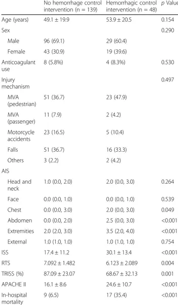
The baseline characteristics of patients are listed in Table
2. Of the 187 patients, 48 underwent hemorrhage control intervention and 139 did not undergo hemorrhage control intervention. The sex ratio was 125:62 (66.8%:33.2%, male/female). There was no significant difference between the hemorrhage control and non-hemorrhage control
intervention groups (p = 0.290). There was no significant difference between the two groups in the use of anticoagu-lants (p = 0.530). When classified according to the injury mechanism, pedestrian trauma injuries caused by motor vehicle accidents were the most frequent in both groups, followed by falls, which had the second highest frequency. However, there was no statistically significant difference between the groups that received hemorrhage control intervention and those that did not (p = 0.497).
When the AIS values of the two groups were com-pared, both systems, such as the abdomen (p < 0.001) and the extremity (p < 0.001), showed a significant dif-ference. However, there were no significant differences in other systems. Furthermore, there were significant dif-ferences in the ISS (p < 0.001), RTS (p = 0.004), TRISS (p = 0.001), and APACHE II score (p < 0.001) between the two groups.
Clinical variables comparison
The vital signs and laboratory variables of patients are shown in Table 3. Systolic blood pressure (p = 0.008)
and body temperature (p < 0.001) in the hemorrhage control intervention group were significantly lower than
Table 1 OTA/AO classification of pelvic fracture
Type Description
Type A: Stable– posterior arch is intact A1: Fracture does not involve the pelvic ring (avulsion fracture or fracture of the iliac wing)
- A1.1: Iliac spine - A1.2: Iliac crest - A1.3: Ischial tuberosity
A2: Stable or minimally displaced fracture of the pelvic ring - A2.1: Iliac wing fractures
- A2.2: Unilateral fracture of anterior arch - A2.3: Bifocal fracture of anterior arch A3: Transverse fracture of the sacrum - A3.1: Sacrococcygeal dislocation - A3.2: Sacrum undisplaced - A3.3: Sacrum displaced Type B: Rotationally unstable, vertically stable– incomplete
disruption of the posterior arch
B1: Open book injury (external rotation) - B1.1: Sacroiliac joint, anterior disruption - B1.2: Sacral fracture
B2: Lateral compression injury (internal rotation) - B2.1: Anterior compression fracture, sacrum - B2.2: Partial sacroiliac joint fracture, subluxation - B2.3: Incomplete posterior iliac fracture B3: Bilateral type B fracture
- B3.1: Bilateral open book fracture
- B3.2: Open book fracture and lateral compression - B3.3: Bilateral lateral compression
Type C: Rotationally and vertically unstable– complete disruption of the posterior arch
C1: Unilateral fracture
- C1.1: Fracture of the iliac bone
- C1.2: Sacroiliac dislocation and/or fracture dislocation - C1.3: Sacral fracture
C2: Bilateral fracture with one side type B fracture (rotationally unstable) and one side type C fracture (vertically unstable)
C3: Bilateral fracture with both sides type C fracture (both sides completely unstable)
those in the non-hemorrhage control intervention group. In the arterial gas analysis, the base excess (p = 0.001) was significantly lower in the hemorrhage control intervention group than those in the non-hemorrhage control intervention group, and lactate (p < 0.001) was significantly higher.
Comparison between two groups according to pelvic fracture pattern
According to the classification of posterior pelvic ring stability based on OTA/AO, type B with partial instabil-ity was the most common (n = 26, 54.2%) in the group with hemorrhage control intervention, followed by types A and C. Type A (n = 91, 65.5%) was the most common in the non-hemorrhage control intervention group,
followed by types B and C. Overall, there was significant difference (p < 0.001) between the groups with and with-out hemorrhage control intervention according to the OTA/AO classification (Table4).
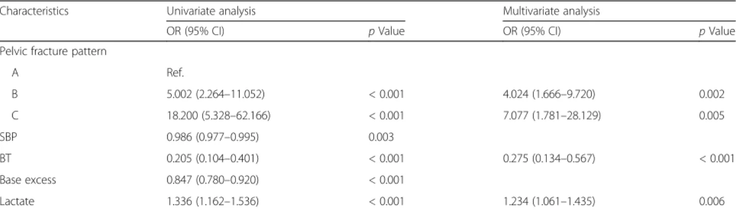
Logistic regression analysis for predictors of hemorrhage control intervention
The results of the univariate and multivariate regression analysis models are shown in Table5. As a result, in the pelvic bone fracture pattern according to OTA/AO clas-sification, types B (OR = 4.024, 95% CI = 1.666–9.720, p = 0.002) and C (OR = 7.077, 95% CI = 1.781–28.129, p = 0.005) were identified as predictors of hemorrhage control intervention. Among the clinical parameters, body temperature (OR = 0.275, 95% CI = 0.134–0.567, p < 0.001) and lactate (OR = 1.234, 95% CI = 1.061–1.435, p = 0.006) were identified as predictors. Furthermore, al-though univariate analysis revealed that the two groups had no significant differences in the use of anticoagu-lants, we additionally conducted a multivariable logistic regression analysis including “anticoagulant use” as co-variate, given its proven clinical significance. There was no difference in the results (Additional file1).
Table 3 Comparison of clinical parameters between two groups No hemorrhage control intervention (n = 139) Hemorrhagic control intervention (n = 48) p Value Vital sign SBP 118.8 ± 33.7 98.8 ± 46.2 0.008 HR 88.7 ± 23.3 96.1 ± 31.1 0.132 RR 18.7 ± 4.5 17.3 ± 7.2 0.223 BT 36.4 ± 0.5 35.9 ± 0.7 < 0.001 GCS 12.9 ± 4.0 11.4 ± 4.8 0.057 ABGA pH 7.38 ± 0.08 7.33 ± 0.16 0.057 BE -3.48 ± 3.30 -6.71 ± 5.79 0.001 Lactate 2.99 ± 1.89 5.42 ± 4.26 < 0.001
Values are presented as mean ± SD.
ABGA arterial blood gas analysis, SBP systolic blood pressure, HR heart rate, RR respiration rate, BT body temperature, GCS Glasgow Coma Scale, BE base excess
Table 2 Baseline characteristics of patients
No hemorrhage control intervention (n = 139) Hemorrhagic control intervention (n = 48) p Value Age (years) 49.1 ± 19.9 53.9 ± 20.5 0.154 Sex 0.290 Male 96 (69.1) 29 (60.4) Female 43 (30.9) 19 (39.6) Anticoagulant use 8 (5.8%) 4 (8.3%) 0.530 Injury mechanism 0.497 MVA (pedestrian) 51 (36.7) 23 (47.9) MVA (passenger) 11 (7.9) 2 (4.2) Motorcycle accidents 23 (16.5) 5 (10.4) Falls 51 (36.7) 16 (33.3) Others 3 (2.2) 2 (4.2) AIS Head and neck 1.0 (0.0, 2.0) 2.0 (0.0, 3.0) 0.264 Face 0.0 (0.0, 1.0) 0.0 (0.0, 1.0) 0.539 Chest 0.0 (0.0, 3.0) 2.0 (0.0, 3.0) 0.049 Abdomen 0.0 (0.0, 2.0) 2.5 (0.0, 3.0) <0.001 Extremities 2.0 (2.0, 3.0) 3.5 (2.0, 4.0) <0.001 External 1.0 (1.0, 1.0) 1.0 (1.0, 1.0) 0.754 ISS 17.4 ± 11.2 30.1 ± 13.4 <0.001 RTS 7.092 ± 1.482 6.123 ± 2.089 0.004 TRISS (%) 87.09 ± 23.07 68.67 ± 32.13 0.001 APACHE II 16.1 ± 8.6 24.6 ± 10.7 <0.001 In-hospital mortality 9 (6.5) 17 (35.4) <0.001
Values are presented as mean ± SD or n (%).
MVA motor vehicle accident, AIS Abbreviated Injury Scale, ISS Injury Severity Score, RTS Revised Trauma Score, TRISS Trauma and Injury Severity Score, APACHE Acute Physiology and Chronic Health Evaluation
Table 4 Comparison via pelvic fracture pattern of patients between two groups (OTA/AO)
No hemorrhage control intervention (n = 139) Hemorrhagic control intervention (n = 48) p Value Pelvic fracture pattern <0.001 A 91 (65.5) 11 (22.9) B 43 (30.9) 26 (54.2) C 5 (3.6) 11 (22.9)
OTA/AO Orthopedic Trauma Association/Arbeitsgemeinschaft fur Osteosynthesefragen
Discussion
Blunt pelvic injuries from high-energy mechanisms are often associated with pelvic fractures and injuries to the rectum and genitourinary tract [1–4]. The seriousness of blunt pelvic fractures lies in the possible occurrence of retroperitoneal hematomas and hemorrhagic shock [5,
6]. Unstable pelvic fractures are associated with massive hemorrhage [22], which is the leading cause of death in patients with major pelvic fractures [23, 24]. In the present study, patients with pelvic bleeding had signifi-cantly higher in-hospital mortality rates than those with-out pelvic bleeding. Moreover, among patients with pelvic bone fractures, trauma-related severity scores such as the GCS, ISS, RTS, and TRISS were significantly higher in the hemorrhage control intervention group than in the non-hemorrhage control intervention group. Therefore, trauma patients in need of emergent inter-vention or surgery for ongoing hemorrhage have in-creased chances of survival if the elapsed time between traumatic injury and bleeding control intervention is minimized [5,10,16,25].
The recent evolution of rapid pelvic stabilization by ex-ternal fixation or pelvic binding and of hemostasis by angiographic embolization, resuscitative endovascular bal-loon occlusion, or preperitoneal pelvic packing has signifi-cantly decreased the mortality rates of devastating pelvic injuries. However, early detection of bleeding is not easy in blunt pelvic fractures [14–20]. Furthermore, despite on-going bleeding in a severely injured patient arriving at a hospital, the vital signs of the patient may not show typical changes in the immediate and early periods after injury [26, 27]. In blunt pelvic trauma with hemodynamic in-stability, it is difficult to achieve adequate hemostasis due to rapid exsanguination. Therefore, early and quick pre-diction of the need for hemorrhage control interventions for pelvic injuries is important.
In the present study, type B and C fractures according to the OTA/AO classification were revealed as inde-pendent factors predicting the need for hemorrhage
control intervention in patients with blunt pelvic trauma. Type B and C fractures show pelvic bone frac-ture patterns including posterior pelvic ring instability. Type B fracture is a result of rotational forces that cause partial disruption of the posterior sacroiliac complex [28,
29]. Complete disruption of the posterior complex oc-curs in type C fractures, which are both rotationally and vertically unstable [28, 29]. In this study, the need for early hemorrhage control interventions was 4 and 7 times higher for type B and C fractures than for type A fractures, respectively.
Although patients with high-grade pelvic ring injuries may not have significant bleeding, the bleeding risk gener-ally increases with the degree of instability of the posterior pelvic ring [30,31]. Manson et al. [24] reported that trans-fusion requirements and mortality were significantly higher in the posterior ring instability pattern than in the pelvic bone fracture pattern without involvement of the posterior structures, and this suggests that stretching and tearing of soft tissues, like artery and vein, around the pos-terior pelvic ring showed greater hemorrhagic instability in lateral compression III, anterior-posterior compression III, and vertical shear. Costantini et al. [32] similarly con-cluded that there is a higher need for hemorrhage control intervention in the posterior pelvic ring instability pat-terns, such as anterior-posterior compression III or open pelvic fracture. In the current guidelines, markers of pelvic hemorrhage also include anterior-posterior and vertical shear deformations on standard roentgenograms [33–35].
In the present study, patterns of pelvic fracture were evaluated with pelvic radiography. To date, CT has re-placed radiography in classifying pelvic fractures [35]. Contrast-enhanced CT also helps diagnose pelvic hematoma and active extravasation of contrast [7]. Mul-tidetector CT has short acquisition times and allows for rapid identification and assessment of pelvic hemorrhage [36]. However, CT cannot be performed for all patients and is dependent on the situation, such as hemodynamic instability or absence of resources in each institution [36].
Table 5 Multivariable regression analysis according to OTA/AO classification
Characteristics Univariate analysis Multivariate analysis
OR (95% CI) p Value OR (95% CI) p Value
Pelvic fracture pattern
A Ref. B 5.002 (2.264–11.052) < 0.001 4.024 (1.666–9.720) 0.002 C 18.200 (5.328–62.166) < 0.001 7.077 (1.781–28.129) 0.005 SBP 0.986 (0.977–0.995) 0.003 BT 0.205 (0.104–0.401) < 0.001 0.275 (0.134–0.567) < 0.001 Base excess 0.847 (0.780–0.920) < 0.001 Lactate 1.336 (1.162–1.536) < 0.001 1.234 (1.061–1.435) 0.006
SBP systolic blood pressure, BT body temperature, OR odds ratio, CI confidence interval, OTA/AO Orthopedic Trauma Association/Arbeitsgemeinschaft fur Osteosynthesefragen
Although evaluation of the sacrum and sacroiliac joints is sometimes limited on a portable anteroposterior pelvic radiograph, pelvic radiography is one of the tools that can easily and quickly reveal the pelvic bone fracture pattern, and it is generally performed as an initial examination in the trauma bay [37,38]. Furthermore, pelvic radiography in hemodynamically unstable patients helps in identifying life-threatening pelvic ring injuries [39].
The patterns of pelvic fracture were classified as types A, B, and C using the OTA/AO classification. The OTA/ AO classification was based on fracture stability, especially the stability of the posterior lesion [28, 40–42]. Unstable pelvic fractures are more frequently associated with hemorrhage [4,8]. The OTA/TA classification is easier to use in classifying patterns of pelvic fracture through pelvic radiography than the Young-Burgess classification, which is based on mechanism of injury [38, 42]. Furthermore, the Young-Burgess classification scheme for pelvic ring in-jury basically cannot be used to guide transfusion require-ments and the need for angiography and embolization in individual cases [30].
In the multivariable regression analysis, the body temperature was significantly low in the hemorrhage control intervention group. In other words, hypothermia was a predictor of the need for hemorrhage control intervention. Hypothermia is common in trauma victims and is associated with an increased risk of severe bleed-ing and increased mortality [43, 44]. In the study of Gentilello et al. [45], the group of trauma patients with a mean body temperature of 34.5°C showed a mortality of 100% when they failed to be rewarmed to 36°C. There-fore, warming and euthermia in a trauma patient with pelvic bone fracture are crucial.
The current guidelines recommend either serum lactate or base deficit measurements as sensitive tests to estimate and monitor the extent of bleeding and shock [46–49]. Additionally, serial measurement of these parameters can be used to monitor the response to therapy [48]. The amount of lactate produced by anaerobic glycolysis is an indirect marker of oxygen debt, tissue hypoperfusion, and severity of hemorrhagic shock [47,50]. Similarly, base def-icit values derived from arterial blood gas analysis provide an indirect estimation of global tissue acidosis due to im-paired perfusion [50, 51]. Moreover, the lactate and base deficit have been mentioned in many studies as predictive values related to bleeding in pelvic bone fractures in trauma patients [52–55]. In this study, serum lactate was identified as an independent predictor of the need for hemorrhage control intervention. In other previous stud-ies, both initial serum lactate and lactate clearance after 6 hours were identified as independent risk factors for mor-tality in trauma patients [56]. In addition, an increased serum lactate level is associated with massive hemorrhage in pelvic ring fractures [57], and the serum lactate level
measured in the pre-hospital period was found to be asso-ciated with the clinical outcome in trauma patients [58].
We additionally conducted a multivariable logistic re-gression analysis including“anticoagulant use” as potential predictor. It is indisputable that anticoagulants may worsen bleeding in a trauma patient. However, despite the clinical significance of anticoagulants, the results have not changed whether anticoagulant use is included or not in the multivariable logistic regression analysis. Therefore, our results should be carefully interpreted and used strictly in a wider context of the patient’s clinical condi-tion, clinical setting, and individual included factors.
There are several limitations to the present study. First, it is a retrospective study. Second, it may be diffi-cult to generalize the results of this study, as it is a single-center study. Third, the statistical power is insuffi-cient because of the small number of subjects. There-fore, multicenter studies are needed to overcome these limitations.
Conclusion
OTA/AO type B and C fractures, hypothermia, and in-creased lactate level are independent factors predicting the need for hemorrhage control intervention in patients with blunt pelvic traumas. Type B and C fractures are more likely to be associated with vascular injuries than are type A fractures. Hypothermia is well known to worsen coagulopathy. Lactate is a marker of systemic tis-sue perfusion and is elevated in cases of hypoperfusion of the tissues, such as hemorrhagic shock. These three factors may reflect the severity and occurrence of pelvic bleeding in patients with blunt pelvic trauma. Therefore, the predictors can be helpful in making decisions about management of pelvic bone fractures with hemorrhage.
Additional file
Additional file 1:Multivariable regression analysis according to OTA/AO classification. (DOCX 14 kb)
Abbreviations
AIS:Abbreviated Injury Scale; APACHE: Acute Physiology and Chronic Health Evaluation; BE: Base excess; BT: Body temperature; GCS: Glasgow Coma Scale; HR: Heart rate; ISS: Injury Severity Score; LOS: Length of stay; MV: Mechanical ventilation; MVA: Motor vehicle accident; RR: Respiration rate; RTS: Revised Trauma Score; SBP: Systolic blood pressure; TRISS: Trauma and Injury Severity Score Injury Severity Score
Acknowledgments
The authors thank JE Moon for providing assistance with the statistical analyses, and we also thank all of the coordinators for their valuable help and cooperation.
Funding Not applicable.
Availability of data and materials
The datasets used and/or analyzed during the current study are available from the corresponding author on reasonable request.
Authors’ contributions
SHL designed the study. MJK and SHL were responsible for data acquisition. SHL and MJK analyzed and interpreted the data with JGL. MJK wrote the article. All authors read and approved the final manuscript.
Ethics approval and consent to participate
The study was approved by the Institutional Review Board of Severance Hospital, Yonsei University Health System (4-2017-0545), which waived the requirement for informed consent because of the retrospective nature of the study.
Consent for publication Not applicable.
Competing interests
The authors declare that they have no competing interests.
Publisher’s Note
Springer Nature remains neutral with regard to jurisdictional claims in published maps and institutional affiliations.
Received: 7 August 2018 Accepted: 5 November 2018
References
1. Demetriades D, Karaiskakis M, Toutouzas K, Alo K, Velmahos G, Chan L. Pelvic fractures. epidemiology and predictors of associated abdominal injuries and outcomes. J Am Coll Surg. 2002;195:1–10.
2. Snyder HS. Blunt pelvic trauma. Am J Emerg Med. 1988;6:618–27. 3. Trunkey DD, Chapman MW, Lim RC Jr, Dunphy JE. Management of pelvic
fractures in blunt trauma injury. J Trauma. 1974;14:912–23.
4. Durkin A, Sagi HC, Durham R, Flint L. Contemporary management of pelvic fractures. Am J Surg. 2006;192:211–23.
5. Heetveld MJ, Harris I, Schlaphoff G, Sugrue M. Guidelines for the management of haemodynamically unstable pelvic fracture patients. ANZ J Surg. 2004;74:520–9.
6. Geeraerts T, Chhor V, Cheisson G, Martin L, Bessoud B, Ozanne A, et al. Clinical review. initial management of blunt pelvic trauma patients with haemodynamic instability. Crit Care. 2007;11:204.
7. Huittinen VM, Slatis P. Postmortem angiography and dissection of the hypogastric artery in pelvic fractures. Surgery. 1973;73:454–62.
8. White CE, Hsu JR, Holcomb JB. Haemodynamically unstable pelvic fractures. Injury. 2009;40:1023–30.
9. Sathy AK, Starr AJ, Smith WR, Elliott A, Agudelo J, Reinert CM, et al. The effect of pelvic fracture on mortality after trauma: an analysis of 63,000 trauma patients. J Bone Joint Surg Am. 2009;91:2803–10.
10. Costantini TW, Coimbra R, Holcomb JB, Podbielski JM, Catalano R, Blackburn A, et al. Current management of hemorrhage from severe pelvic fractures. Results of an American Association for the Surgery of Trauma multi-institutional trial. J Trauma Acute Care Surg. 2016;80:717–23 discussion 723-15. 11. Chu CH, Tennakoon L, Maggio PM, Weiser TG, Spain DA, Staudenmayer KL. Trends in the management of pelvic fractures, 2008-2010. J Surg Res. 2016; 202:335–40.
12. Chiara O, di Fratta E, Mariani A, Michaela B, Prestini L, Sammartano F, et al. Efficacy of extra-peritoneal pelvic packing in hemodynamically unstable pelvic fractures, a propensity score analysis. World J Emerg Surg. 2016;11:22. 13. Starr AJ, Griffin DR, Reinert CM, Frawley WH, Walker J, Whitlock SN, et al.
Pelvic ring disruptions: prediction of associated injuries, transfusion requirement, pelvic arteriography, complications, and mortality. J Orthop Trauma. 2002;16:553–61.
14. Burlew CC, Moore EE, Smith WR, Johnson JL, Biffl WL, Barnett CC, et al. Preperitoneal pelvic packing/external fixation with secondary angioembolization: optimal care for life-threatening hemorrhage from unstable pelvic fractures. J Am Coll Surg. 2011;212:628–35 discussion 635-7. 15. Tang J, Shi Z, Hu J, Wu H, Yang C, Le G, et al. Optimal sequence of surgical procedures for hemodynamically unstable patients with pelvic fracture. A network meta-analysis. Am J Emerg Med. 2018;S0735-6757(18):30489–3.
16. Matsushima K, Piccinini A, Schellenberg M, Cheng V, Heindel P, Strumwasser A, et al. Effect of door-to-angioembolization time on mortality in pelvic fracture. Every hour of delay counts. J Trauma Acute Care Surg. 2018;84: 685–92.
17. Fu CY, Chan SY, Wang SY, Hsieh CH, Liao CH, Huang JF, et al. The effect of angioembolization for life-threatening retroperitoneal hemorrhage in patients with pelvic fracture. Am J Emerg Med. 2018;S0735-6757(18): 30507–2.
18. Tesoriero RB, Bruns BR, Narayan M, Dubose J, Guliani SS, Brenner ML, et al. Angiographic embolization for hemorrhage following pelvic fracture. Is it “time” for a paradigm shift? J Trauma Acute Care Surg. 2017;82:18–26. 19. Burlew CC, Moore EE, Stahel PF, Geddes AE, Wagenaar AE, Pieracci FM, et al.
Preperitoneal pelvic packing reduces mortality in patients with life-threatening hemorrhage due to unstable pelvic fractures. J Trauma Acute Care Surg. 2017;82:233–42.
20. Marzi I, Lustenberger T. Management of bleeding pelvic fractures. Scand J Surg. 2014;103:104–11.
21. Balogh Z, Caldwell E, Heetveld M, D’Amours S, Schlaphoff G, Harris I, et al. Institutional practice guidelines on management of pelvic fracture-related hemodynamic instability: do they make a difference? J Trauma. 2005;58: 778–82.
22. Ertel W, Keel M, Eid K, Platz A, Trentz O. Control of severe hemorrhage using C-clamp and pelvic packing in multiply injured patients with pelvic ring disruption. J Orthop Trauma. 2001;15:468–74.
23. Smith W, Williams A, Agudelo J, Shannon M, Morgan S, Stahel P, et al. Early predictors of mortality in hemodynamically unstable pelvis fractures. J Orthop Trauma. 2007;21:31–7.
24. Manson T, O’Toole RV, Whitney A, Duggan B, Sciadini M, Nascone J. Young-Burgess classification of pelvic ring fractures: does it predict mortality, transfusion requirements, and non-orthopaedic injuries? J Orthop Trauma. 2010;24:603–9.
25. Salim A, Teixeira PG, DuBose J, Ottochian M, Inaba K, Margulies DR, et al. Predictors of positive angiography in pelvic fractures: a prospective study. J Am Coll Surg. 2008;207:656–62.
26. Frank M, Schmucker U, Stengel D, Fischer L, Lange J, Grossjohann R, et al. Proper estimation of blood loss on scene of trauma: tool or tale? J Trauma. 2010;69:1191–5.
27. Guly HR, Bouamra O, Spiers M, Dark P, Coats T, Lecky FE, Trauma Audit and Research Network. Vital signs and estimated blood loss in patients with major trauma: testing the validity of the ATLS classification of hypovolaemic shock. Resuscitation. 2011;82:556–9.
28. Osterhoff G, Scheyerer MJ, Fritz Y, Bouaicha S, Wanner GA, Simmen HP, et al. Comparing the predictive value of the pelvic ring injury classification systems by Tile and by Young and Burgess. Injury. 2014;45:742–7. 29. Nuchtern JV, Hartel MJ, Henes FO, Groth M, Jauch SY, Haegele J, et al.
Significance of clinical examination, CT and MRI scan in the diagnosis of posterior pelvic ring fractures. Injury. 2015;46:315–9.
30. Sarin EL, Moore JB, Moore EE, Shannon MR, Ray CE, Morgan SJ, et al. Pelvic fracture pattern does not always predict the need for urgent embolization. J Trauma. 2005;58:973–7.
31. Kimbrell BJ, Velmahos GC, Chan LS, Demetriades D. Angiographic embolization for pelvic fractures in older patients. Arch Surg. 2004;139:728– 32 discussion 732-3.
32. Costantini TW, Coimbra R, Holcomb JB, Podbielski JM, Catalano RD, Blackburn A, et al. Pelvic fracture pattern predicts the need for hemorrhage control intervention– results of an AAST multi-institutional study. J Trauma Acute Care Surg. 2017;82:1030–8.
33. Miller PR, Moore PS, Mansell E, Meredith JW, Chang MC. External fixation or arteriogram in bleeding pelvic fracture: initial therapy guided by markers of arterial hemorrhage. J Trauma. 2003;54:437–43.
34. Hagiwara A, Minakawa K, Fukushima H, Murata A, Masuda H, Shimazaki S. Predictors of death in patients with life-threatening pelvic hemorrhage after successful transcatheter arterial embolization. J Trauma. 2003;55:696–703. 35. Coccolini F, Stahel PF, Montori G, Biffl W, Horer TM, Catena F, et al. Pelvic trauma: WSES classification and guidelines. World J Emerg Surg. 2017;12:5. 36. Kos S, Gutzeit A, Hoppe H, Liu DM, Jacob AL. Diagnosis and therapy of
acute hemorrhage in patients with pelvic fractures. Semin Musculoskelet Radiol. 2013;17:396–406.
37. Burgess AR, Eastridge BJ, Young JW, Ellison TS, Ellison PS Jr, et al. Pelvic ring disruptions: effective classification system and treatment protocols. J Trauma. 1990;30:848–56.
38. Dalal SA, Burgess AR, Siegel JH, Young JW, Brumback RJ, Poka A, et al. Pelvic fracture in multiple trauma: classification by mechanism is key to pattern of organ injury, resuscitative requirements, and outcome. J Trauma. 1989;29: 981–1000 discussion 1000-2.
39. Agri F, Bourgeat M, Becce F, Moerenhout K, Pasquier M, Borens O, et al. Association of pelvic fracture patterns, pelvic binder use and arterial angio-embolization with transfusion requirements and mortality rates; a 7-year retrospective cohort study. BMC Surg. 2017;17:104.
40. Furey AJ, O’Toole RV, Nascone JW, Sciadini MF, Copeland CE, Turen C. Classification of pelvic fractures: analysis of inter- and intraobserver variability using the Young-Burgess and Tile classification systems. Orthopedics. 2009;32:401.
41. Alton TB, Gee AO. Classifications in brief: Young and Burgess classification of pelvic ring injuries. Clin Orthop Relat Res. 2014;472:2338–42.
42. Koo H, Leveridge M, Thompson C, Zdero R, Bhandari M, Kreder HJ, et al. Interobserver reliability of the young-burgess and tile classification systems for fractures of the pelvic ring. J Orthop Trauma. 2008;22:379–84.
43. Peng RY, Bongard FS. Hypothermia in trauma patients. J Am Coll Surg. 1999; 188:685–96.
44. Watts DD, Trask A, Soeken K, Perdue P, Dols S, Kaufmann C. Hypothermic coagulopathy in trauma: effect of varying levels of hypothermia on enzyme speed, platelet function, and fibrinolytic activity. J Trauma. 1998;44:846–54. 45. Gentilello LM, Jurkovich GJ, Stark MS, Hassantash SA, O’Keefe GE. Is
hypothermia in the victim of major trauma protective or harmful? A randomized, prospective study. Ann Surg. 1997;226:439–47 discussion 447-9. 46. Bilkovski RN, Rivers EP, Horst HM. Targeted resuscitation strategies after
injury. Curr Opin Crit Care. 2004;10:529–38.
47. Baron BJ, Scalea TM. Acute blood loss. Emerg Med Clin North Am. 1996;14: 35–55.
48. American College of Surgeons Committee on Trauma. ATLS ® Student Manual. 10th ed. Chicago, IL: American College of Surgeons; 2018. 49. Davis JW, Parks SN, Kaups KL, Gladen HE, O’Donnell-Nicol S. Admission base
deficit predicts transfusion requirements and risk of complications. J Trauma. 1996;41:769–74.
50. Wilson M, Davis DP, Coimbra R. Diagnosis and monitoring of hemorrhagic shock during the initial resuscitation of multiple trauma patients: a review. J Emerg Med. 2003;24:413–22.
51. Porter JM, Ivatury RR. In search of the optimal end points of resuscitation in trauma patients: a review. J Trauma. 1998;44:908–14.
52. Abramson D, Scalea TM, Hitchcock R, Trooskin SZ, Henry SM, Greenspan J. Lactate clearance and survival following injury. J Trauma. 1993;35:584–8 discussion 588-9.
53. Lavery RF, Livingston DH, Tortella BJ, Sambol JT, Slomovitz BM, Siegel JH. The utility of venous lactate to triage injured patients in the trauma center. J Am Coll Surg. 2000;190:656–64.
54. Claridge JA, Crabtree TD, Pelletier SJ, Butler K, Sawyer RG, Young JS. Persistent occult hypoperfusion is associated with a significant increase in infection rate and mortality in major trauma patients. J Trauma. 2000;48:8– 14 discussion 14-5.
55. Toth L, King KL, McGrath B, Balogh ZJ. Factors associated with pelvic fracture-related arterial bleeding during trauma resuscitation: a prospective clinical study. J Orthop Trauma. 2014;28:489–95.
56. Odom SR, Howell MD, Silva GS, Nielsen VM, Gupta A, Shapiro NI, et al. Lactate clearance as a predictor of mortality in trauma patients. J Trauma Acute Care Surg. 2013;74:999–1004.
57. Ohmori T, Matsumoto T, Kitamura T, Tamura R, Tada K, Inoue T, et al. Scoring system to predict hemorrhage in pelvic ring fracture. Orthop Traumatol Surg Res. 2016;102:1023–8.
58. Guyette F, Suffoletto B, Castillo JL, Quintero J, Callaway C, Puyana JC. Prehospital serum lactate as a predictor of outcomes in trauma patients: a retrospective observational study. J Trauma. 2011;70:782–6.