저작자표시-비영리-변경금지 2.0 대한민국 이용자는 아래의 조건을 따르는 경우에 한하여 자유롭게 l 이 저작물을 복제, 배포, 전송, 전시, 공연 및 방송할 수 있습니다. 다음과 같은 조건을 따라야 합니다: l 귀하는, 이 저작물의 재이용이나 배포의 경우, 이 저작물에 적용된 이용허락조건 을 명확하게 나타내어야 합니다. l 저작권자로부터 별도의 허가를 받으면 이러한 조건들은 적용되지 않습니다. 저작권법에 따른 이용자의 권리는 위의 내용에 의하여 영향을 받지 않습니다. 이것은 이용허락규약(Legal Code)을 이해하기 쉽게 요약한 것입니다. Disclaimer 저작자표시. 귀하는 원저작자를 표시하여야 합니다. 비영리. 귀하는 이 저작물을 영리 목적으로 이용할 수 없습니다. 변경금지. 귀하는 이 저작물을 개작, 변형 또는 가공할 수 없습니다.
Increased airway surface liquid and
decreased mucin expression in
pendrin-deficient human airway
epithelia
Hyun Jae Lee
Department of Medical Science
The Graduate School, Yonsei University
Increased airway surface liquid and
decreased mucin expression in
pendrin-deficient human airway
epithelia
Directed by Professor Jae Young Choi
The Doctoral Dissertation
submitted to the Department of Medical Science,
the Graduate School of Yonsei University
in partial fulfillment of the requirements for the
degree of Doctor of Philosophy
Hyun Jae Lee
This certifies that the Doctoral
Dissertation of Hyun Jae Lee is
approved.
---Thesis Supervisor: Jae Young Choi
---Thesis Committee Member #1: Min Goo Lee
---Thesis Committee Member #2: Chul Hoon Kim
---Thesis Committee Member #3: Jinwoong Bok
---Thesis Committee Member #4: Sung Huhn Kim
The Graduate School
Yonsei University
ACKNOWLEDGEMENTS
학위과정 동안 많은 가르침과 도움을 주신 모든 분들께 감사의 말씀을 전합니다. 먼저, 지도교수님으로서 많은 조언과 가르침을 주신 최재영 교수님께 감사 드립니다. 연구자로서 가야 할 길과 목표를 제시해 주신 윤주헌 교수님, 이비인후과 발전을 위해 힘쓰시는 김창훈 교수님, 가까이서 격려와 조언을 해주신 이상남 선생님, 유지환 선생님 모두 감사 드립니다. 바쁘신 와중에도 학위논문에 대한 검토와 실험적 조언을 해주신 이민구 교수님, 김철훈 교수님, 복진웅 교수님, 김성헌 교수님께도 감사 드립니다. 지난 학위과정 동안 많은 시간을 함께하며 고생했던 실험실 동료들에게도 감사의 인사를 전합니다. 힘이 들 때 옆에서 힘이 되어 주고, 기쁠 때 같이 기쁨을 나눠 주었던 친구들에게도 고맙다는 말을 전합니다. 마지막으로 사랑하는 가족들. 긴 시간 동안 공부할 수 있도록 무한한 사랑과 격려를 아끼지 않으셨던 아버지, 어머니. 많은 관심 주시고 함께 걱정해 주신 장인어른, 장모님. 바쁘다는 핑계로 소홀했던 저의 역할을 대신해준 동생. 짧지 않은 시간 동안 참고 기다려준 아내와 눈에 넣어도 아프지 않을 딸 소윤이에게 감사하다는 말을 전합니다. 이제 또 다른 시작이라는 마음으로 겸손한 태도를 가지고 좀 더 발전해 나아가는 연구자가 되도록 노력하겠습니다. 2015. 07 이현재TABLE OF CONTENTS
ABSTRACT
···
1
I. INTRODUCTION
···
3
II. MATERIALS AND METHODS
···
6
1. Subjects and tissue harvesting
···
6
2. Cell culture and IL-13 treatment
···
7
3. Quantitative Real-time PCR
···
7
4. Measurement of ASL thickness
···
8
5. Short circuit current
···
9
6. Measurement of anion channel activity
···
9
7. Histology and periodic acid-Schiff (PAS) staining
···
10
8. Statistics
···
11
1. ASL thickness; DFNB4 patients vs. controls
···
12
2. Expression pattern of mRNAs encoding Na
+and Cl
-channels
···
13
3. Anion exchanger expression and activity
···
16
4. MUC5AC expression and goblet cell differentiation
···
19
IV. DISCUSSION
···
24
V. CONCLUSION
···
28
REFERENCES
···
29
ABSTRACT (IN KOREAN)
···
34
LIST OF FIGURES
Figure 1. ASL thickness in human nasal epithelia from
patients harboring SLC26A4 mutations (DFNB4) and
controls···12
Figure 2. Expression of epithelial sodium channels (ENaC)
and Cl
-channels (CFTR and ANO-1) in human nasal
epithelial cells from patients harboring SLC26A4 mutations
(DFNB4) and control···14
Figure 3. Changes in ion channel expression in response to
IL-13 treatment···15
Figure 4. Changes in ion channel activity in response to
IL-13 treatment
···16
Figure 5. Anion exchanger expression in cultured human
nasal epithelia from controls···18
Figure 6. Anion exchange activity in human nasal epithelia
cultured from patients with SLC26A4 mutations (DFNB4)
and controls···19
Figure 7. MUC5AC expression and goblet cell differentiation
in cultured human nasal epithelial cells···20
Figure 8. PAS staining of inferior turbinates···21
Figure 9. The hypothetical role of pendrin in the regulation of
airway surface liquid (ASL)···22
LIST OF TABLES
ABSTRACT
Increased airway surface liquid and decreased mucin expression
in pendrin-deficient human airway epithelia
Hyun Jae Lee
Department of Medical Science
The Graduate School, Yonsei University
(Directed by Professor Jae Young Choi)
Pendrin is an anion exchanger whose mutations are known to cause hearing loss. However, recent data support the linkage between pendrin expression and airway diseases, such as asthma. To evaluate the role of pendrin in the regulation of the airway surface liquid (ASL) volume and mucin expression, we investigated the function and expression of pendrin and ion channels and anion exchangers.
Human nasal epithelial cells were cultured from 16 deaf patients carrying pendrin mutations (DFNB4) and 17 controls. The cells were treated with IL-13 to induce mucus hypersecretion. ASL thickness was measured and real-time polymerase chain reaction was performed targeting various transporters
and MUC5AC. Anion exchanger activity was measured using a pH-sensitive fluorescent probe. Periodic acid-Schiff staining was performed on the cultured cells and inferior turbinate tissues.
The ASL layer of the nasal epithelia from DFNB4 subjects was thicker than the controls, and the difference became more prominent following IL-13 stimulation. There was no difference in anion exchange activity after IL-13 treatment in the cells from DFNB4 patients, while it increased in the controls. Goblet cell metaplasia induced by IL-13 treatment seen in the controls was not observed in the DFNB4 cells. Furthermore, the periodic acid-Schiff staining-positive area was lesser in the inferior turbinate tissues from DFNB4 patients that those from controls.
Pendrin plays a critical role in ASL volume regulation and mucin expression as pendrin-deficient airway epithelial cells are refractory to stimulation with IL-13. Specific blockers targeting pendrin in the airways may therefore have therapeutic potential in the treatment of allergic airway diseases.
Increased airway surface liquid and decreased mucin expression
in pendrin-deficient human airway epithelia
Hyun Jae Lee
Department of Medical Science
The Graduate School, Yonsei University
(Directed by Professor Jae Young Choi)
I. INTRODUCTION
Pendrin, encoded by SLC26A4, is a membrane protein that exchanges anions such as HCO3- and I-for Cl-.1 The protein is predominantly expressed in the inner ear, thyroid and kidney.2-5 Pendrin presumably acts as an HCO3 -/Cl-exchanger in the inner ear, and mutations cause prelingual hearing loss with enlarged vestibular aqueducts (DFNB4).6-8 In the thyroid, pendrin is essential for I- uptake, and defects in this protein may cause goiter and possibly hypothyroidism. Goiter combined with hearing loss is known as Pendred syndrome.9,10
Pendrin is also expressed in the airway epithelia11-16and recent data suggest that pendrin expression is associated with airway diseases. Pendrin expression
is up-regulated in airway tissues of patients with asthma and allergic rhinitis.11-14,17,18 Moreover, recent study showed that patients harboring bi-allelic SLC26A4 mutations do not experience asthma,14 although the prevalence of asthma among these patients is not significantly lower compared to controls due to the low numbers of subjects. Animal studies also showed the role of pendrin in the pathogenesis of asthma. Ovalbumin-induced airway hyperresponsiveness and inflammation are attenuated in pendrin-knockout mice13,19 and the overexpression of pendrin in airway epithelia of mice resulted in increased airway hyperresponsiveness.13 These findings suggest that pendrin may play an important role in the development of asthma. Homeostasis of the volume and composition of the airway surface liquid (ASL) is essential for maintaining the mucociliary clearance system.20,21ASL volume is regulated by the coordinated interactions of various ion transporters, such as epithelial sodium channels (ENaC) and cystic fibrosis transmembrane conductance regulators (CFTR). A recent report13 indicates that pendrin is involved in ASL volume regulation. ASL thickness was shown to increase following allergic cytokine, interleukin (IL)-13, stimulation in the airway epithelia of pendrin-knockout mice. Another interesting discovery is that pendrin may strongly participate in the secretion of airway mucus. Overexpression of pendrin induces mucus hypersecretion in mice, and pendrin has been shown to be a key modulator of IL-13-induced mucus secretion.14
Based on these recent findings, pendrin may be a potential new drug target for asthma and other inflammatory airway diseases. However, before deciding on the value of pendrin as a new drug target, further elucidation of the physiological role of pendrin in human airways is necessary because the inner ear and the thyroid phenotypes of pendrin knockout mice have shown to be different from those of patients harboring SLC26A4 mutations and the physiology underlying ASL homeostasis and mucus secretion in human airways is quite different from that in animal models.
In this study, we compare the ASL thickness, mucin expression and the response to stimulation with IL-13 in nasal epithelial cells cultured from patients with SLC26A4 mutations (DFNB4) to those in controls.
II. MATERIALS AND METHODS
1. Subjects and tissue harvesting
This study was approved by the Institutional Review Board of the Yonsei University Health System. Human nasal mucosa was obtained from the inferior turbinates of 16 deaf patients harboring bi-allelic SLC26A4 mutations (DFNB4) during cochlear implantation. Nasal mucosa was also harvested in 17 controls whom underwent septoplasty for correction of deviated nasal septum. These controls had no history of allergic rhinitis or chronic sinusitis, and a genetic study confirmed they did not carry any SLC26A4 mutations. A genetic analysis confirmed the presence of the bi-allelic SLC26A4 mutation in all 16 deaf patients. The genotypes of the DFNB4 patients were as follows: H723R homozygote (n=6), H723R/IVS7-2A>G (n=6), IVS7-2A>G homozygote (n=2), IVS7-2A>G/M147V (n=1) and IVS7-2A>G/I302K (n=1). (Table 1) shows the characteristics of the subjects.
Table 1. Subject Characteristics
N Age Male (no. [%]
DFNB4* 16 18 ± 12 8 (50)
Control 17 25 ± 7 10 (59)
2. Cell culture and IL-13 treatment
Primary cultures of human nasal epithelial (HNE) cells were obtained as previously described.22 Passage-2 cells were seeded into a Transwell-Clear culture insert with a 0.45-μm pore size (Costar Co., Cambridge, MA, USA) at a density of 2 x 10⁵cells/cm2. The cells were maintained in a 1:1 mixture of Dulbecco’s modified Eagle’s medium (Lonza, Walkersville, MD, USA) and bronchial epithelial growth medium (Lonza), supplemented with the following growth factors according to the manufacturer’s instructions: hydrocortisone (0.5 μg/ml), insulin (5 μg/ml), transferrin (10 μg/ml), epinephrine (0.5 μg/ml), triiodothyronine (6.5 μg/ml), gentamicin (50 μg/ml), amphotericin B (50 μg/ml), retinoic acid (15 ng/ml), bovine pituitary extract (50 μg/ml), bovine serum albumin (1.5 μg/ml) and epidermal growth factor (0.5 ng/ml). The cells were maintained submerged for the first 7 days, after which they were exposed to the apical air interface for the remainder of the culture period. The cells were used between 14 and 21 days after the establishment of the air-liquid interface. At all stages of culture, the cells were maintained at 37°C under 5% CO2in an air incubator. To stimulate the cells, they were treated basolaterally with 10 ng/ml of recombinant human IL-13 (R&D Systems, Minneapolis, MN, USA) for 7 days.
Total RNA was isolated from primary cultures of HNE cells from five patients harboring bi-allelic SCL26A4 mutations and four controls. RNA was isolated using the TRIzol reagent (Invitrogen, San Diego, CA, USA)
according to the manufacturer's protocol. Purified RNA samples were reverse
transcribed with a cDNA Synthesis Kit (Applied Biosystems, Foster City, CA) according to the manufacturer’s instructions. Quantitative Real-time PCR for pendrin, epithelial sodium channel (ENaC) α, β, γ, CFTR, ANO-1, Anion exchanger (AE) 2, AE3, AE4 and SLC26A3, SLC26A6, SLC26A7, SLC26A8, SLC26A9, SLC26A11, MUC5AC was performed using the 7300 Real-Time PCR System (Applied Biosystems) and the DyNAmoHSSYBR Green qPCR Kit (Finnzymes, Espoo, Finland). The primers employed for real time PCR are listed in Table S1.
4. Measurement of ASL thickness
The thickness of the ASL was measured in HNE cells using a modified version of the method described by Terran et al.21 Briefly, the cells were washed, and 20 μl of PBS containing 0.2% (v/v) Texas Red-dextran (Invitrogen) was added onto the apical cell surface to label the ASL layer. A 100-μl aliquot of perfluorocarbon (Fluorinert FC-770; 3M, St. Paul, MN, USA)was then added to the apical surface to prevent evaporation of the ASL. After 12 hr, the cultures were transferred to the stage of an inverted confocal
microscope (LSM 700; Carl Zeiss MicroImaging Inc.), and the height of the ASL was measured at five predetermined points in the cultures (one central, four circumferential) via XZ scans. Images were reconstructed 3-dimensionally and analyzed using Imaris 7.1 software (Bitplane Co, Zurich, Switzerland).
5. Short circuit current
Human nasal epithelial cells were mounted in Ussing chambers (Physiologic Instruments, San Diego, CA). Amiloride, CFTRinh-172, ATP were added to the apical solution, and an equal volume of vehicle was added at the same time to the basolateral solution. Symmetrical HCO3--buffered solutions (120 mM NaCl, 5 mM KCl, 1 mM MgCl2,1mM CaCl2, 10 mM D-glucose, 5 mM HEPES, and 25 mM NaHCO3, pH 7.4)were used for human nasal epithelial cells. Cells were bathed for a 10 min stabilization period and aerated with 95% O2/5% CO2 at 37 °C or room temperature. Short circuit current was measured using an EVC4000 MultiChannel V/I Clamp (World Precision Instruments, Sarasota, FL) and recorded using PowerLab/8sp (AD Instruments, Castle Hill, Australia).
Measurements of the pHi in the HNE cells were performed with a pH-sensitive fluorescent probe, bis-carboxyethyl-carboxyfluorescein (BCECF)
(Invitrogen, San Diego, CA, USA), as described previously.23 Briefly, the measurements were performed in PBS- or IL-13-treated cells, in which a cluster of cells showing green fluorescent protein (GFP) fluorescence was exposed to BCECF, and the pHi was monitored. After adding BCECF, the cells were perfused with HCO3-buffered solution (120 NaCl, 5 KCl, 1 MgCl2, 1 CaCl2, 10 D-glucose, 5 HEPES and 25 NaHCO3 in mmol/l, pH 7.4), and BCECF fluorescence was recorded at excitation wavelengths of 490 nm and 440 nm at a resolution of 2 sec on a recording setup (Delta Ram; PTI Inc., Birmingham, NJ, USA). The Cl-/HCO3- exchange activities were estimated from the initial rate of the pHiincrease as a result of Cl-removal in the HCO3- -containing buffer (25 mM HCO3-with 5% CO2). The intrinsic buffer capacity (βi) was measured as described previously.23
7. Histology and periodic acid-Schiff (PAS) staining
Periodic acid-Schiff (PAS) staining was performed with a PAS staining kit (Sigma, St. Louis, MO, USA) according to the manufacturer's protocol. The harvested nasal mucosa and HNE cells on the transwell inserts were gently washed with PBS and fixed with 4% paraformaldehyde. After deparaffinization and hydration, the slides were immersed in periodic acid
solution for 5 min at room temperature. After being rinsed in distilled water, the slides were immersed in Schiff’s reagent for 15 min at room temperature, and then washed in running tap water for 5 min. Slides were counterstained in Hematoxylin Solution for 90 sec and then rinsed in running tap water,
dehydrated, cleared and permanently mounted. For morphometric analysis,
images of the PAS-stained slides (five different slides of tissue from each patient) were acquired with a Leica DM750 LED microscope and PAS-positive cells were counted.
8. Statistics
Statistical analysis was performed with SPSS software for PC, version 19 (SPSS Inc., Chicago, IL). Differences between groups regarding anion exchange activities and the rates of pendrin positivity were evaluated via one-way analysis of variance. A p-value of less than 0.05 was considered statistically significant.
III. RESULTS
1. ASL thickness; DFNB4 patients vs. controls
First, we measured the ASL thickness in HNE cells from patients harboring
SLC26A4 mutations (DFNB4) and controls. Twelve hours after PBS/Texas
Red-dextran loading, the ASL thickness in the DFNB4 patients (14.1 ± 2.9 μm) was thicker than that in the controls (8.0 ± 2.1 μm, p<0.05). The increase in ASL thickness following IL-13 stimulation was more prominent in DFNB4 cells (31.3 ± 7.6 μm) than in controls (13.5 ± 2.8 μm) (Figure 1).
Figure 1. ASL thickness in human nasal epithelia from patients harboring
SLC26A4 mutations (DFNB4) and controls. Confocal images (A) and
summary of ASL thickness measurements (B) at 12 hr after the apical addition of 20 μl of PBS containing Texas Red-dextran to primary nasal epithelial cultures from DFNB4 patients (n=5) and controls (n=4) in the absence or presence of IL-13 (10 ng/ml). Scale bars, 10 μm. * p<0.05, ** p<0.01,compared with the controls. Error bars, S.E.
2. Expression pattern of mRNAs encoding Na+and Cl-channels
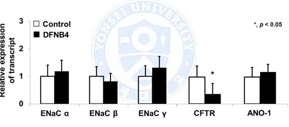
We compared the mRNA expression patterns of ion transporters involved in ASL volume regulation to rule out the possibility that the difference in ASL thickness between the pendrin mutants and controls stemmed from the differential expression of these transporters. The expression patterns of ENaC subtypes α, β, and γ and ANO-1 were similar between DFNB4 patients and controls. However, CFTR showed lower expression in the DFNB4 patients (0.36 ± 0.25 fold of that in the controls) (Figure 2).
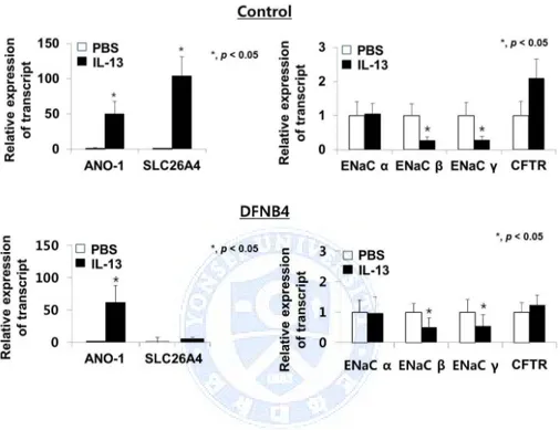
When the HNE cells from controls were treated with IL-13, ENaC subtypes β and γ were down-regulated, whereas ANO-1 (49 ± 18 fold of PBS) was up-regulated. As previously reported, pendrin expression was also greatly increased by 104 ± 27 fold following treatment with IL-13. The response of the pendrin-deficient epithelial cells to IL-13 treatment was similar to the
response in the controls: ENaC subtypes β and γ were down-regulated, and ANO-1 was up-regulated (61 ± 26 fold) (Figure 3).
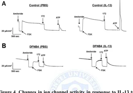
Short circuit current measurements in HNE cells showed these expression patterns. IL-13-treated HNE cells from control showed decreased amiloride sensitive ENaC current and increased CaCC current. In DFNB4 cells, IL-13 treatment had no effect on ENaC and CFTR current, but had increased CaCC current following ATP stimulation (Figure 4).
Figure 2. Expression of epithelial sodium channels (ENaC) and Cl -channels (CFTR and ANO-1) in human nasal epithelial cells from patients harboring SLC26A4 mutations (DFNB4) and controls. The expression patterns in the cells from patients with DFNB4 (n=3) are similar to those in controls (n=3), except for CFTR, which shows low levels of
expression in the epithelia from DFNB4 patients. * indicates p<0.05, compared to controls.
Figure 3. Changes in ion channel expression in response to IL-13 treatment (10 ng/ml). In both the control (n=3) and SLC26A4 mutant (DFNB4) (n=3) nasal epithelia, ENaC β and γ expression are suppressed, and ANO-1 expression is greatly increased (by more than 50 fold) following
IL-13 treatment. In the control epithelia, pendrin is also upregulated (approximately 100 fold) following IL-13 treatment. * indicates p<0.05.
Figure 4. Changes in ion channel activity in response to IL-13 treatment (10 ng/ml). (A) Short circuit current measured in HNE cells from control. Amiloride (10 µM)-sensitive Na+ current was decreased by IL-13 treatment. CFTR was activated by 10 µM forskolin and inhibited by CFTRinh-172 (10 µM). ATP-induced CaCC current was increased by IL-13. (B) Short circuit current measured in HNE cells from DFNB4. ENaC and CFTR currents were not different between PBS and IL-13 treatment. But, ATP-induced CaCC current was increased by IL-13 treatment.
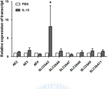
3. Anion exchanger expression and activity
In addition to SLC26A4, various anion exchangers, including AE2, AE3,
were found to be expressed in the HNE cells of the controls. When the cells were treated with IL-13, SLC26A3 was up-regulated (by 8.2 ± 5.4 fold compared to PBS treatment) along with SLC26A4. The other anion exchangers did not respond to IL-13 stimulation (Figure 5). We also examined anion channel activity by measuring the Cl-/HCO3- exchange ratio. The anion channel activity in epithelia from the patients harboring SLC26A4 mutations (DFNB4) (0.12 ± 0.07 ΔpH/min) was not different from that in the controls (0.15 ± 0.06 ΔpH/min) under basal conditions. When the HNE cells from the controls were treated with IL-13 for 7 days, the anion channel activity was significantly increased (0.23 ± 0.17 ΔpH/min, P<0.05), but not in the epithelia from patients with SLC26A4 mutations (0.18 ± 0.06 ΔpH/min) (Figure 6). The molecular and functional data indicate that pendrin is a major anion exchanger that responds to IL-13 and that the intracellular and ASL pH may be regulated by various anion exchangers other than pendrin under basal conditions.
Figure 5. Anion exchager expression in cultured human nasal epithelia from controls. Along with pendrin, various anion exchangers (AE2, AE3 and
AE4) and SLC26A family transporters (SLC26A3, SLC26A6, SLC26A7, SLC26A8, SLC26A9 and SLC26A11) are expressed in nasal epithelia from
controls (n=5). Only SLC26A3 is increased by IL-13 (10 ng/ml). * indicates p<0.05 compared with the PBS-treated cells.
Figure 6. Anion exchange activity in human nasal epithelia cultured from patients with SLC26A4 mutations (DFNB4) and controls. (A) Representative tracings of anion channel activity. (B) Measurements of anion exchanger activity showing significantly increased activity following IL-13 treatment (10 ng/ml) in the controls (n=4) but not in epithelia from patients harboring SLC26A4 mutations (DFNB4, n=5). * indicates p<0.05 compared with the PBS-treated cells.
4. MUC5AC expression and goblet cell differentiation
We also examined the role of pendrin in mucus secretion in the airway epithelia. Quantitative real-time PCR data showed that the expression of
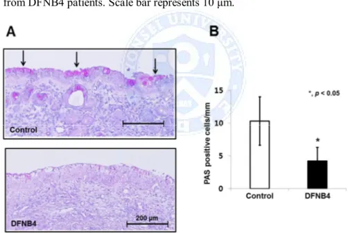
cultured cells from controls. In contrast, MUC5AC expression was comparatively low in cultured cells from DFNB4 patients, and it was not up-regulated by IL-13 treatment (Figure 7A). PAS staining revealed goblet cell hyperplasia in the control HNE cells following IL-13 treatment, but this was not observed in the epithelia from DFNB4 subjects (Figure 7B). Histologic examination of the inferior turbinates also revealed that there was a much lower number of PAS-positive cells (3.7 ± 2.1/mm) in the nasal mucosa from DFNB4 patients compared with that from controls (10.3 ± 3.7/mm) (Figure 8).
Figure 7. MUC5AC expression and goblet cell differentiation in cultured human nasal epithelial cells. (A) Real-time PCR analysis showing up-regulation of MUC5AC by IL-13 (10 ng/ml) in controls (n=3). In contrast,
MUC5AC is weakly expressed and does not respond to IL-13 in epithelia from
patients carrying SLC26A4 mutations (DFNB4, n=3). * indicates p<0.05. (B) PAS staining showing goblet cell hyperplasia in nasal epithelial cells from controls following IL-13 treatment (10 ng/ml) for 7 days, but not in epithelia from DFNB4 patients. Scale bar represents 10 μm.
Figure 8. PAS staining of inferior turbinates. (A) Representative images shows a much lower amount of PAS staining in turbinate tissue from patients harboring SLC26A4 mutations (DFNB4) compared with the tissues from
controls (bars indicate 200 μm). (B) Quantitative analysis of the abundance of PAS-positive cells in 6 DFNB4 and 6 controls. * indicates p<0.05.
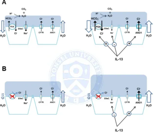
Figure 9. The hypothetical role of pendrin in the regulation of airway surface liquid (ASL). (A) Pendrin regulates ASL volume via Cl- and subsequent water absorption. The secreted HCO3-will evaporate, thus leading to net anion movement into the cell. Under inflammatory conditions, regulated pendrin compensates for the over-secreted fluid caused by up-regulated Cl- channels. (B) In pendrin-deficient airways, ASL thickness is
increased due to the lack of Cl- absorption by pendrin under basal conditions and cannot compensate for the over-accumulated fluid.
IV. DISCUSSION
A recent report13indicates that pendrin is involved in the regulation of ASL thickness in mice. Although the ASL thickness did not differ in cultured tracheal epithelial cells between wild-type and pendrin-deficient mice under basal conditions, the ASL became much thicker in pendrin-deficient mice compared to the cells from wild-type mice after stimulation with an allergic cytokine.13However, pendrin regulates the pH of the ASL but has little effect on fluid secretion in Calu-3 cells, which are human serous airway epithelial cells.24 Therefore, it was unclear whether pendrin regulates ASL volume in airway epithelia in humans. In our data, the expression pattern of ion transporters in airway epithelia was similar between the patients with
SLC26A4 mutations and controls except for the weak expression of CFTR in
patients with SLC26A4 mutations. However, the ASL layer was thicker in
HNE cells from patients with SLC26A4 mutations compared to controls. These data indicate that pendrin is involved in ASL volume regulation even in basal conditions. This finding is not consistent with data from the mouse airway. This discrepancy may stem from species differences in the role of ion transporters in airway epithelia. For instance, the expression patterns of ion channels are quite different between mice and humans as CFTR expression is barely detectable in mouse airway epithelia. Another possible reason for the
conflicting results between reports may be attributable to a methodological problem. Nakagami et al.13 measured ASL thickness 3 min after PBS/dye loading, before ASL thickness had reached a steady state.
The mechanism by which pendrin regulates ASL is puzzling because this electro-neutral transporter cannot generate an osmotic gradient. One possible mechanism underlying this phenomenon is the evaporation of secreted HCO3-, which results in net anion movement into the lumen and subsequent Na+ secretion, via ENaC, to generate osmotic pressure (Figure 9A). Interestingly, the difference of ASL height between normal and pendrin-deficient airway epithelia became much more prominent with IL-13 treatment. IL-13 induces airway fluid secretion and as a result, the ASL thickness is increased. In normal airway epithelial cells, the up-regulation of pendrin (~ 100 fold) following IL-13 treatment compensates for the thickened ASL via Cl -absorption. However, presumably since this compensatory mechanism does not exist in the pendrin mutants, it results in an overaccumulation of ASL (Figure 9B).
Pendrin seems to be not only involved in ASL regulation, but also mucus secretion. Enforced expression of pendrin in NCI-H292 cells or mouse airway epithelia induces the up-regulation of MUC5AC.14 However, pendrin– deficient mice exhibit impaired airway hyperresponsiveness and eosinophilic inflammation, but not mucus production. Our data showed that MUC5AC was
expressed at very low levels in the nasal epithelia from SLC26A4 mutants and was not up-regulated by IL-13 treatment, which contrasts with our findings in the cells of the controls. We also revealed that the goblet cell differentiation in basal condition and hyperplasia induced by IL-13 was minimal in the epithelia from patients with SLC26A4 mutations. Furthermore, we demonstrated that the proportion of goblet cells in the nasal mucosa from patients with SLC26A4 mutation is much lower compared to that in controls. These data all together indicate that pendrin is a major participant in mucous cell differentiation and hyperplasia induced by allergic cytokines in airway epithelia. Although the exact role of pendrin in mucus secretion in the airways is unclear, pendrin may be involved in the release of inflammatory cytokines or may activate transcription factors for the mucin gene, leading to mucus secretion, as suggested by Rose & Voynow.25
An interesting phenomenon is the linkage between the pathogenesis of asthma and pendrin. Pendrin knockout mice show less of an allergic response to OVA stimulation than their wild-type counterparts.13 Pendrin expression is up-regulated after IL-4 stimulation in cultured airway epithelia16and disease tissues, such as those from asthma, COPD, and rhinitis patients.12,18,26 Moreover, patients with pendrin mutations had a tendency to be resistant to asthma.14 Based on our data, we postulate that the increase in ASL thickness observed in the airways of pendrin-deficient patients under basal conditions
may decrease the chance of allergens coming into contact with the airway epithelia, and therefore hinder the initiation of allergic sensitization. Moreover, the dramatic increase in ASL thickness induced by allergic cytokines could further protect the airway epithelia against exposure to allergens in pendrin-deficient airways. Another possible mechanism explaining the lower incidence of asthma in patients harboring pendrin mutations is a defect in mucus secretion, although the exact molecular mechanism underlying this process is unclear.
In summary, we showed that the ASL layer was thicker in human airway epithelial cells from patients with SLC26A4 mutation compared to controls, and this phenomenon was more prominent in allergic conditions. Pendrin seems to be essential in mucin expression and goblet cell development. These findings may explain the low incidence of allergic airway diseases in patients with SLC26A4 mutation. Specific blockers targeting pendrin in the airway epithelium may represent promising candidate drugs for the treatment of allergic airway diseases such as asthma and allergic rhinitis.
V. CONCLUSION
In summary, we showed that the ASL layer was thicker in human airway epithelial cells from patients with SLC26A4 mutation compared to controls, and this phenomenon was more prominent in allergic conditions. Pendrin seems to be essential in mucin expression and goblet cell development. These findings may explain the low incidence of allergic airway diseases in patients with SLC26A4 mutation. Specific blockers targeting pendrin in the airway epithelium may represent promising candidate drugs for the treatment of allergic airway diseases such as asthma and allergic rhinitis.
REFERENCES
1. Mount DB, Romero MF. The SLC26 gene family of multifunctional anion exchangers. Pflügers Archiv 2004;447:710-21.
2. Lacroix L, Mian C, Caillou B, Talbot M, Filetti S, Schlumberger M, et al. Na (+)/I (-) symporter and Pendred syndrome gene and protein expressions in human extra-thyroidal tissues. European journal of endocrinology 2001;144:297-302.
3. Royaux IE, Suzuki K, Mori A, Katoh R, Everett LA, Kohn LD, et al. Pendrin, the protein encoded by the Pendred syndrome gene (PDS), is an apical porter of iodide in the thyroid and is regulated by thyroglobulin in FRTL-5 cells. Endocrinology 2000;141:839-45. 4. Royaux IE, Wall SM, Karniski LP, Everett LA, Suzuki K, Knepper
MA, et al. Pendrin, encoded by the Pendred syndrome gene, resides in the apical region of renal intercalated cells and mediates bicarbonate secretion. Proceedings of the National Academy of Sciences 2001;98:4221-6.
5. Scott DA, Wang R, Kreman TM, Sheffield VC, Karniski LP. The Pendred syndrome gene encodes a chloride-iodide transport protein. Nature genetics 1999;21:440-3.
6. Albert S, Blons H, Jonard L, Feldmann D, Chauvin P, Loundon N, et al. SLC26A4 gene is frequently involved in nonsyndromic hearing impairment with enlarged vestibular aqueduct in Caucasian populations. European journal of human genetics 2006;14:773-9. 7. Nakaya K, Harbidge DG, Wangemann P, Schultz BD, Green ED, Wall
SM, et al. Lack of pendrin HCO3− transport elevates vestibular endolymphatic [Ca2+] by inhibition of acid-sensitive TRPV5 and TRPV6 channels. American Journal of Physiology-Renal Physiology 2007;292:F1314-F21.
8. Yang J-J, Tsai C-C, Hsu H-M, Shiao J-Y, Su C-C, Li S-Y. Hearing loss associated with enlarged vestibular aqueduct and Mondini dysplasia is caused by splice-site mutation in the PDS gene. Hearing research 2005;199:22-30.
9. Morgans M, Trotter W. ASSOCIATION OF CONGENITAL DEAFNESS WITH GOITRE THE NATURE OF THE THYROID DEFECT. The Lancet 1958;271:607-9.
10. Reardon W, Coffey R, Chowdhury T, Grossman A, Jan H, Britton K, et al. Prevalence, age of onset, and natural history of thyroid disease in Pendred syndrome. Journal of medical genetics 1999;36:595-8. 11. Di Valentin E, Crahay C, Garbacki N, Hennuy B, Guéders M, Noël A,
and chronic asthma. American Journal of Physiology-Lung Cellular and Molecular Physiology 2009;296:L185-L97.
12. Kuperman DA, Lewis CC, Woodruff PG, Rodriguez MW, Yang YH, Dolganov GM, et al. Dissecting asthma using focused transgenic modeling and functional genomics. Journal of Allergy and Clinical Immunology 2005;116:305-11.
13. Nakagami Y, Favoreto S, Zhen G, Park S-W, Nguyenvu LT, Kuperman DA, et al. The epithelial anion transporter pendrin is induced by allergy and rhinovirus infection, regulates airway surface liquid, and increases airway reactivity and inflammation in an asthma model. The Journal of Immunology 2008;181:2203-10.
14. Nakao I, Kanaji S, Ohta S, Matsushita H, Arima K, Yuyama N, et al. Identification of pendrin as a common mediator for mucus production in bronchial asthma and chronic obstructive pulmonary disease. The Journal of Immunology 2008;180:6262-9.
15. Nofziger C, Vezzoli V, Dossena S, Schönherr T, Studnicka J, Nofziger J, et al. STAT6 links IL-4/IL-13 stimulation with pendrin expression in asthma and chronic obstructive pulmonary disease. Clinical Pharmacology & Therapeutics 2011;90:399-405.
16. Pedemonte N, Caci E, Sondo E, Caputo A, Rhoden K, Pfeffer U, et al. Thiocyanate transport in resting and IL-4-stimulated human bronchial
epithelial cells: role of pendrin and anion channels. The Journal of Immunology 2007;178:5144-53.
17. Izuhara K, Ohta S, Shiraishi H, Suzuki S, Taniguchi K, Toda S, et al. The mechanism of mucus production in bronchial asthma. Current medicinal chemistry 2009;16:2867-75.
18. Lewis CC, Yang JYH, Huang X, Banerjee SK, Blackburn MR, Baluk P, et al. Disease-specific gene expression profiling in multiple models of lung disease. American journal of respiratory and critical care medicine 2008;177:376-87.
19. Madeo AC, Manichaikul A, Pryor SP, Griffith AJ. Do mutations of the Pendred syndrome gene, SLC26A4, confer resistance to asthma and hypertension? Journal of medical genetics 2009;46:405-6.
20. Boucher R. Regulation of airway surface liquid volume by human airway epithelia. Pfluegers Archiv 2003;445:495-8.
21. Tarran R, Button B, Picher M, Paradiso AM, Ribeiro CM, Lazarowski ER, et al. Normal and cystic fibrosis airway surface liquid homeostasis The effects of phasic shear stress and viral infections. Journal of Biological Chemistry 2005;280:35751-9.
22. Yoon J-H, Kim K-S, Kim HU, Linton JA, Lee J-G. Effects of TNF-a and IL-1 ß on Mucin, Lysozyme, IL-6 and IL-8 in passage-2 Normal
Human Nasal Epithelial Cells. Acta oto-laryngologica 1999;119:905-10.
23. Yoon JS, Park H-J, Yoo S-Y, Namkung W, Jo MJ, Koo SK, et al. Heterogeneity in the processing defect of SLC26A4 mutants. Journal of medical genetics 2008;45:411-9.
24. Garnett JP, Hickman E, Burrows R, Hegyi P, Tiszlavicz L, Cuthbert AW, et al. Novel role for pendrin in orchestrating bicarbonate secretion in cystic fibrosis transmembrane conductance regulator (CFTR)-expressing airway serous cells. Journal of Biological Chemistry 2011;286:41069-82.
25. Rose MC, Voynow JA. Respiratory tract mucin genes and mucin glycoproteins in health and disease. Physiological reviews 2006;86:245-78.
26. Ishida A, Ohta N, Suzuki Y, Kakehata S, Okubo K, Ikeda H, et al. Expression of pendrin and periostin in allergic rhinitis and chronic rhinosinusitis. Allergology International 2012;61:589-95.
ABSTRACT (in Korean)
펜드린 결핍 인간 기도 상피세포에서
증가된 기도 표면 액체와 감소된 mucin 발현
<지도교수 최재영>
연세대학교 대학원 의과학과
이현재
펜드린은 음이온 교환 단백이며 이것의 돌연변이는 난청을 유발한다고 알려져 있다. 그러나 최근 기도 표면 액체의 양과 mucin 발현의 조절에 펜드린의 역할을 밝혀내기 위한 펜드린의 발현과 천식 같은 기도 질환의 관한 연구가 진행되었다. 16 명의 펜드린 돌연변이를 가진 난청 환자와 17 명의 돌연변이를 가지고 있지 않은 환자로부터 얻은 인간 코 상피세포 배양하였다. 세포에 Interleukin 13 을 처리하여 점액 과분비를 유도하였다. 기도 표면 액체의 높이 측정과 실시간 중합효소 연쇄 반응을 이용하여 다양한 이온 운반체와 MUC5AC 를 발현량을 확인 하였다. pH 민감성 형광 프로브를 이용하여 음이온 교환 능력을 측정하였다. 배양된 세포와 하위 갑개 조직에서 과아이오딘산 시프 염색을 시행하였다. 펜드린 돌연변이 세포의 기도 표면 액체는 실험군의기도 표면 액체보다 더 두꺼웠고, 이러한 차이는 Interleukin 13 을 처리하였을 때 현저하게 나타났다. 실험군에서 Interleukin 13 처리 후 음이온 교환 능력이 증가한 반면 돌연변이 군에서는 차이를 보이지 않았다. 배상세포 변질형성은 실험군에서 나타났지만 돌연변이에서는 나타나지 않았다. 게다가 과아이오딘산 시프 염색에 양성인 부분은 돌연변이 군에서 더 적게 나타났다. 펜드린은 기도 표면 액체의 양과 mucin 발현 조절에 중요한 역할을 한다. 기도에 존재하는 펜드린을 표적으로 하는 특이 차단제는 알레르기성의 기도 질환 치료제로의 가능성을 가질 것이다. ---핵심되는 말: SLC26A4, 천식, 음이온 통로
PUBLICATION LIST
1. Cho H-J, Lee HJ, Kim SC, Kim K, Kim YS, Kim C-H, et al. Protease-activated receptor 2–dependent fluid secretion from airway submucosal glands by house dust mite extract. Journal of Allergy and Clinical Immunology 2012;129:529-35. e5.
2. Lee HJ, Yang Y-M, Kim K, Shin DM, Yoon J-H, Cho H-J, et al. Protease-activated receptor 2 mediates mucus secretion in the airway submucosal gland. PloS one 2012;7:e43188.
3. Lee HJ, Yang WS, Park HW, Choi HS, Kim SH, Kim JY, et al. Expression of Anion Exchangers in Cultured Human Endolymphatic Sac Epithelia. Otology & Neurotology 2012;33:1664-71.
4. Lee H, Jung J, Shin J, Song M, Kim S, Lee JH, et al. Correlation between genotype and phenotype in patients with bi‐allelic SLC26A4 mutations. Clinical genetics 2014;86:270-5.